
Joined June 2019
- Tweets 114
- Following 41
- Followers 100
- Likes 53
48 Photos and videos


Pinned Tweet

2 Jun 2020
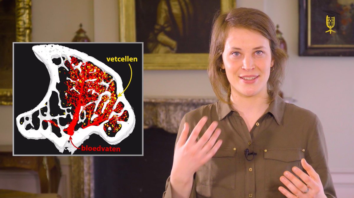
@Sarah_Vgb, one of our PhD students, did a fantastic job in explaining what contrast-enhanced CT (CE-CT) is all about! Watch the video below to learn the basics of contrast agents and their use in CE-CT 🦴

Wat hebben beenmerg, de lever & een tumor gemeen? Ze bestaan uit zgn 'zacht' #weefsel dat zich niet gemakkelijk via CT-scan laat visualiseren. @Sarah_Vgb @KU_Leuven @UCLouvain_be ontwikkelt nieuwe #contraststoffen om zacht weefsel 'in beeld te brengen' 🎥👉wetenschapuitgedokterd.be/we…

2
26 Aug 2025
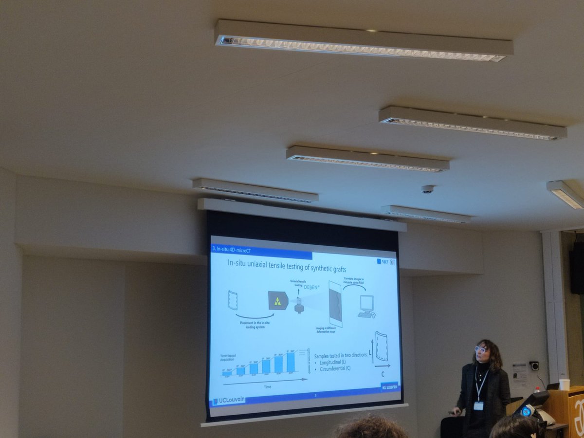
A new article co-authored by members of the Contrast Team (Maïté Pétré and @GreetKerckhofs) unravelling the link between aortic microstructure and mechanical behaviour using CECT for more flexible modelling
doi.org/10.1016/j.compbiomed…
1
1
62
14 Jul 2025
A new article co-authored by members of the Contrast Team (Tim Balcaen and @GreetKerckhofs) on yet another application for CECT: imaging of biofilm for water treatment. doi.org/10.1016/j.watres.202…
1
1
46
18 Apr 2025
A new review article authored by members of the Contrast Team (Lara Mazy and @GreetKerckhofs) on combined in situ testing and X-ray microfocus computed tomography for biological application.
doi.org/10.1016/j.tmater.202…
1
3
118
14 Apr 2025
A new article co-authored by members of the Contrast Team (Delia Hoffmann, Tim Balcaen, Sarah Vangrunderbeeck, Arne Maes, Grzegorz Pyka and @GreetKerckhofs) on 3D histology of Soft Tissues by Contrast-Enhanced X-Ray Microfocus Computed Tomography.
doi.org/10.1093/mam/ozaf013
1
4
80
14 Feb 2025
A new article co-authored by members of the ContrasTTeam (Pierre Schneidewind, Grzegorz Pyka, and @GreetKerckhofs ) on Unravelling fascia lata intrinsic vascular architecture using microfocus X-ray Computed Tomography.sciencedirect.com/science/ar…
1
1
54
3 Feb 2025
A new review article by members of the contrasTTeam (Tim Balcaen, Sarah Vangrunderbeeck, @GreetKerckhofs) on contrast-enhancing staining agents (CESAs) for ex vivo Contrast-Enhanced Computed Tomography. sciencedirect.com/science/ar…
1
82
22 Jan 2025
Another article, co-authored by a member of the Contrast Team (@GreetKerckhofs), where CECT was used to analyze the vasculature in the bone marrow of murine bones. sciencedirect.com/science/ar…
1
103
10 Jan 2025
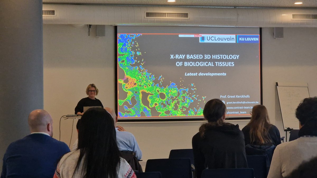
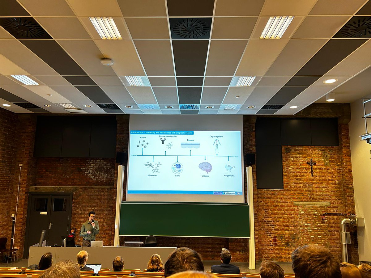
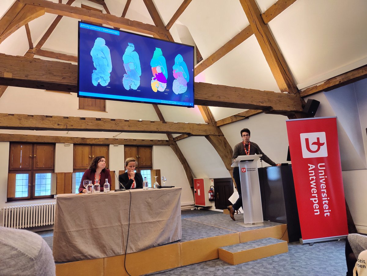
During our annual symposium, fascinating seminars on X-ray based 3D histology of tissues were presented. Various types of biomedical research enabled by the X-ray microfocus computed tomography (microCT) machine were showcased. The event concluded with a visit of our lab.
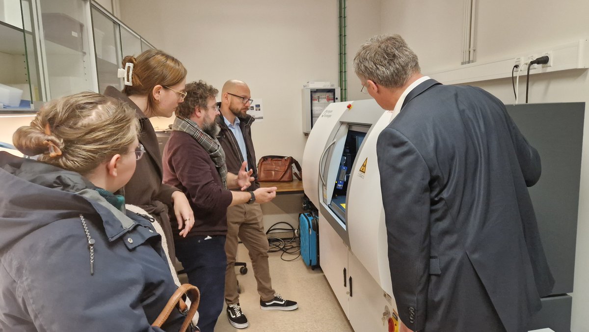

1
38
2 Dec 2024
Another publication with author and co-authors from the Contrast Team (Lisa Leyssens, Tim Balcaen, @GreetKerckhofs ) sciencedirect.com/science/ar…
1
120
19 Nov 2024
Another publication with co-authors from the Contrast Team exploring the biological effects of copper alloying in Zn-based biodegradable arterial implants. sciencedirect.com/science/ar…
1
1
64
12 Aug 2024
Another publication from the Contrast Team (Lisa Leyssens, Grzegorz Pyka, @GreetKerckhofs ), Exploring the biodegradability of metallic intravascular stent materials using microCT. dx.doi.org/10.1002/jbm.b.354…
3
160
10 Jul 2024
Another publication, co-authored by members of the Contrast Team (Maïté Pétré, Tim Balcaen, Pierre Schneidewind, Lara Mazy, Grzegorz Pyka,@GreetKerckhofs), assessing the effect on microstructural and mechanical properties of six different staining agents. sciencedirect.com/science/ar…
3
157
1 Jul 2024
A new publication from the ContrasT Team (Tim Balcaen, @GreetKerckhofs ), where several contrast-enhancing staining agents have been explored for studying adipose tissue through contrast-enhanced computed tomography.jlr.org/article/S0022-2275%2…
1
1
88
19 Jun 2024
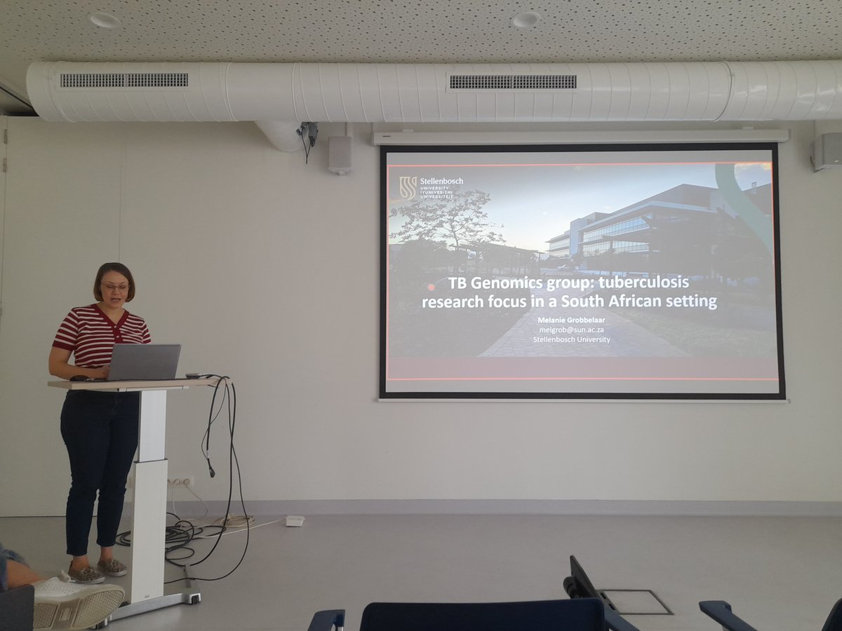
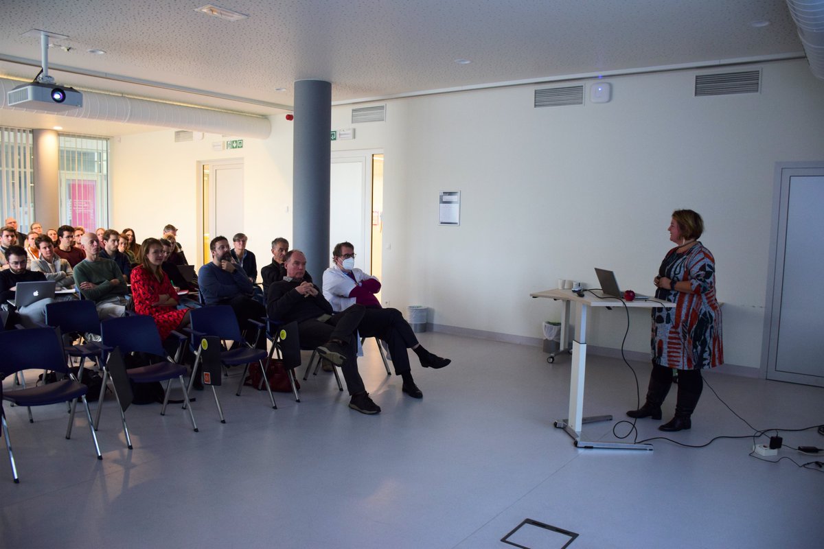
This morning, Dr. Melanie Grobbelaar presented a nice seminar on tuberculosis research focused on a South African setting, which is conducted within the Tuberculosis Genomics (TB Genomics) research group.
1
164
17 May 2024
And yet another publication with co-authors from the ContrasT Team (@GreetKerckhofs , Arne Maes) showing how X-ray-based 3D histology on human biopsies could reveal blood vessel ingrowth in the donor fascia.
journals.lww.com/transplanta…
1
2
109
13 May 2024
Another publication, co-authored by members of the Contrast team (@GreetKerckhofs, Grzegorz Pyka), focusing on the use of micro-CT for imaging large ex-vivo human samples in anatomical and forensic research : sciencedirect.com/science/ar…
1
4
127
2 Apr 2024
Another publication featuring a co-author from the ContrasT Team (@GreetKerckhofs ) on the use of CECT imaging to measure cranial development 🎉 disq.us/t/4nmahjm
1
5
142
2 Apr 2024
Another publication featuring a co-author from the ContrasT Team (@GreetKerckhofs ) on the use of the staining Hf-WD POM to visualize bone marrow vasculature with Contrast-Enhanced Computer Tomography. 🥳disq.us/t/4ndfzi6
1
3
106
21 Feb 2024
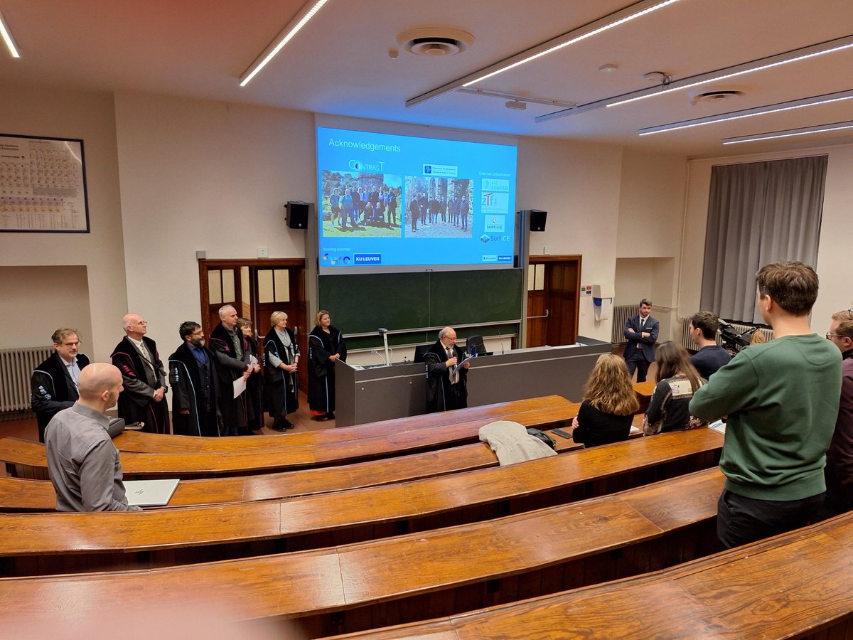
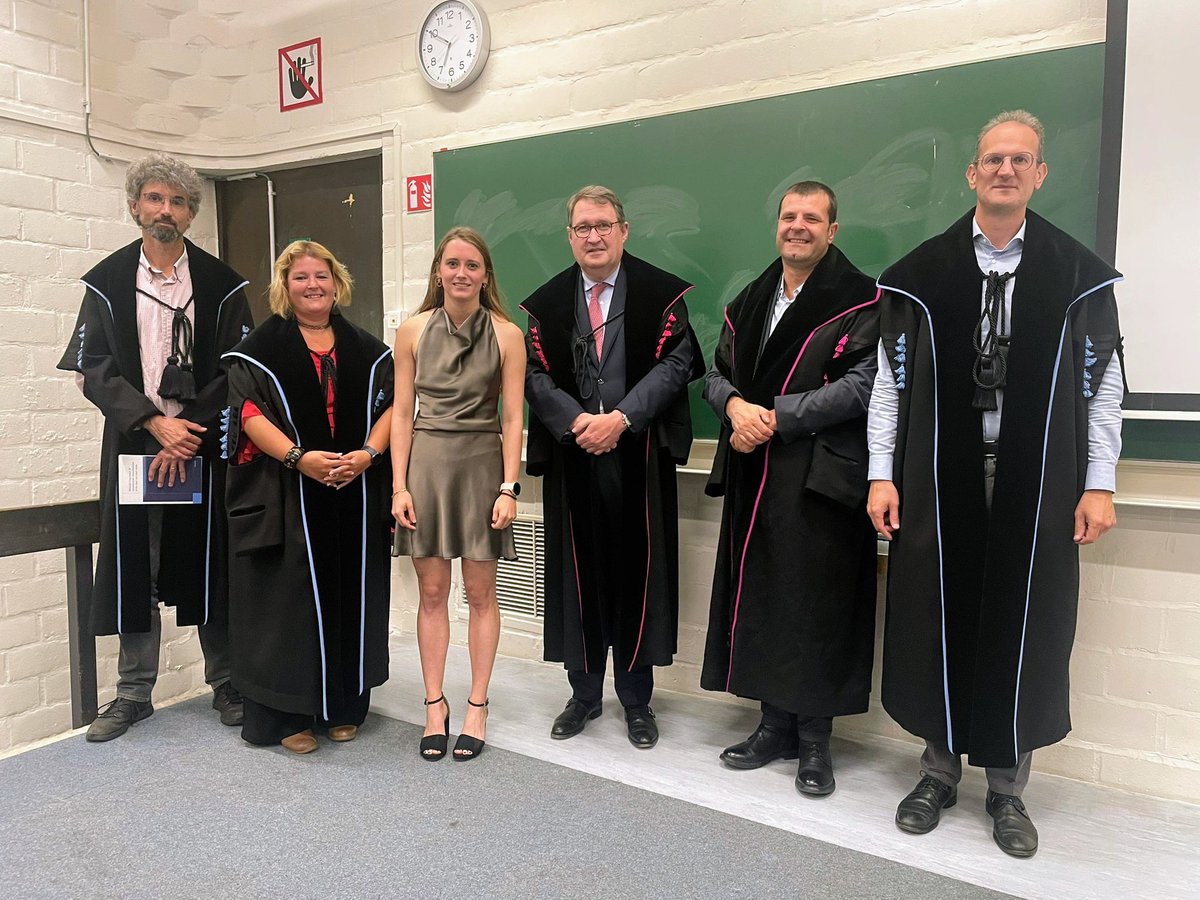
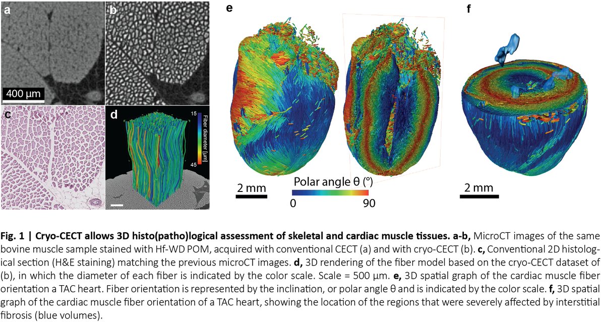
Yesterday, Arne Maes successfully defended his PhD thesis, introducing a novel technique called cryogenic contrast-enhanced microCT,
Congratulations, Dr. Maes!
1
4
397
2 Feb 2024
Another great publication featuring co-authors from the ContrasT Team 🎉 sciencedirect.com/science/ar…
1
4
158