
Jun 12
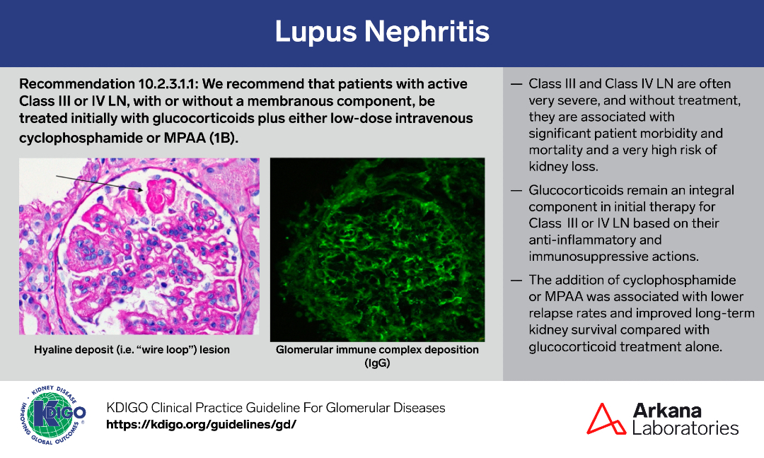
A sample from the pathology teaching slide set on Lupus Nephritis, jointly prepared by Arkana Laboratories and @goKDIGO. KDIGO.org/guidelines/gd/
#KDIGO #pathology #renal #kidneypath

4
7
882
Jun 11
#KidneyQuiz In patients with glomerulonephritis (GN) due to infective endocarditis (IE) what percentage can have positive ANCA antibodies?
#nephX #kidneypath #pathX #nephtwitter
0%
A. 0%
50%
B. 5-10%
50%
C. 10-20%
0%
D. 20 -30%
14 votes • Final results
1
4
1,323
Jun 11
Here is today’s #eyeSCANdy! Macula densa basement membrane tunnels.
Photo courtesy of Dr. Stephen Bonsib. #renal #pathology #kidneypath
2
299
Angel Panizo MD, PhD 🇪🇸 🔬 retweeted

4
10
36
3,667
This photomicrograph shows a high power view of the renal interstitium with histiocytic inflammation and numerous histocytes with PAS positive material within their cytoplasm. Within the center of the field is a small, target shaped structure—a Michalis-Gutmann (MG) body. These findings are consistent with Malakoplakia which often forms mass lesions mimicking a neoplasm. Malakoplakia results from dysregulated lysosomal breakdown and processing of bacteria (typically E. coli) with resulting foamy macrophages with PAS positive intracellular accumulations. Partially digested bacteria can also form a nidus for calcium and iron deposition, thus MG-body formation. Malakoplakia is an unusual finding and is not expected in an otherwise healthy person. It is frequently associated with an altered immune status such as HIV/AIDS, underlying malignancy, or immunosuppressive therapy.
#renalpath #kidneypath #pathology #renal #pathtwitter

3
11
1,245
The differential diagnosis of kidney injury in CLL/SLL patients.
There are many causes of decreased kidney function in patients with chronic lymphocytic leukemia/small lymphocytic lymphoma (CLL/SLL), and a renal biopsy may provide a definitive diagnosis.
Prerenal etiologies include poor oral intake, sepsis, and heart failure. Intrarenal causes include diseases of the glomeruli (e.g. minimal change disease, MPGN pattern glomerulopathy, membranous glomerulopathy), tubules (e.g. toxic or ischemic tubular injury/necrosis, light chain cast nephropathy), interstitium (e.g. acute interstitial nephritis caused by infection or drugs), and vasculature (e.g. TMA). The biopsy shown in this image illustrates infiltration of the kidney parenchyma by the lymphoid neoplasm (characteristic immunophenotypic studies not shown). Postrenal causes include obstruction (e.g. extrarenal tumor mass, lymphadenopathy) and uric acid nephropathy related to tumor lysis syndrome, among others.
Reference
Rimda Wanchoo, et al. Renal involvement in chronic lymphocytic leukemia. Clinical Kidney Journal, sfy026, doi.org/10.1093/ckj/sfy026 (Advance article).
#TeachingPoints #kidneypath #renal #pathology

6
21
998
Here is today’s #eyeSCANdy! Glomerulus obliterated by a fibrous crescent.
Photo courtesy of Dr. Stephen Bonsib. #renal #pathology #kidneypath
2
12
549
#KidneyQuiz The histopathologic changes seen in FSGS secondary to adaptive response can be:
A) Glomerulomegaly
B) Perihilar adhesions
C) Segmental FPE
D) All of the above
#nephX #kidneypath #pathX #nephtwitter
0%
A
0%
B
0%
C
100%
D
3 votes • Final results
4
979
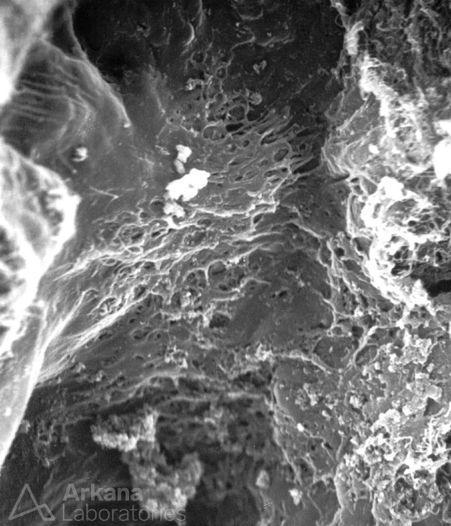
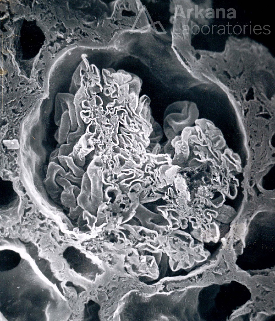
A sample from the pathology teaching slide set on Lupus Nephritis, jointly prepared by Arkana Laboratories and @goKDIGO. KDIGO.org/guidelines/gd/
#KDIGO #pathology #renal #kidneypath

3
7
321
May 29
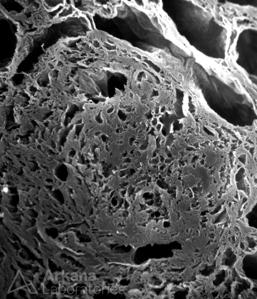
A sample from the pathology teaching slide set on Lupus Nephritis, jointly prepared by Arkana Laboratories and @goKDIGO. KDIGO.org/guidelines/gd/
#KDIGO #pathology #renal #kidneypath
3
12
1,485
May 28
Here is today’s #eyeSCANdy! Acellular scanning EM showing a glomerulus in cross-section though the macular densa and showing the origin of the proximal tubule from Bowman’s capsule.
Photo courtesy of Dr. Stephen Bonsib. #renal #pathology #kidneypath
2
10
442
May 27
The image shows extensive effacement of the normal tubulointerstitial architecture and replacement by a dense, plasma cell rich infiltrate. While this finding is consistent with a chronic, active tubulointerstitial nephritis the image also shows a unique pattern of fibrosis. Within the inflammation, bands of collagen are present that create the effect of a nesting pattern or what some have argued is a storiform or ‘birds-eye maple’ pattern. This unique pattern of fibrosis is characteristic for IgG4-related disease as is true in this case (see IgG4 immunohistochemical stain below). Of note, IgG4 is a patchy disease which can often be mass forming. This is important to remember as small biopsy samples often show near total fibrosis. However, this finding may not be representative of the kidneys as a whole and these patients often respond well to treatment.
#renalpath #kidneypath #pathology #renal #pathtwitter
May 26
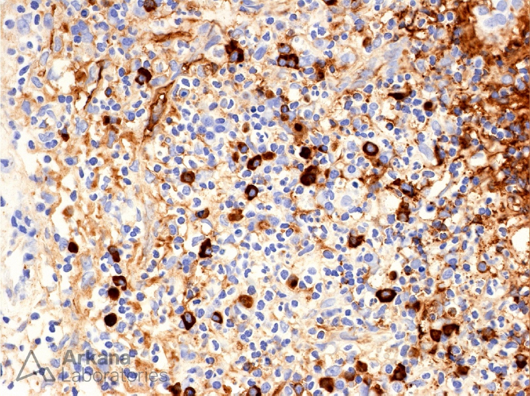

3
10
1,240
May 27
One of the many patterns of tubular atrophy in the kidney is the aptly named “thyroidization” pattern because of its resemblance to normal thyroid gland follicles. The dilated tubules contain abundant protein, resembling thyroid colloid, which is surrounded by flattened epithelial cells. This pattern of tubular atrophy is non-specific, although it is often more frequently encountered in the setting of chronic pyelonephritis and reflux nephropathy.
Reference
Lusco MA, Fogo AB, Najafian B, Alpers CE. AJKD Atlas of Renal Pathology: Tubular Atrophy. Am J Kidney Dis. 2016 Jun; 67(6):e33-4.
#TeachingPoints #kidneypath #renal #pathology

12
32
1,295