Mr Boundless retweeted

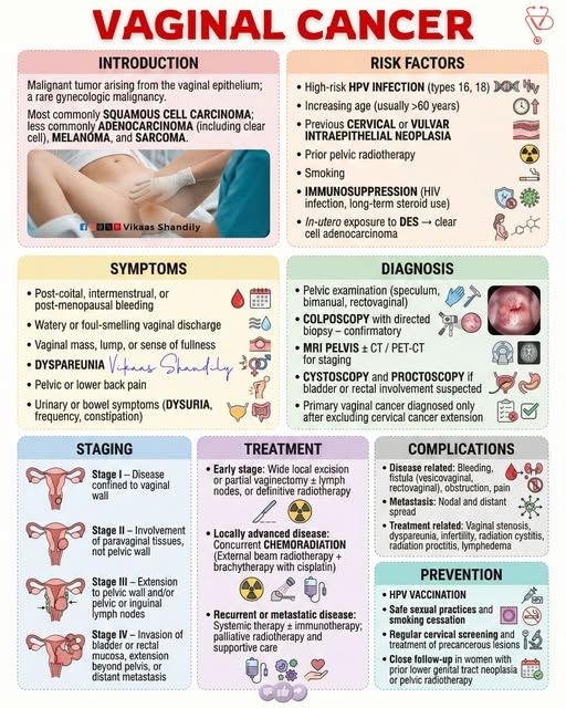
VAGINAL CANCER
What is it?
A rare cancer that begins in the vaginal lining, most commonly Squamous Cell Carcinoma.
Risk Factors
• HPV infection (types 16 & 18)
• Age >60 years
• Smoking
• Previous cervical/vulvar disease
• Immunosuppression
• Prior pelvic radiotherapy
Symptoms
• Abnormal vaginal bleeding
• Unusual discharge
• Vaginal lump/fullness
• Pain during sex
• Pelvic pain
• Urinary or bowel changes
Diagnosis
• Pelvic exam
• Biopsy (confirmatory)
• MRI/CT for staging
Treatment
• Early stage: Surgery or radiotherapy
• Advanced stage: Chemoradiation
• Recurrent cases: Systemic treatment & supportive care
Prevention
• HPV vaccination
• Safe sexual practices
• Stop smoking
• Regular screening
Early detection improves treatment outcomes.
2
4
6
88

Akeso and Summit’s ivonescimab extends survival in squamous cell lung cancer monkeylink.co/209c47 via @statnews #healthcare #lungcancer
1

hahahahaha that's awesome. not to be confused with squamous which is a type of skin cell. or squeamish.
1
2
217
Cristina Zannori retweeted

🎯 NRG1 Fusions in NSCLC.
In a real-world cohort of 1,536 non-squamous NSCLC cases evaluated with paired DNA/RNA NGS, NRG1 fusions were detected in 0.8%, higher than historical estimates.
Most cases were invasive mucinous adenocarcinomas, PD-L1 negative and TMB-low. Metastatic patients without access to targeted therapy had a median OS of only 7.9 months.
RNA sequencing matters.
📖 Lung Cancer
DOI 👉🏻 doi.org/10.1016/j.lungcan.20…
#CánCare #NSCLC #lcsm #NRG1 #thoraciconcology #precisiononcology


1
4
8
647

American sunscreen just got its first real upgrade in over two decades and it's long overdue.
The FDA has approved bemotrizinol (BEMT), the first new sunscreen ingredient cleared in the U.S. since 1999. While Europe and Asia have used it for 20 years, U.S. regulations, which classify sunscreen as an over-the-counter drug rather than a cosmetic, slowed its arrival here significantly. Europe currently allows roughly 30 sunscreen filters. The U.S. allowed just over a dozen, until now.
BEMT isn't just "new." It offers broader UV protection (covering both UVA and UVB), lasts longer on skin, causes less irritation, and critically, doesn't leave that greasy white cast Americans hate. It also sits on top of skin with lower absorption than older chemical filters.
Dermatologists say daily sunscreen cuts melanoma risk by 50% and squamous cell carcinoma by 40%. Yet only 13% of American adults use it daily. Better formulas might actually change that.
Products could hit shelves as early as this summer.
#TheLolgic #Sunscreen #Bemotrizinol

6

🎧 #PressRelease:
"A randomized double-blind placebo-controlled phase I/II clinical trial of a human papillomavirus therapeutic vaccine, PepCan, for reducing head and neck squamous cell carcinoma recurrence"
soundcloud.com/oncotarget/hp…
#cancer #podcast #audio #SoundCloud #press
16

**Cannabidiol (CBD) Exhibits Dose-Dependent Anti-Cancer Effects Across Multiple Cancer Types**
Research demonstrates that cannabidiol (CBD) consistently produces dose-dependent reductions in cancer cell viability, proliferation, migration, invasion, and tumor growth while inducing apoptosis and cell cycle arrest in various preclinical models. Below is a compilation of key studies highlighting these effects across different cancers, with specific dose ranges and outcomes.
### Breast Cancer
**CBD Induces Concentration-Dependent Apoptosis and Reduces Viability in Breast Cancer Cell Lines**
CBD treatment (0–10 μM for 24 hours) significantly decreased cell viability in both estrogen receptor-positive (e.g., MCF-7, T-47D) and triple-negative (e.g., MDA-MB-231) breast cancer lines in a concentration-dependent manner. It triggered apoptosis (measured by Annexin V staining) and inhibited survival pathways independently of CB1/CB2 receptors at lower doses, with partial CB2 involvement at higher concentrations. Higher doses enhanced pro-apoptotic proteins like p53, Caspase-3, and Bax.
**CBD and THC Mixture Shows Dose- and Time-Dependent Anti-Proliferative and Pro-Apoptotic Effects in Breast Cancer**
In MDA-MB-231 and MCF-7 cells, CBD/THC (3:1 ratio, 12.5–17.5 μg/mL) inhibited proliferation dose- and time-dependently over 24–48 hours, reducing viability by up to 86% at higher doses. It increased apoptotic cells (Annexin V/PI staining) and altered cell cycle progression, with stronger effects in triple-negative lines.
### Colorectal Cancer
**CBD Represses Viability and Induces G1 Arrest and Apoptosis in Colorectal Cancer Cells**
CBD (0–40 μM for 24–48 hours) reduced viability in multiple human colorectal cancer lines (e.g., HCT116, SW480) in a dose-dependent manner, with IC50 values ranging from 4.7–20 μM. It caused G1-phase cell cycle arrest, elevated apoptosis markers, and endoplasmic reticulum stress.
**CBD Reduces Viability and Enhances Apoptosis in p53-Dependent Colorectal Cancer Models**
In HCT116 (p53 wild-type and knockout) and other lines, CBD (5–20 μM for 24–48 hours) lowered viability dose-dependently (IC50 7.9–24.3 μM). Sensitivity was higher in p53 wild-type cells, with increased ROS, mitochondrial disruption, and caspase activation at higher doses.
### Glioma/Glioblastoma
**CBD Is Cytotoxic and Perturbs Mitochondrial Function in Glioma Cells**
CBD (0–20 μg/mL for 96 hours) inhibited proliferation and induced cytotoxicity in human (U87MG, U373MG) and canine glioma lines in a dose-dependent manner (IC50 4.9–8.2 μg/mL). It reduced oxygen consumption and ATP production dose-dependently within 2 hours, leading to swollen mitochondria and apoptosis (rescued by apoptosis inhibitors).
**CBD Suppresses Glioma Cell Viability in Combination Contexts**
CBD and BCNU each produced dose-dependent suppression of T98G glioma cell viability, with CBD enhancing overall anti-tumor activity through apoptotic pathways.
### Prostate Cancer
**CBD Decreases Viability and Induces Apoptotic Cell Death in Prostate Cancer Cells**
In PC3 cells, CBD (0.03–10 μM for 48 hours) reduced viability dose-dependently (up to 37% at higher doses) and activated caspase-3/7 pathways, increasing DNA fragmentation, ROS, and pro-apoptotic Bax while altering mitochondrial potential.
### Lung Cancer and Other Solid Tumors
**CBD Inhibits Long-Term Survival in Multiple Cancer Cell Lines Including Lung Carcinoma**
CBD showed dose-dependent killing in large cell lung carcinoma (H460, IC50 8.47 μM), metastatic breast (MDA-MB-231, IC50 8.35 μM), melanoma (A375, IC50 2.45 μM), cervical carcinoma, and osteosarcoma lines after 7–10 days. Apoptosis was confirmed via PARP cleavage, with ER stress as a key mechanism.
**CBD Reduces Proliferation, Migration, and Invasion in Head and Neck Squamous Cell Carcinoma**
CBD treatment decreased HNSCC cell viability, migration, and invasion dose- and t
1
14
Saint v. Sinner 🦬 retweeted

"Most impressive of all is Ivermectin Cream will completely clear skin Cancers...Basal Cell Carcinoma, Squamous Cell Carcinoma & Melanoma will heal & fall off."
"Topical Ivermectin will heal any inflammatory or Autoimmune skin condition including Rosacea, Cystic Acne & Eczema."
~Dr. William Makis, radiologist, oncologist & cancer researcher.
Ivermectin has anti-inflammatory, anti-viral, anti-bacterial & anti-tumor properties.
Ivermectin has amazing topical applications...skin cancers disappearing after a few weeks of applying it twice a day...the skin cancer literally falls off.
People with the worst type of cystic acne are completely clearing that lifelong painful debilitating condition.
Ivermectin is a pretty fascinating veritable wonder drug. It’s primarily known as a broad-spectrum antiparasitic agent with multiple mechanisms of action.
The story of its discovery, in the 1960s when Satoshi Ōmura stumbled upon a unique soil bacteria. This bacteria produced something called avermectin, & ivermectin is essentially a synthetic derivative of this component.
Ivermectin Cream Treats These Dermatology Conditions:
Rosacea
Eczema
Psoriasis
Facial Mites
Scabies
Demodex Skin Mites
Perioral Dermatitis
Hookworms
Lice
Basal Cell Carcinoma
Squamous Cell Carcinoma
Melanoma
Moles
Warts
Skin Tags
Ringworm
Candida
Athlete's Foot
Purchasing Topical Ivermectin Cream In 1 of 2 Ways:
#1: Doctor Rx Prescription for 1% cream...pharmacy filled & purchased after doctor visit or diagnosis.
#2: Over The Counter private purchase of 1.87% cream at any veterinary, pet or farm supply store for immediate use. Available for immediate purchase online on sites like Amazon & Major online pet sites. Most creams & pastes sell for under $10 & are ready for immediate use without a doctor visit, copay or pharmacy visit.
Dosage For Over The Counter Private Purchase:
Twice per day, massage in a pea sized/pencil eraser sized amount onto affected area or lesion.
264
4,429
12,346
407,497

Online now in GIE’s Articles in Press: "Long-term outcome after endoscopic submucosal dissection for entire circumferential cT1aN0M0 esophageal squamous cell carcinoma" by Atsushi Inaba et al. giejournal.org/article/S0016…
#GITwitter

1
189
🎥 #OncotargetShort:
"A randomized double-blind placebo-controlled phase I/II clinical trial of a human papillomavirus therapeutic vaccine, PepCan, for reducing head and neck squamous cell carcinoma recurrence" @UAMS_COM
youtube.com/watch?v=oh0MNrrP…
#cancer #HPV #HNSCC #openaccess
40

New Research: Unusual denture-associated oral squamous papilloma with koilocytosis: a case report and literature review frontiersin.org/articles/10.… #FrontiersIn #DentalMedicine
5
Bob Willhite retweeted

Jun 14
I had a squamous cell cancer erupt on my right jawline three weeks ago. It was 8mm in diameter and 4mm high by the end of the week (pic on L). I got some “horse paste” at a local equine center and one week later (pic on R) it’s almost gone.
8 years ago, I had a nearly identical one erupt on the side of my nose. I had Mohs surgery and now look like a former NHLer with the scar.
My brother, who got jabbed, has been diagnosed w/ stage 4 turbo prostate cancer. He’s undergoing chemo and surgery. He won’t even read the research. My heart is broken. Please pray for him.

4
13
41
1,053

STUDY FINDS SUNSCREEN USE LINKED TO DRAMATICALLY HIGHER RISK OF MULTIPLE SKIN CANCERS A UK Biobank analysis of 470,000 people found sunscreen users faced significantly higher risk of: - MELANOMA: 292% - BASAL CELL CARCINOMA: 140% - SQUAMOUS CELL CARCINOMA: 126% This is what happens when you slather rapidly absorbed hormone disrupting chemicals all over your body while blocking vitamin D — one of the body’s key defenses against cancer. These dramatic cancer signals remained even after accounting for major skin cancer risk factors: age, sex, skin type, tanning ability, sunburn history, sunlamp use, and time spent outdoors.

1
2
507

Knockdown of IGF2BP2 Inhibits THBS1 in Regulating the Progression of Oral Squamous Cell Carcinoma: An Integrative Analysis
dl.begellhouse.com/journals/…
#OralCancer #OSCC #EpigeneticsResearch
1
🔆 #PaperSpotlight:
"A randomized double-blind placebo-controlled phase I/II clinical trial of a human papillomavirus therapeutic vaccine, PepCan, for reducing head and neck squamous cell carcinoma recurrence"
#PressRelease ⬇️
oncotarget.net/2026/06/15/hp…
#cancer #press #oa
1
23