
CEMAS is the center at The Ohio State University that breaks through the current limitations in medicine, environmental science, energy materials and beyond.
Joined April 2013
- Tweets 832
- Following 129
- Followers 705
- Likes 476
224 Photos and videos


New Ohio State study shows how a protein orchestrates step‑by‑step assembly of a gene‑regulation complex. Using cryo‑EM at CEMAS, researchers reveal how cells control gene activation, unlocking a long‑standing “black box.”
news.osu.edu/unsealing-cells…
2
5
506
Ohio State researchers captured unprecedented snapshots of a DNA repair protein tied to BRCA‑driven cancers. Using state‑of‑the‑art cryo‑EM at CEMAS, they revealed how it repairs DNA, insights that could guide targeted cancer drugs.
news.osu.edu/best-snapshots-…
1
5
91
CEMAS recently welcomed Krishna Chinthalapudi, Associate Professor of Physiology and Cell Biology, as the new Associate Director of Biological Sciences! This new strategic role supports the growing demand for advanced biological imaging and cryo‑EM.
cemas.osu.edu/news/2026/04/c…
4
112
New insight from CEMAS 📷
Using HRSTEM HAADF imaging, researchers can see the nucleation of a complex carbide within a microtwin in the fcc matrix of an additively manufactured Ni‑based superalloy. Postdoc Andreas Bezold was able to capture this image on CEMAS' Themis Z S/TEM.
1
4
141
Recent work published in Nature Communications reports high‑resolution structural characterization of the human cardiac sodium channel Nav1.5, enabled by advanced cryo‑electron microscopy conducted at CEMAS.
Read the full article: nature.com/articles/s41467-0…
1
3
79
Congratulations to Gopal Viswanathan on receiving a university Accelerator Grant 🎊His project proposes an accelerated, high-throughput method to assess creep behavior of Titanium alloys by combining cantilever bending with digital image correlation.
research.osu.edu/fall-2025-a…
1
1
3
53
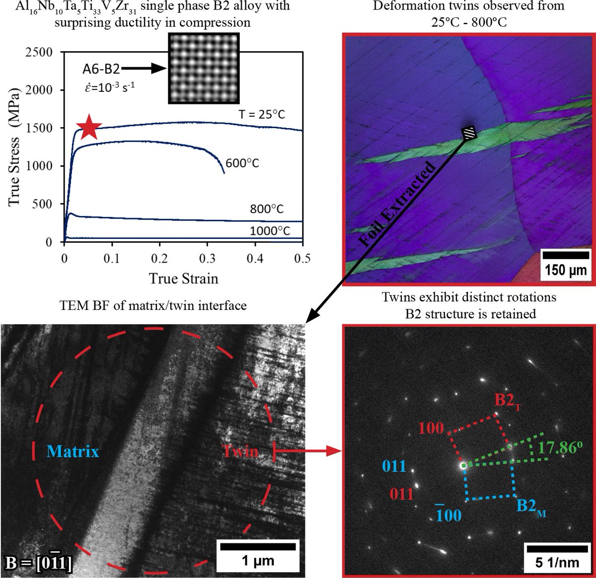
Researchers have uncovered how a NbTaTiV high‑entropy alloy deforms under room‑temperature compression. CEMAS’ advanced electron microscopy continues to enable this level of atomic‑scale insight, supporting breakthroughs in alloy characterization.
cemas.osu.edu/news/2026/03/r…
1
4
82
⏰ Early bird pricing ends today! Learn SEM fundamentals and advanced imaging techniques in CEMAS’ On the Scope course, featuring expert‑led online content and hands‑on training 🔬
Learn more & register: go.osu.edu/ots
1
87
Happy #MicroscopyMonday 🔬 New research from Ohio State shows that simulated lunar dirt can be turned into extremely durable structures. Researchers utilized CEMAS' scanning electron microscopes to characterize the microstructures of the printed products.
news.osu.edu/using-moon-dirt…
1
3
46
🔬 New research enabled by CEMAS:
Work from Gerald Frankel and Irem Efe shows how simple maintenance practices can significantly reduce corrosion product buildup and improve surface passivation aluminum and low‑carbon steel.
Read the study: content.ampp.org/corrosion/a…
1
2
28
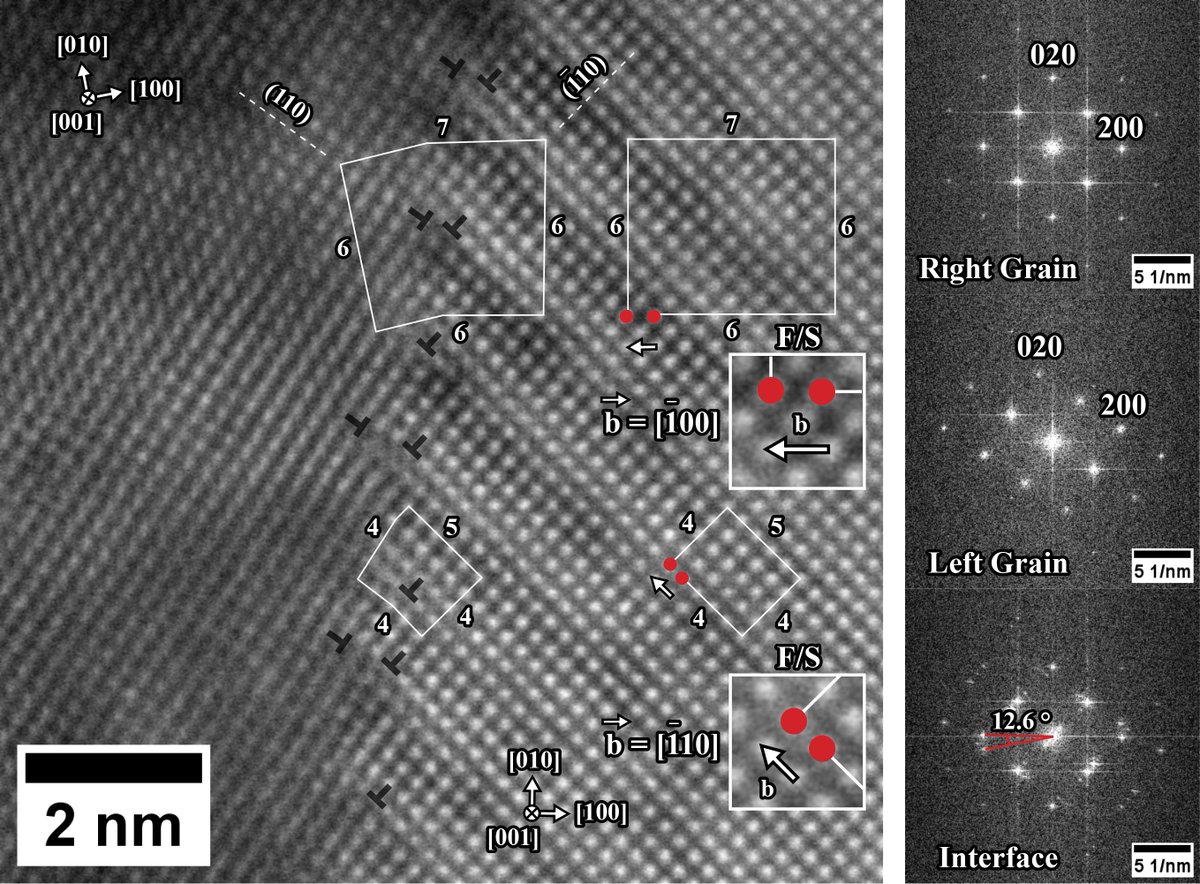
Happy #MicroscopyMonday 🔬 Here is a HRSTEM HAADF micrograph captured by Bryan Crossman, showing a low-angle grain boundary (LAGB) in a BCC Ta₅₅Re₄₅ (at.%) refractory alloy. This boundary is primarily a tilt boundary, formed by a ~12.6° rotation about the [001] zone axis.
2
72

Our friends at @CEMAS_OSU are hosting the annual On The Scope – A Masterclass in Practical Scanning Electron Microscopy - April 1, 2026. If you would like more information or to register for the course, please visit: ow.ly/YSfF50U9wNy
#Microscopy #Microanalysis
1
2
64
🔬 Ready to take your microscopy skills to the next level?
The On The Scope masterclass is back, offering engineers, researchers and industry professionals a hands-on deep dive into practical scanning electron microscopy.
🔗 Learn more & register: professionals.engineering.os…
2
33
🔬 #SeeMoreWithCEMAS: Recent research enabled by CEMAS investigates micro- and nanoplastics in treated drinking water and bottled water. CEMAS' scanning electron microscopes allowed the team to detect and identify particles down to their smallest parts.
engineering.osu.edu/news/202…
5
74
Interested in learning scanning electron microscopy techniques? On The Scope is a training program covering the fundamentals of SEM operation. Participants do a combination of online learning and hands-on labs to learn advanced imaging techniques.
professionals.engineering.os…
1
37
Happy #MicroscopyMonday 🔬 In-situ electron backscatter diffraction (EBSD) experiments were performed in the scanning electron microscope (SEM) during heating of a heavily deformed magnesium alloy.
1
3
86
These experiments were performed by graduate student Rogine Gomez in the Leonard research group in MSE with CEMAS Senior Research Associate Daniel Veghte. Recrystallization and growth are observed from the heavily deformed areas (initially non-indexable areas).
1
42
The paper demonstrated that alloying Mg with Ca shows promise in weakening deformation and recrystallization texture that can be associated with increased ductility and formability.
doi.org/10.1016/j.jma.2025.1…
27
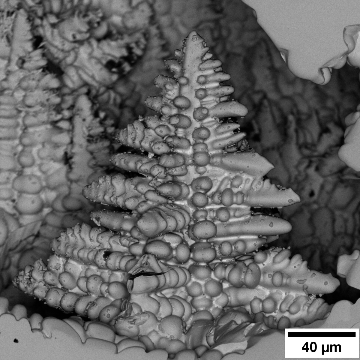
Check out the research SEM image that turned out unexpectedly artistic when a mouse appeared, taken by graduate student Nicole Hudak! Captured on Apreo 1/2 using standard conditions, it’s a BSE image of a titanium alloy. It takes a bit of imagination to see the tiny creature 🔬

1
80
What appear to be the “ears” are actually regions outlined by colloidal silica left behind during polishing and final cleaning. The “eye” is a void, and the rest of the “mouse” takes shape along the alloy’s grain boundaries.
27