
Online Journal Club for gastrointestinal, hepatobiliary and pancreatic pathology #GIPathJC by Pallavi A. Patil @PAPatilMD
Joined August 2020
- Tweets 598
- Following 109
- Followers 652
- Likes 306
13 Photos and videos


DigestivePathJC retweeted

21 Feb 2023
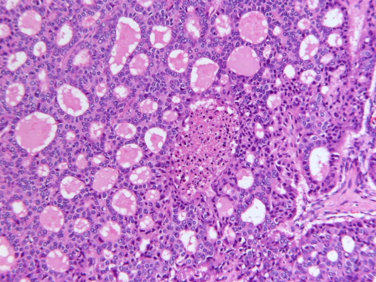
Liver lesion. Chromogranin, synaptophysin, cytokeratin positive. Ki67 ~20%. Questions? #gipath #pathologist #pathology #liver
14
73
239
39,701

21 Dec 2022
21 Dec 2022

Since pathologists do not categorize dysplasia as conventional or non-conventional dysplasia, it is difficult to answer this question at this time.
1
483
21 Dec 2022
21 Dec 2022
The majority of missed dysplastic lesions were non-conventional subtypes with hypermucinous dysplasia being the most common subtype.
1
461
21 Dec 2022
21 Dec 2022
I don’t recall finding a case of “false positive” (i.e., completely normal colon was called as dysplasia or non-conventional dysplasia).
1
370
21 Dec 2022
21 Dec 2022
In our experience, non-conventional dysplasia was often reported as indefinite for dysplasia or had a descriptive diagnosis (such as ‘glandular atypia,’ ‘crypt atypia’, ‘atypia,’ ‘favor reactive atypia,’ etc) without a definite diagnosis of dysplasia.
1
464
21 Dec 2022
#GIPathJC
Thank you @DorukBahceci for joining!
We wrap our journal club at 9:30 pm EST, until we meet next time!
1
373
21 Dec 2022
21 Dec 2022
However, in our analysis of 207 consecutive total colectomy or proctocolectomy specimens of IBD patients, we found a total of 49 missed dysplastic lesions in 27 patients (13%).
156
21 Dec 2022
21 Dec 2022
So, I would say that “false negative” cases are not uncommon, as many non-conventional dysplasia cases were ignored or missed as negative for dysplasia.
141
21 Dec 2022
#GIPathJC During retrospective review, did the authors encounter any false positives or false negatives ?
2
428
21 Dec 2022
21 Dec 2022
One more thing. Aberrant p53 expression was also noted in up to 50% of hypermucinous dysplasia. It can be useful in this setting as well.
1
264
21 Dec 2022
21 Dec 2022
In addition, it is reported that SATB2 loss could be potentially useful in identifying IBD-associated dysplasia; however, in our experience, SATB2 is often patchy and weak (even in normal colon) and difficult to interpret, so we don’t recommend this stain at this time.
164
21 Dec 2022
21 Dec 2022
We do not regularly perform p53 immunostaining in IBD-associated dysplasia cases. A diagnosis of dysplasia should be based on morphologic findings.
1
234
21 Dec 2022
21 Dec 2022
However, wild-type staining pattern does not exclude a diagnosis of dysplasia, and pathologists should not be deterred from making a diagnosis of dysplasia even if p53 is weak or patchy (wild-type)
159
21 Dec 2022
21 Dec 2022
If p53 staining is weak or patchy in a potential crypt cell dysplasia case, we often use a descriptive diagnosis (e.g., “crypt cell atypia”) or a diagnosis of indefinite for dysplasia.
1
225
21 Dec 2022
21 Dec 2022
However, in challenging situations like crypt cell dysplasia (mild enlargement and hyperchromasia of round/mildly irregular, non-stratified nuclei limited to the crypt base, no surface involvement), strong and diffuse p53 staining can be potentially helpful.
121
21 Dec 2022
#GIPathJC
- Undetected dysplastic lesions were often associated with non-conventional dysplasia, flat/invisible gross appearance, and a smaller number of biopsies per colonoscopy
- Study recommends increased random biopsy sampling
97
21 Dec 2022
#GIPathJC
Greater proportion of the undetected (19%) or previously detected (23%) dysplasia group had concurrent PSC compared with only 3% in the group without dysplasia
88
21 Dec 2022
#GIPathJC
@DorukBahceci
Did the authors find any use for p53 to determine dysplasia?
In which situations would it be most useful?
1
1
1
682
21 Dec 2022
#GIPathJC
- Three (11%) patients in the undetected dysplasia group also had undetected CRC, of which two (67%) were found in the same colonic segment as nonconventional dysplasia
93