
Monthly publication links you to new peer-reviewed research esp. lameness, laminitis, racing, farriery, anatomy, equine podiatry. biomechanics. Subscribe now!
Joined June 2017
- Tweets 440
- Following 225
- Followers 228
- Likes 595
76 Photos and videos


HoofSearch retweeted

The U.S. FDA has approved Gastrobim (omeprazole) oral paste for the treatment of gastric ulcers and prevention of gastric ulcer recurrence in horses and foals four weeks of age and older. bit.ly/4csF2Fp
1
277
HoofSearch retweeted

Apr 5
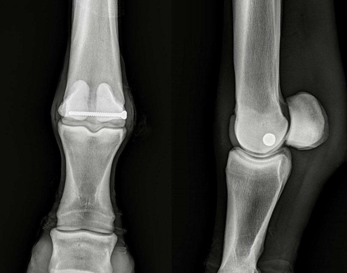
The use of advanced imaging - both CT and PET scanning - has added a whole new level of 'smarts' to equine care and is helping to make us all much better veterinarians!
Case in point - you've heard the common term cannon bone or fetlock "bone bruising" that has sidelined many a racehorse. It is a frustrating and painful condition related to the way bone remodels in response to applied stress (i.e. training) - the cannon bones are a common site for this issue and it can lead to debilitating chronic lameness, attrition, and in some cases a condylar fracture. Besides prolonged rest, treatment options have been few and the incidence of recurrence is high, especially as a horse ages out in its racing career.
In trying to find answers to this problem, we have been applying targeted compression screws (using the imaging information that CT can provide) to chronic, recurrent cases to encourage this bruised/sclerotic bone to mobilize and 'shore itself up'. This approach, combined with rest, seems to help these recurring cases and the results have been encouraging.
This TB colt struggled with chronic "bone-bruising" issues in both hind ankles, yet he was very talented and still had room for a promising career. Using CT imaging to clearly map the pathology, we placed precise compression screws in both hind ankles this Fall.
After proper time off he successfully returned to his winning ways in February and then this past weekend, impressively stepped up and won a very competitive Stakes race 🏆 - nice to see science in action...💪


21
77
5,404

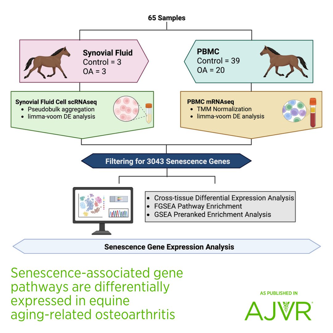
Therapeutic strategies targeting senescent cells may be a disease-modifying strategy to treat #equine #osteoarthritis. Open access article: jav.ma/oa @csuvetmedbiosci #senescence #senotherapeutics #senolytics
3
3
188
HoofSearch retweeted

Apr 2
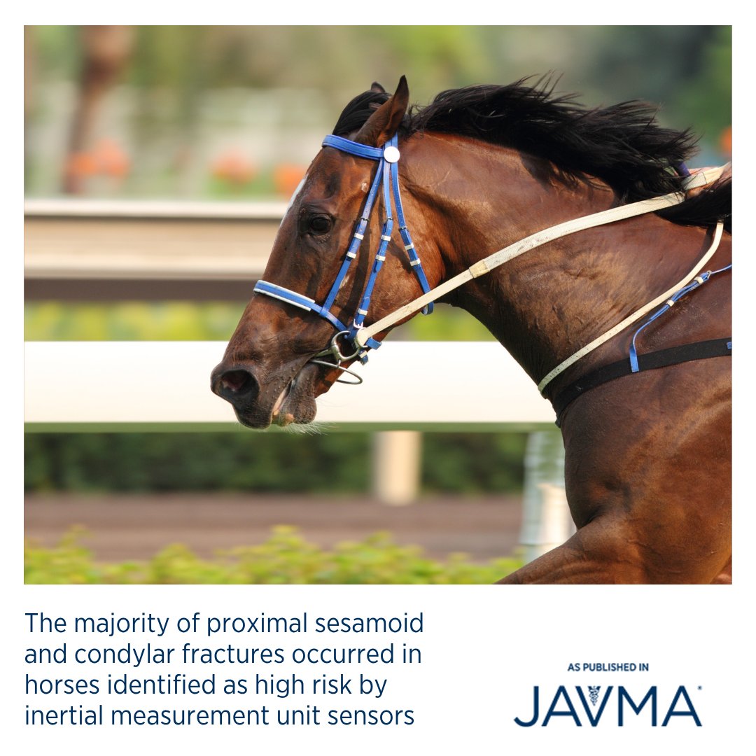
Fracture-specific algorithms can help identify horses at high risk of condylar or proximal sesamoid bone fractures, supporting veterinarians in their clinical evaluation and decision-making. Open access article: jav.ma/horsefracture @wsuvetmed #catastrophicinjury #sesamoids
4
3
276
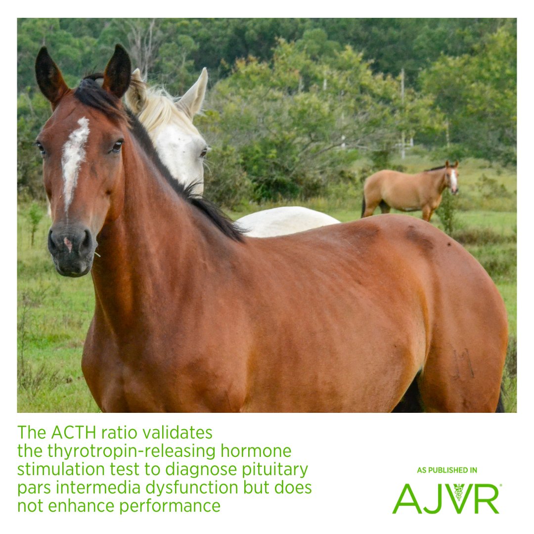
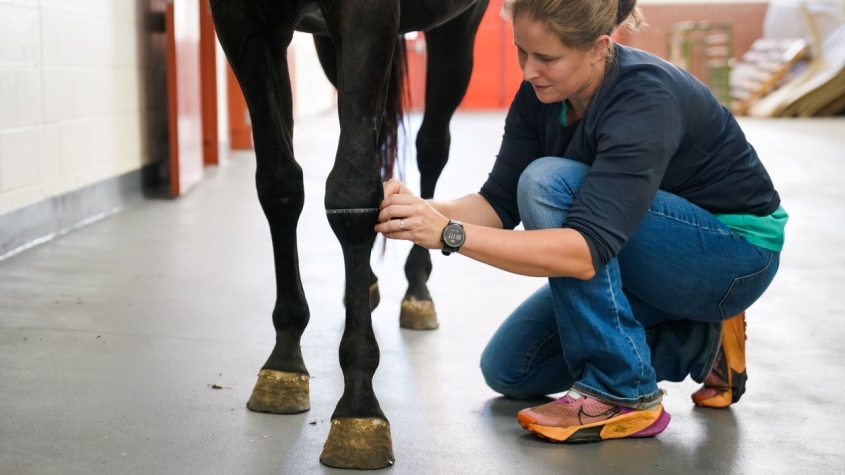
The thyrotropin-releasing hormone (TRH) stimulation test is recommended for evaluation of the pars intermedia of the equine pituitary gland in the context of diagnosing PPID: jav.ma/trh @pucvm #horse #endocrinology #laminitis #geriatric
1
52
HoofSearch retweeted
16 Aug 2025
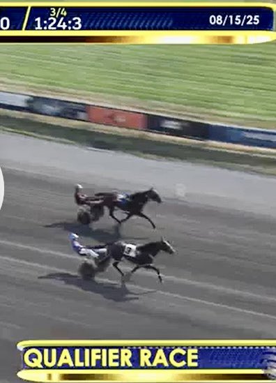
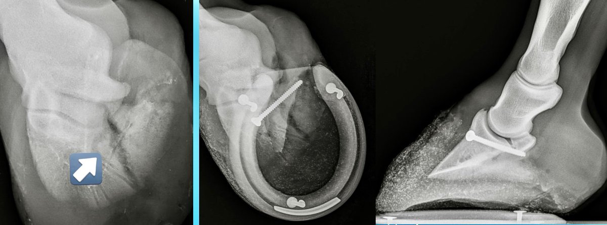
A true testimony to the patience and confidence in this very talented horse by the owner/trainer. This top STB stallion broke his LF Coffin bone over 2 years ago and was repaired at another clinic. He had much difficulty healing, screw became infected and was finally removed. He struggled with a nonunion and never made it back to the racetrack/ the owner did not want to give up so in January he was referred to us for his last chance - we repaired this difficult nonunion and remarkably he healed beautifully! Yesterday he won his qualifier in an impressive 1:51, last quarter in 26!🤩. He is on his way 🙌🏻🙌🏻🙌🏻 what a champ!
7 Feb 2025

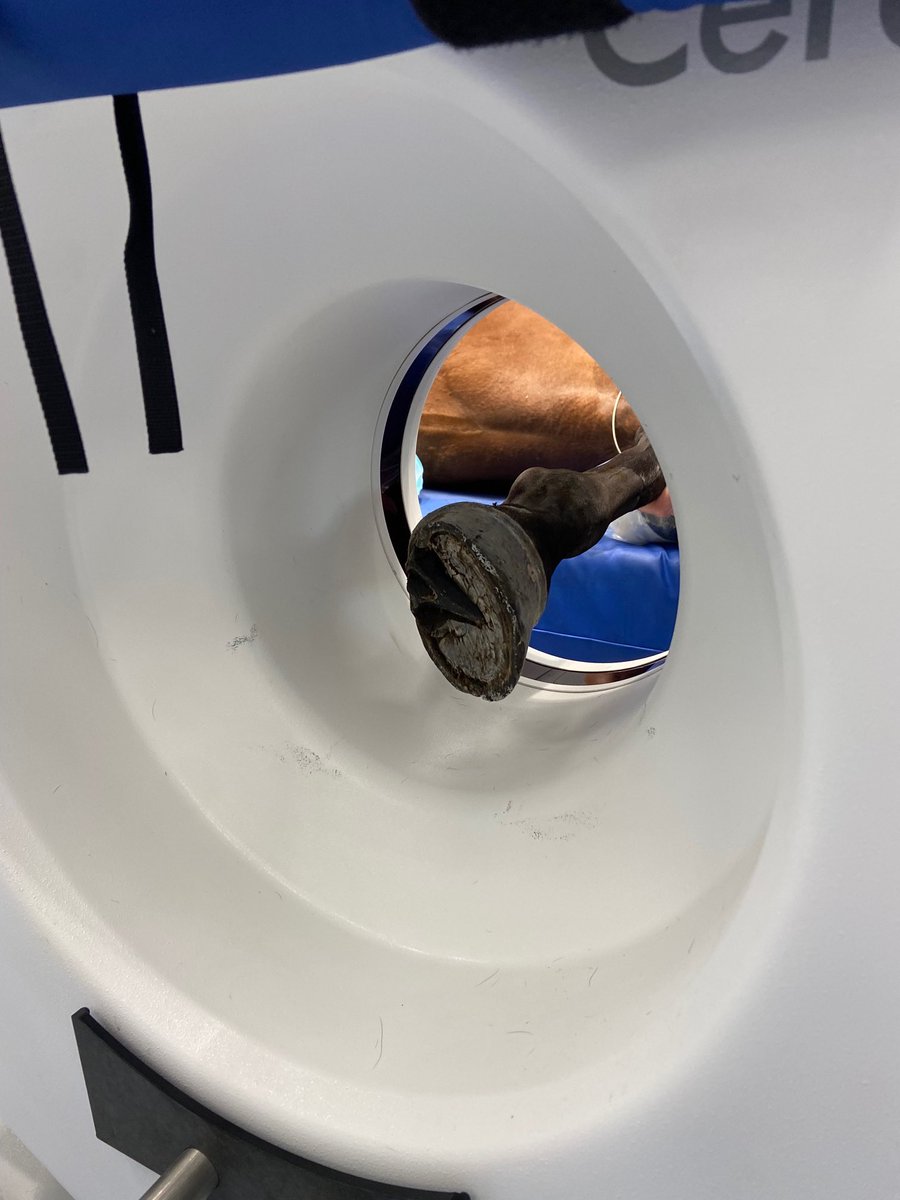
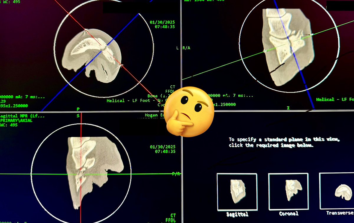
Computed Tomography (CT) is a game-changer for imaging in vet med. CT allowed for precise repair of this very complicated chronic coffin bone (P3) fx thru a tiny hole in the hoof. Using CT mapping w/markers, we could safely place screw as close to joint as possible for best result and see true alignment/proper compression of joint in 3-D 🤓

9
18
85
4,408
HoofSearch retweeted

16 Jun 2025
Clinical use of triamcinolone acetonide in the horse (205 cases) and the incidence of glucocorticoid-induced laminitis associated with its use eurekamag.com/research/004/0…
2
55
HoofSearch retweeted

16 Jun 2025
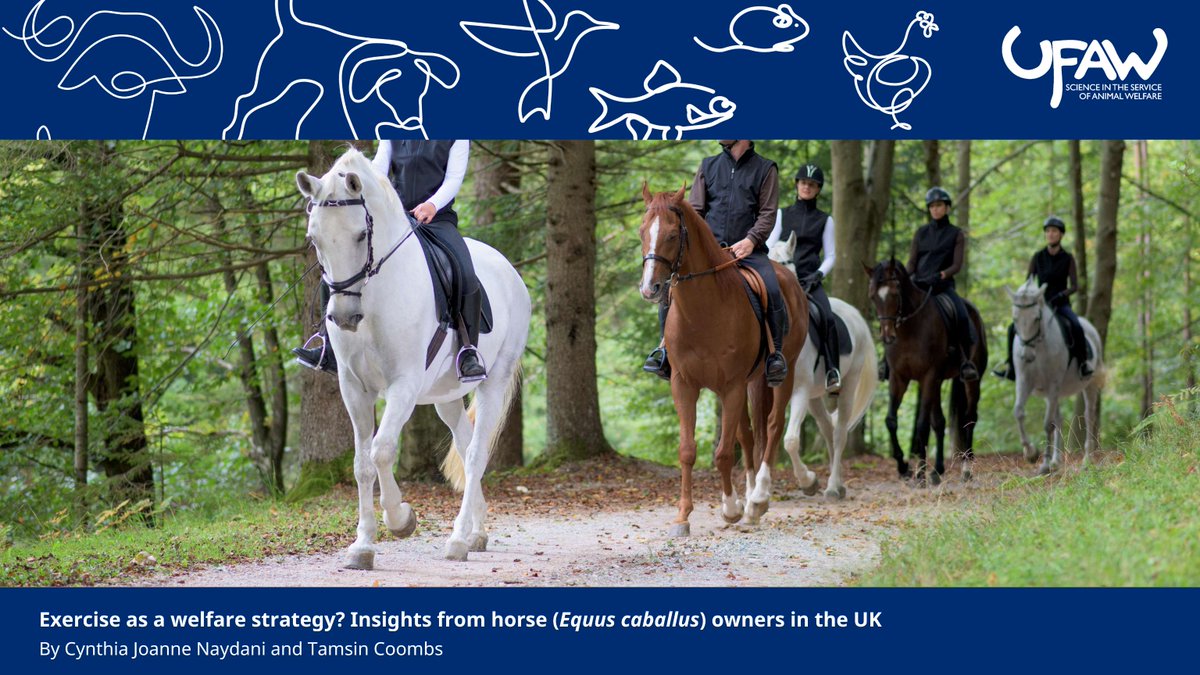
🔍 NEW study explores how exercise impacts #equinewelfare in the UK. Surveying 804 horse owners, the research found that while exercise is linked to reduced risk of laminitis & Equine Metabolic Syndrome, many owners face barriers to providing exercise.
🔗 ow.ly/1c6m50W9V2U
1
2
44

What a wonderful Phi Zeta Research Day at the LSU School of Veterinary Medicine. Very proud of our LECOR summer scholar Ralph Delgado for his presentation on his work on 3D imaging of hoof tissues to create models for laminitis research. #LSU #LSUVetMed #LECOR #EHSP

1
2
93
HoofSearch retweeted

20 Jun 2025
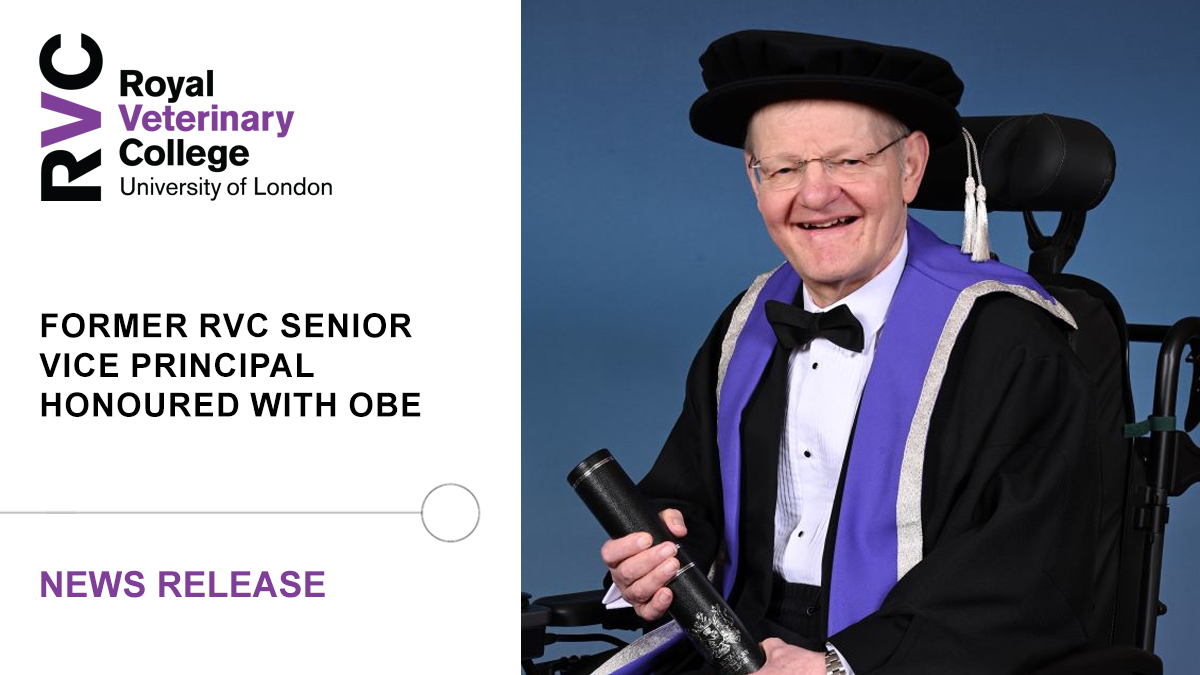
🎖️ Professor Stephen May, former Senior Vice Principal at the RVC, has been appointed Officer of the Order of the British Empire (OBE) in the King’s Birthday Honours
➡️ Read more at: rvc.uk.com/stephen-may-obe
2
7
326
HoofSearch retweeted

New study pinpoints key proteins in #horse joint fluid that may help detect & treat #equine #osteoarthritis early. Using advanced proteomics, CVM researchers found markers like alpha-2-macroglobulin differed between healthy & #arthritic joints. Learn more: vet.cornell.edu/about-us/new…
6
13
877
HoofSearch retweeted

6 Apr 2025
JOURNALISM (Curlin x Mopotism) was an impressive winner of the G1 Santa Anita Derby yesterday and sits as antepost favourite for the Kentucky Derby in May.
Here he is parading at the 2023 Saratoga Select Sale, where he was a 95.53 / 100 [A] rated yearling on biomechanics 📊

8
34
225
35,363
HoofSearch retweeted

1 Mar 2025
#UGAVetMed's Dr. Kelsey Hart, and co-investigator Dr. Shune Kimura, have received a research grant from @AVMFvets and the Veterinary Pharmacology Research Foundation. Their study will explore intravenous metformin in horses with experimental SIRS.
--
ugavetmed.com/43fJ1kJ
1
151
HoofSearch retweeted

3 Mar 2025
Authors from @NottinghamVets @Bellequine and @UoN_SHS have just published new research on the important roles and experiences of veterinary and charity teams, when contributing to euthanasia and end-of-life decision-making for horses.
Read the paper here: bit.ly/43goI6F

4
5
307
HoofSearch retweeted

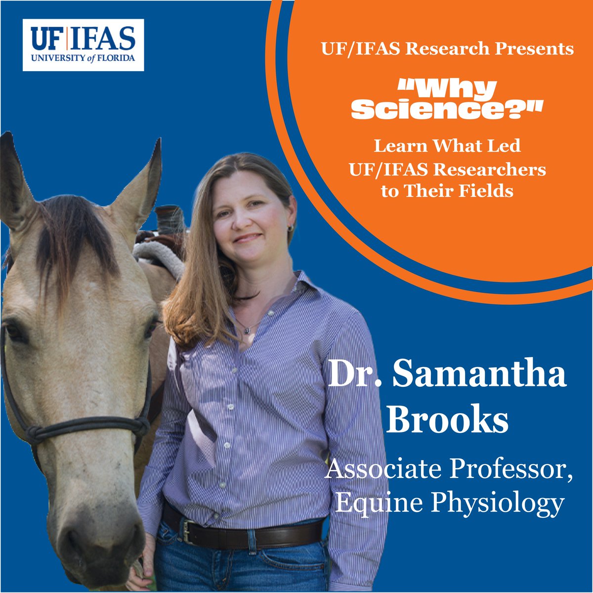
24 Feb 2025
In the next installment in our "Why Science?" series, we'll hear from Dr. Samantha Brooks, an equine geneticist with @uf_ansci. Check out the link below to learn how Dr. Brooks discovered her passion for equine research!
youtube.com/watch?v=WvkrtXju…
1
6
274
HoofSearch retweeted

5 Mar 2025
Free for all to access today is Means et. al's "Vertical pelvic movement asymmetry and lameness location in ipsilateral combined forelimb and hindlimb lameness cases".
Read it here - beva.onlinelibrary.wiley.com…

1
1
90
Immersion of the distal limb in ice and water was most effective for cooling the digital lamellae under clinically relevant conditions. Open access article: jav.ma/lamellae @pennvet @acvim #cryotherapy #laminitis #digitalhypthermia #lamellar #temperature

2
4
161
HoofSearch retweeted

1 Feb 2025
Quarter of leading UK universities cutting staff due to budget shortfalls | University funding | The Guardian theguardian.com/education/20…
4
2
1,250
HoofSearch retweeted
31 Jan 2025
Quam et al's article "Equine bone marrow MSC-derived extracellular vesicles mitigate the inflammatory effects of interleukin-1β on navicular tissues in vitro" looks into new options to treat DDFT and NBF.
Read it here - beva.onlinelibrary.wiley.com…

3
2
194
HoofSearch retweeted

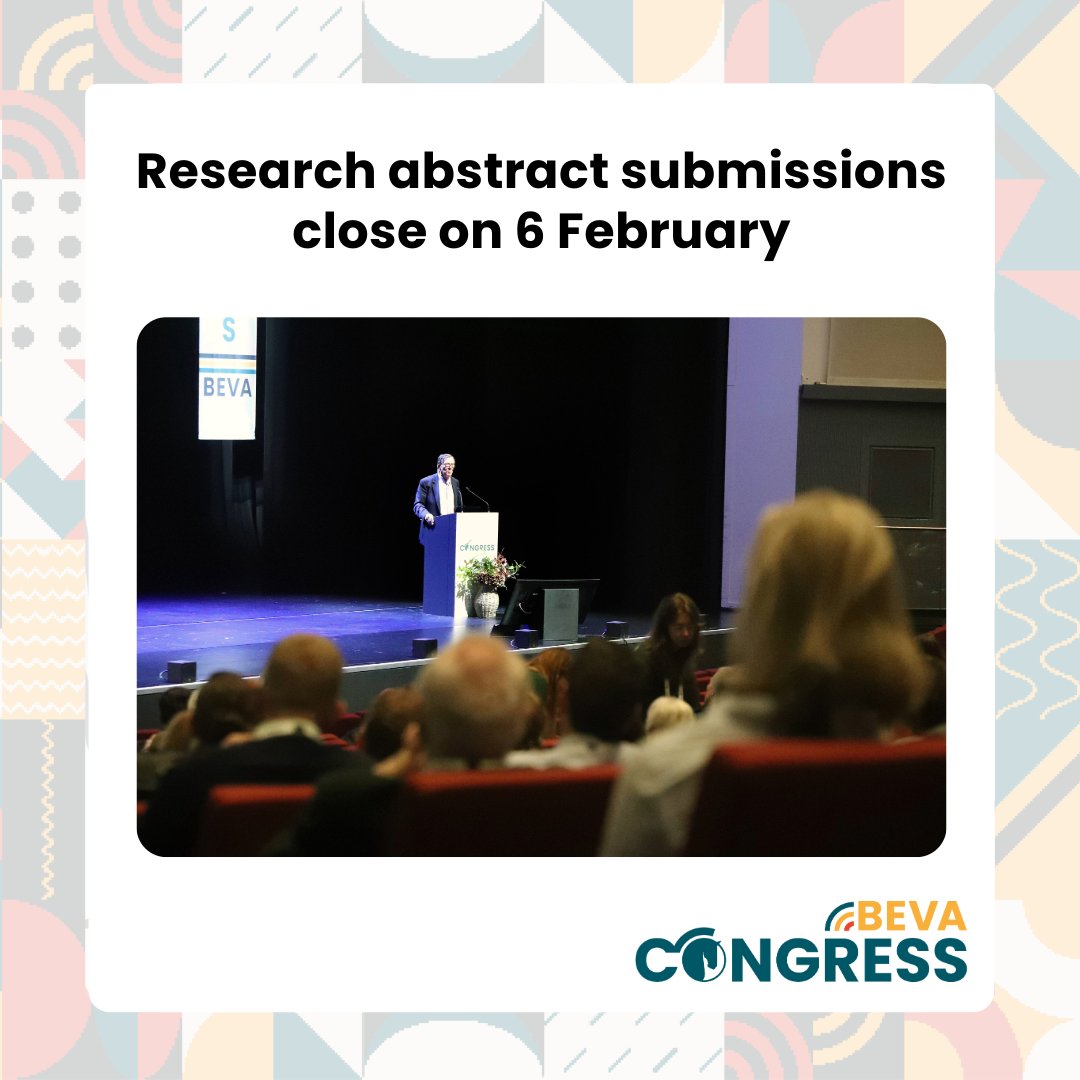
1 Feb 2025
Don't miss your chance to present your research at #BEVA25! Submissions are open until midnight on Thursday 6 February.
Find out more and submit your abstract today - bevacongress.org/copy-of-cli…
3
4
168