The Journal of Eukaryotic Microbiology, the journal of the International Society of Protistologists, publishes original research on all aspects of #protists.
A nice tribute to Frank John Rupert “Max” Taylor by Juan F. Saldarriaga, Rowan Haigh.
Please share your remembrances of him in the comments.
onlinelibrary.wiley.com/doi/…
🚨Sept/Oct Highlight Image!
🔬2 Nanochloropsis gaditana cells visualized by quick-freeze deep-etch electron microscopy.
➡️Goodenough & Roth. Ultrastructure of the Endoplasmic Reticulum in Eukaryotic Microalgae.
doi.org/10.1111/jeu.70030#protists
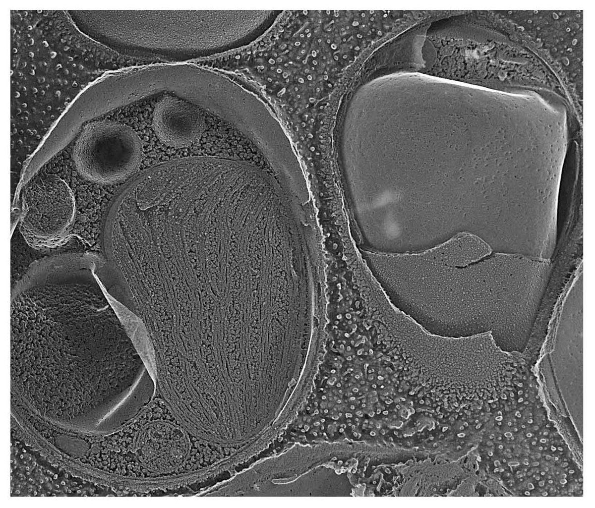
ALT Two Nanochloropsis gaditana cells visualized by quick-freeze deep-etch electron microscopy. Left cell displays a chloroplast containing thylakoids, a nucleus, a mitochondrion, and a fracture face of the plasma membrane. In the right cell, the innermost membrane, marked with pits, is the fractured outer membrane of the chloroplast envelope, above which is the fractured outer membrane of the chloroplast ER, above which is the granular cell wall.
EIC Joel Dacks looks back on the year and shares some of our new initiatives, including "Issue Highlight" image and AI use guidance. Read about these initiatives and more in the Editor's Message onlinelibrary.wiley.com/doi/…
From the arid north to the cold seas of Magallanes, Chilean Labyrinthulomycetes astonish some strains yield up to 34× more DHA in cold,alkaline waters and serve as sources of carotenoids and PUFAs.First Chilean study protist lipids temp gradient @JEukMicrodoi.org/10.1111/jeu.70035
The microscopic world is full of surprises!
Check out the July/August 2025 Featured Article pick from @JEukMicro.
Read it now here and explore our past featured articles too: ow.ly/3siX50WNicT
ALT Banner promoting an Interesting Article for July/August 2025 for 'The Journal of Eukaryotic Microbiology'. The article is '3D Electron Microscopy Reveals the Structural Complexity of the Intravacuolar Membranous Network in Cyrilia lignieresi-inflected Erythrocytes of the Fish Synbranchus marmoratus'
The microscopic world is full of surprises! This stunning SEM reveals the parasite Cyrilia lignieresi living inside a red blood cell of the freshwater fish Synbranchus marmoratus. A rare snapshot of the complexity of life invisible to the naked eye. ➡️doi.org/10.1111/jeu.70031
🥳Congratulations to Monica Santin and colleagues for winning the Trager Award for the most cited paper in the past two-year period.
➡️Check out the paper: Division of Blastocystis ST10 into three new subtypes: ST42-ST44
onlinelibrary.wiley.com/page…
Check out this month's highlighted paper from the Journal of Eukaryotic Microbiology 'Algal Symbiont Diversity and Host Fitness Variation in Amoebozoan Photosymbiosis.'
ow.ly/Hkqf50WakQs@marulab_Utokyo@JEukMicro
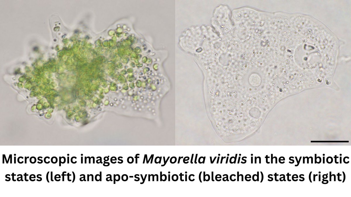
ALT Journal of Eukaryotic Microbiology | Interesting article: Algal Symboint Diversity and Host Fitness Variation in Amoebozoan Photosymbiosis
Daisuke Yamagishi, Ryo Onuma, Sachihiro Matsunaga, Shin-ya Miyagishima, Shinichiro Maruyama
Read Now
🆕 Out now in JEM (@JEukMicro)! Using gene and organelle phylogenies from recovered cysts, we describe Apostichocystis gudetama, an apicomplexan parasite found in the ovaries of the Japanese sea cucumber 🥚🥒🇯🇵 dx.doi.org/10.1111/jeu.70013
Check out exciting protist research in JEM! Holly Moeller et al. explore the feeding habits of Mesodinium rubrum and provide a tool for studying its cell biology and photophysiology in their open access paper in our March/April issue!
doi.org/10.1111/jeu.13066
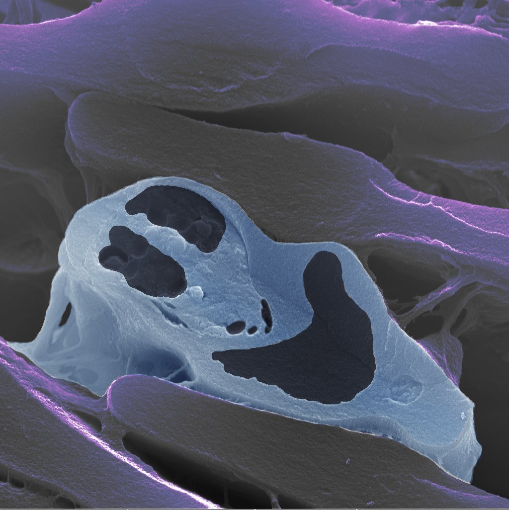
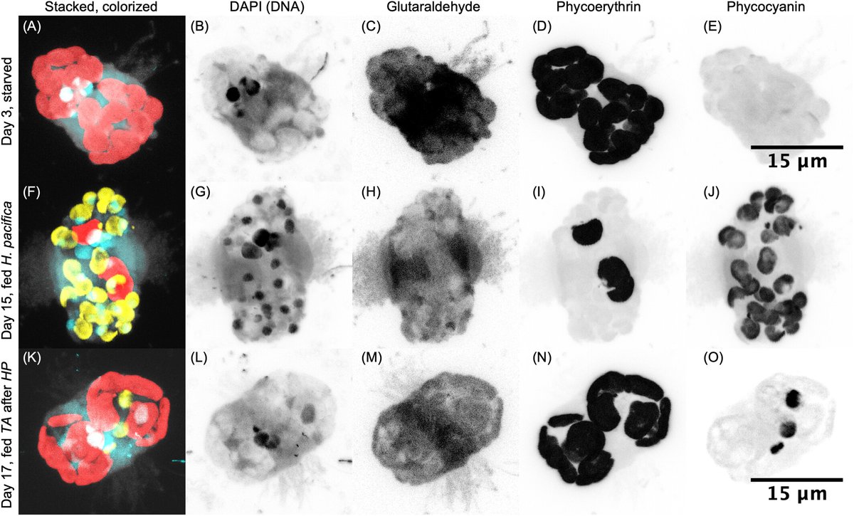
ALT The marine ciliate Mesodinium rubrum (bottom right) is famed for its ability to steal chloroplasts and nuclei from red cryptophyte algae in the Teleaulax/Gemingera clade. But even these prey specialists can make "mistakes," ingesting blue-green Hemiselmis pacifica (upper left) and transiently retaining their plastids. Here, a M. rubrum cell points its feeding tentacles (labeled with an anti-centrin antibody, yellow) at an H. pacifica cell. The cryptophyte's flagella and ciliate's cilia (labeled with an anti-tubulin antibody, purple) and nuclei (labeled with DAPI, blue) are visible in this expansion micrograph taken with a Nikon spinning disk confocal microscope. Numerous stolen prey nuclei are visible inside of the M. rubrum cell. See Moeller et al. https://doi.org/10.1111/jeu.13066
We’ve published incredible #protist research in the first quarter of 2025! Julia Packer et al. describe two new species of free-living bacterivores including Novijibodo darinka pictured here. Check out the open access paper from our Jan/Feb issue. doi.org/10.1111/jeu.13072
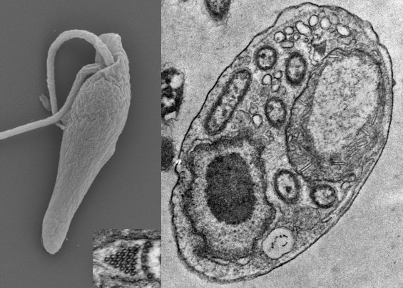
ALT Scanning and transmission electron micrographs of Novijibodo darinka n. gen. n. sp.. Ventral view of cell with flagellar pocket, and cross section of cell with nucleus, mitochondrion containing kinetoplast, microtubular prism (enlarged), and endosymbiotic bacteria. Images taken by J. A. Packer. See Packer et al. https://doi.org/10.1111/jeu.13072
Protistologists work around the world in many languages & alphabets. We want to meet you as you see yourself. Now authors can also include their names in any script they want in published papers at JEM. Learn how in our Checklist for Manuscript Prep: bit.ly/3G3PANH
First published paper! I'm thrilled to be a co-author of this work by Jara-Zapata et al., which reports for the first time the presence of the genus Aplanochytrium on the Chilean coast, expanding its geographical distribution 1/2
doi.org/10.1111/jeu.70007
Morphology and Molecular Phylogeny of Endosymbiotic Ciliates (Peritrichia, Mobilida) of Marine Invertebrates with Descriptions of Two Novel Species Urceolaria clepsydra n. sp. and Urceolaria bratalia n. sp.
Gabrielle Martinez et al.
onlinelibrary.wiley.com/doi/…
Characterization of Allobodo yubaba sp. nov. and Novijibodo darinka gen. et sp. nov., cultivable free-living species of the phylogenetically enigmatic kinetoplastid taxon Allobodonidae
Julia A. Packer et al.
onlinelibrary.wiley.com/doi/…