Glad to announce that Picasso v0.10.0 is out!
- ⚡️ GPUfit built in for localization (Windows)
- New fast fitting methods in SPINNA
- ThunderSTORM .csv import directly in Render
- Vastly extended API
- And many, many more…
Visit our page to find out: github.com/jungmannlab/picas…
In DNA-PAINT, nearby proteins create overlapping localizations. You may see two by eye, but analysis often misses them, wasting precious precision. G5M solves this. Now in @NatureComms: doi.org/10.1038/s41467-026-7… 1/7
G5M is implemented in Picasso since version 0.9.5: github.com/jungmannlab/picas…, although we always recommend using the most recent version! A short documentation: picassosr.readthedocs.io/en/…. We advise reading the whole paper for best results! 6/7
We hope G5M helps push quantitative molecular mapping forward, and we’re excited to see the discoveries it enables. Thanks to @rafalkowalew, @SusanneReinhar3, @IBaudrexel, Shuhan Xu, @l_masu, for developing G5M and the rest of the @JungmannLab for testing and improving it. 7/7
Up to 15-fold improvement in protein labeling for DNA-PAINT 🧬🔬. We are excited to present our latest work published in @angew_chem! onlinelibrary.wiley.com/doi/…
Importantly, this workflow is broadly applicable and compatible with virtually any high off-rate binder. Many “bad” binders currently sitting in lab freezers may now become powerful imaging tools. 5/6
How do you scale super-resolution microscopy to dozens of proteins without linearly scaling imaging time?
We introduce Combi-PAINT: a combinatorial DNA-PAINT strategy that breaks the 1-target-per-round bottleneck of Exchange-PAINT: biorxiv.org/content/10.64898…
1/5
#pictureofthemonth: The pic shows 30 different protein types with spatial resolution in a neuronal "atlas“ created with the imaging method SUM-PAINT.
Find out more: biochem.mpg.de/jungmann
November – institute calendar of @MPI_Biochem and @MPIforBI
Image by Eduard Unterauer
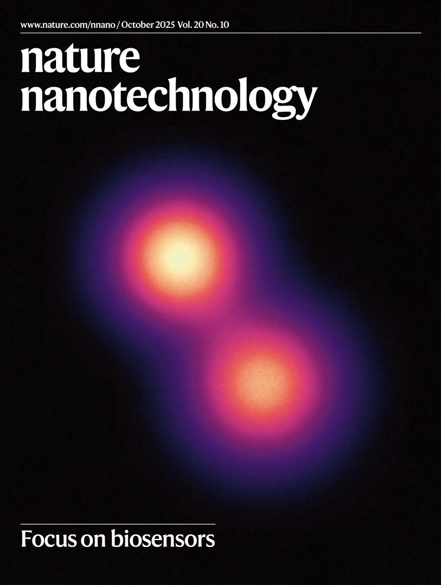
ALT The image on the cover shows two sugars from the same cell-surface glycan separated by 9 Å, visualized with RESI (resolution enhancement by sequential imaging) enabled by metabolic labelling with DNA barcodes.
IMAGE: Luciano A. Masullo, Max Planck Institute of Biochemistry, Germany.
COVER DESIGN: Vanitha Selvarajan
Original paper: Masullo, L.A., et al. Ångström-resolution imaging of cell-surface glycans. Nat. Nanotechnol. 20, 1457–1463 (2025). https://doi.org/10.1038/s41565-025-01966-5
Abstract: Glycobiology is rooted in the study of monosaccharides, ångström-sized molecules that are the building blocks of glycosylation. Glycosylated biomolecules form the glycocalyx, a dense coat encasing every human cell with central relevance—among others—in immunology, oncology and virology. To understand glycosylation function, visualizing its molecular structure is fundamental. However, the ability to visualize the molecular architecture of the glycocalyx has remained challenging. Techniques
Thrilled to announce some big news!
I’ve been awarded a Wellcome Trust Early Career Award to establish my research as a Group Leader in the Glycosciences Program at the Institute of Biology and Experimental Medicine (IBYME) in Buenos Aires, Argentina, starting early 2026.