
Cell biologist studying how a heart grows and dies; also Blebbisomes. Associate Professor at Vanderbilt. Married to @gillianhoo.
Joined January 2015
- Tweets 3,268
- Following 1,268
- Followers 33,371
- Likes 2,088
1,746 Photos and videos


Pinned Tweet

21 Feb 2025
I am excited to finally be able to share with you our work reporting the first Extracellular Vesicle with a personality! This video does not show a cell, it is a Blebbisome! #CellBiology
nature.com/articles/s41556-0…
25
130
611
56,318
A control cell going through cell division with normal membrane blebs (top) and a cell we messed with (bottom). Both were videoed through a DIC microscope. #CellBiology
molbiolcell.org/doi/full/10.…
2
15
1,626
Jun 13
Not all cells do the whole membrane blebbing thing the same. Some of them get weird about it. This is a single cell videoed through a DIC microscope. #CellBiology
1
9
85
8,577
Jun 10
Actin filament-based structures in a cell photographed using structured illumination microscopy. The video starts with the bottom of the cell and steps up through the Z dimension (i.e., going up from the substrate). #CellBiology
rupress.org/jcb/article/205/…
1
5
34
2,275
Jun 4
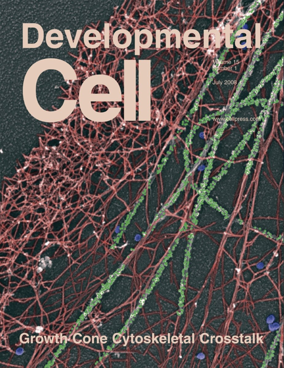
Microtubules (green) fighting retrograde actin flow (magenta) in a neuronal growth cone videoed through a TIRF microscope. #CellBiology
1
6
39
3,487
Jun 2
The newest episode of Nikon's Small World Speaks.
"Dylan Burnette (that's me) shares some of the stories behind one of his favorite images he's submitted to the Nikon Small World competition to date." #CellBiology
1
5
31
2,074
May 26
This was my first journal cover from back in the day. It shows the side of a neuronal growth cone photographed with an electron microscope. The microtubules (green) were labeled with nanoscale particles of colloidal gold bound to antibodies (i.e., immunogold). #CellBiology
2
3
20
836
Dylan Burnette retweeted

May 22
Sometimes blebbisomes like to hitch a ride on cells, like you see here! Extracellular vesicles with a personality indeed! These guys do so many cool and unique things that we will be sharing soon! Stay tuned!#fluorescencefriday #blebbisomes #extracellularvesicle

1
7
1,058
May 19
A dividing cell videoed through a microscope. Hot tip: Don't mess around with myosin II paralogs if you don't want your cells to freak out. #CellBiology
cell.com/cell-reports/fullte…
2
22
170
17,200
May 15
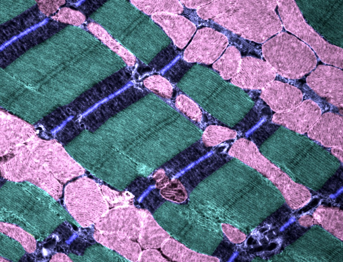
The inside of a heart muscle cell photographed with an electron microscope. The powerhouses/overlords of the cell, mitochondria, are shown in pink. The repeating structures are the basic units of contraction that drive each heart beat (i.e., sarcomeres). #CellBiology
14
59
2,409
May 13
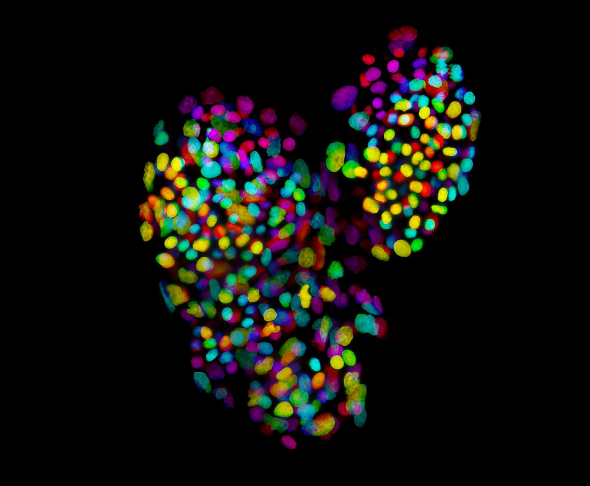
The heart of a zebrafish embryo photographed through a microscope. (Ventricle: Left; Atrium: Right). The nuclei of both the heart cells and blood cells are shown. "What? Red blood cells do not have nuclei!", you say. So human centric of you......
#CellBiology
4
29
1,569
May 5
Immature heart muscle cells photographed through a microscope. Nuclei (yellow/green) and the molecular motor driving muscle contraction, myosin II (purple/blue), are shown. #CellBiology
1
5
39
1,257
Apr 28
Neuronal growth cones at the end of neurites (immature neural extensions that have yet to turn into axons or dendrites) videoed through a microscope. #CellBiology
11
55
2,025
Apr 16
Cover image for "The microtubule GTP-tubulin cap size is modulated during cell division" by Anna Cassidy! I know it says "May 1, 2026", but it is already online so here it is!
molbiolcell.org/doi/10.1091/…
17
764
Mar 6
A cell videoed through a microscope. DNA in the nucleus (cyan), mitochondria (yellow), and the actin filament cytoskeleton (magenta) are shown. #CellBiology
4
36
127
5,368
Mar 2
A cell going through cell division to create two daughter cells videoed through a spinning disk confocal microscope. DNA (red), Golgi apparatus (green), actin filaments (blue) are shown. #CellBiology
3
26
112
5,615
Feb 27
Are there still Zoom-based scientific seminar series? As I plan for a promotion cycle that expects ~10 invited talks/year. I love giving talks and talking with interesting scientists, just not the travel part. I study the growth and function of the heart and large EVs.
2
3
18
3,587
Feb 26
A cancer cell videoed through a microscope. It has 3 nuclei (magenta). The powerhouse/overlords of the cell, mitochondria, are also shown (green). #CellBiology
6
54
257
16,938
Feb 25
Two cells isolated from a fish scale videoed through a DIC microscope. The cells are cool but now all I see are extracellular vesicles floating by in the media and stuck to the substrate....... #CellBiology
3
21
134
17,795