
Joined September 2022
- Tweets 157
- Following 82
- Followers 85
- Likes 294
13 Photos and videos


Scanlab Diagnostics retweeted

Ultrasound speaks. I interpret!
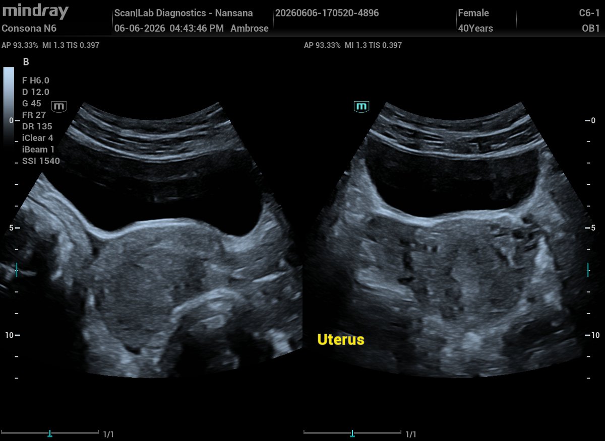
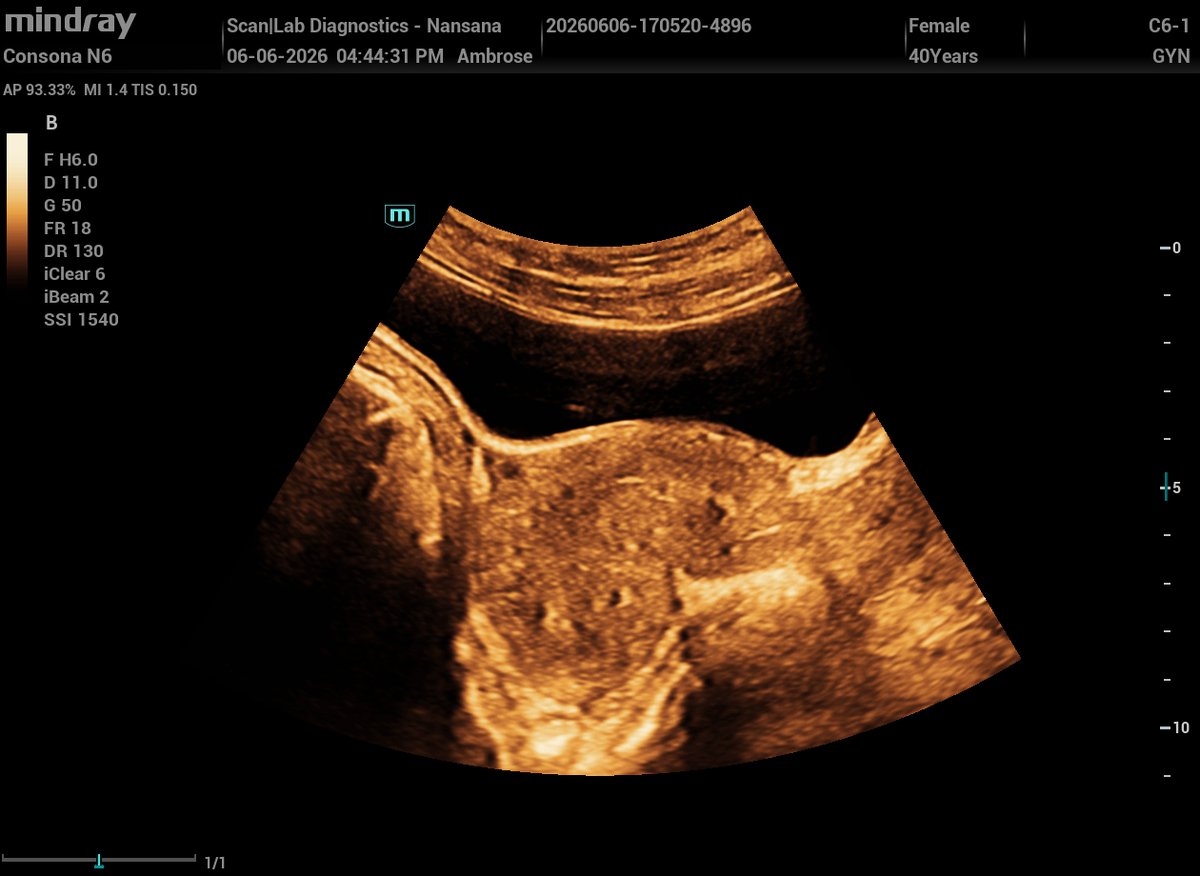
Post‑partum uterus (1 week post C/S)
Ultrasound findings:
Endometrial cavity contains fluid with multiple echogenic foci,
No internal vascularity on color Doppler,
Volume ≈10.5 c.
Are these 'normal' post-partum contents or RPOC?
Sonograms 👇👇
3
2
14
1,591
Scanlab Diagnostics retweeted
Retroverted or Retroflexed? 👇👇
4
2
15
2,005
Scanlab Diagnostics retweeted
Normal trilaminar of proliferative phase
1
1
1
223
Scanlab Diagnostics retweeted
Ultrasound speaks. I interpret!
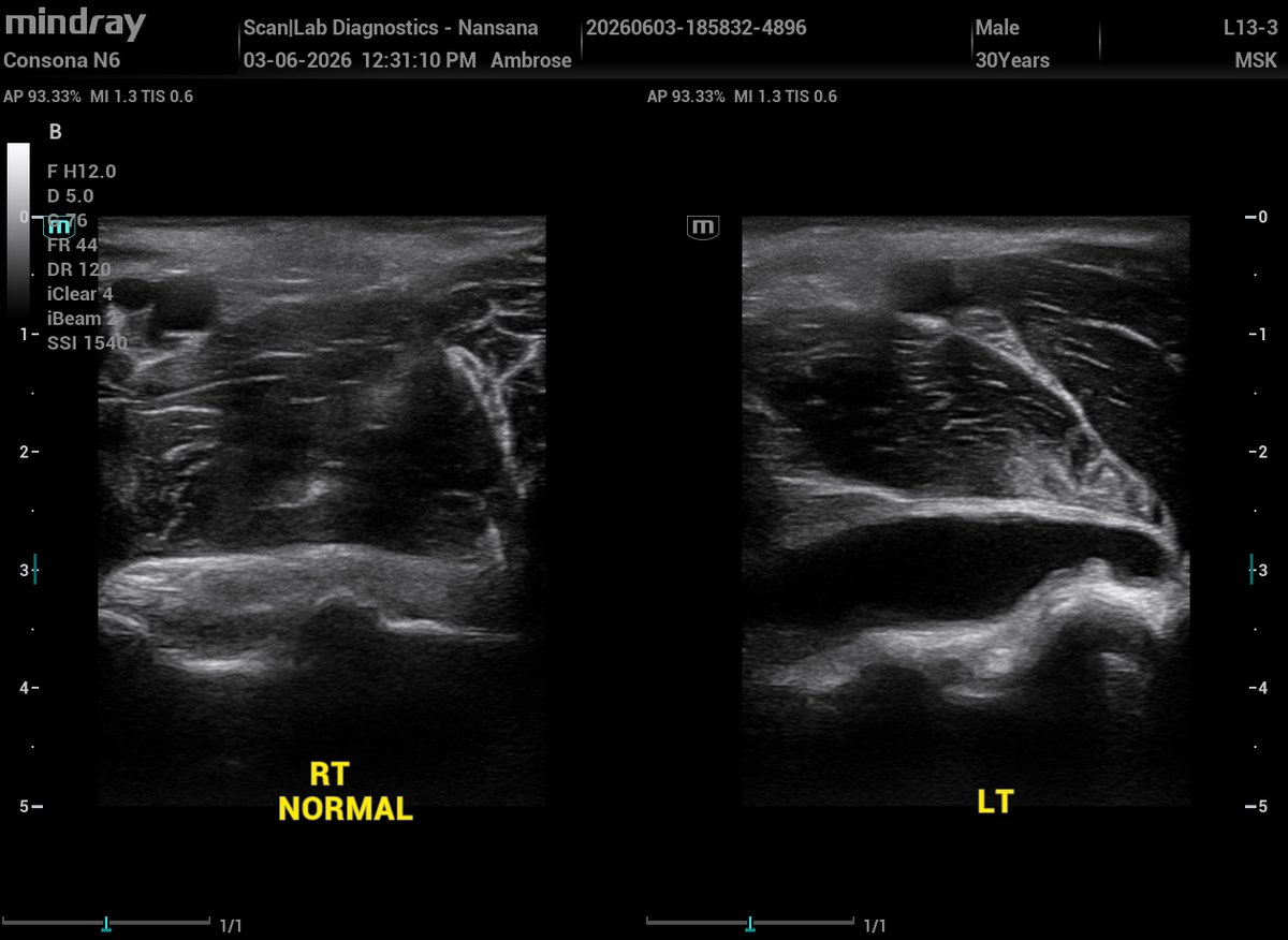
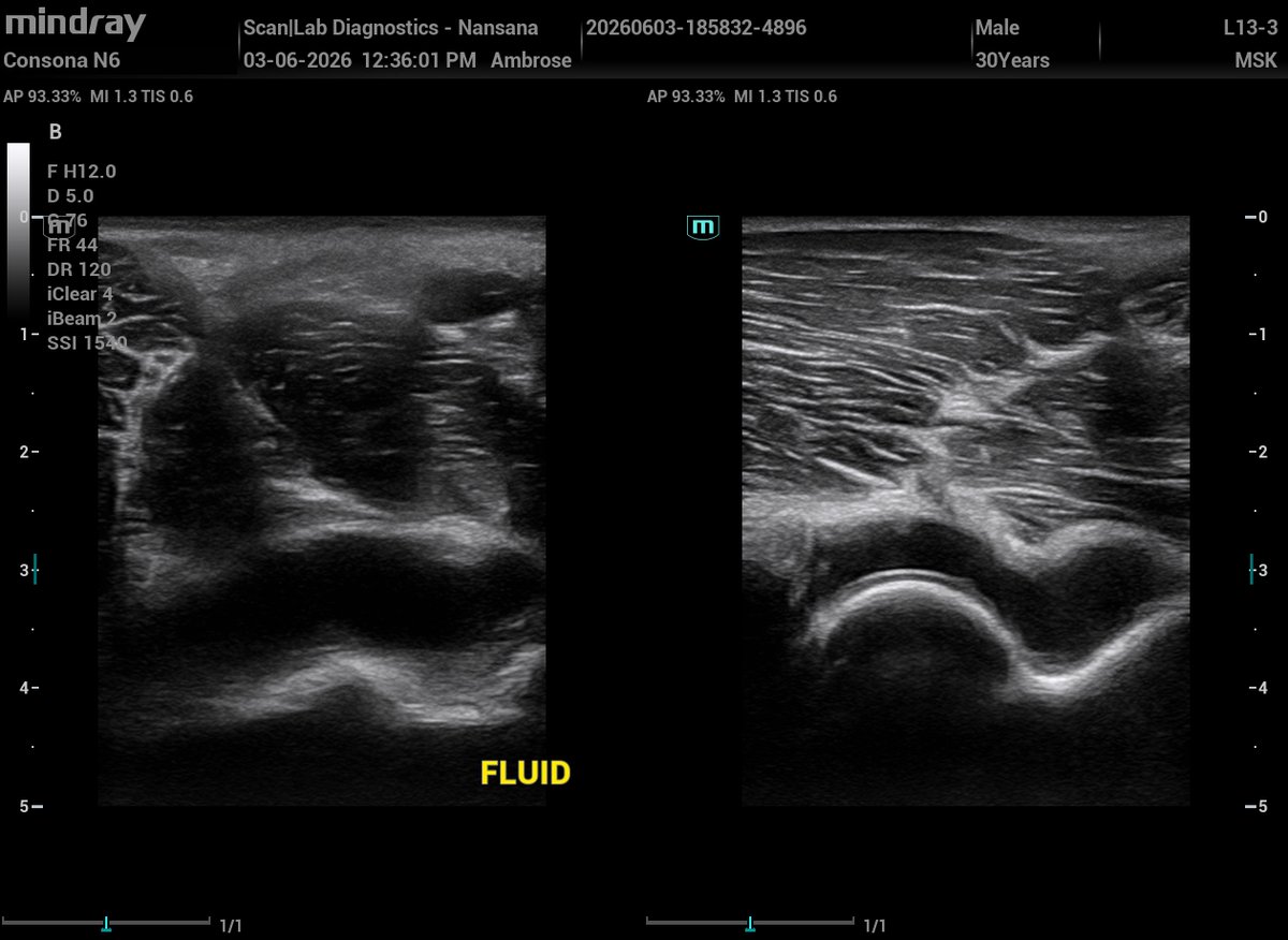
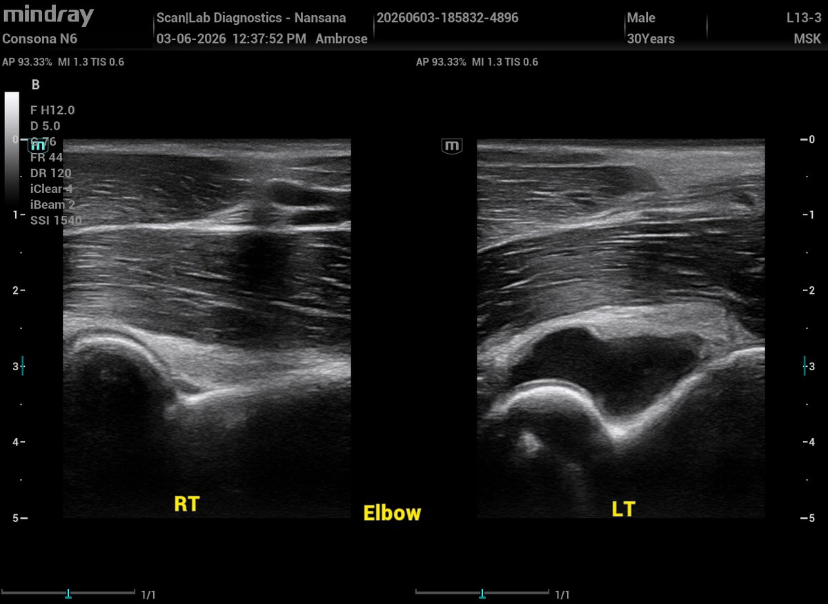
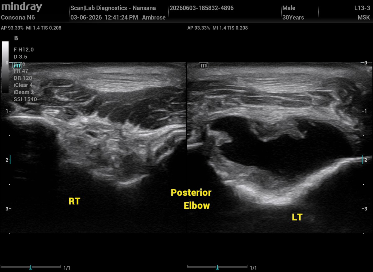
30‑year‑old male presents with 1 month of persistent left‑elbow pain, with no history of trauma.
MSK U/S of the elbow demonstrates multicompartment fluid collections and synovial thickening in the posterior recess.
What pathology does this pattern make you think of and why?
NOTE: Consider joint recesses, and synovial hypertrophy.
Here are the sonograms 👇👇
2
4
26
845

Scanlab Diagnostics retweeted
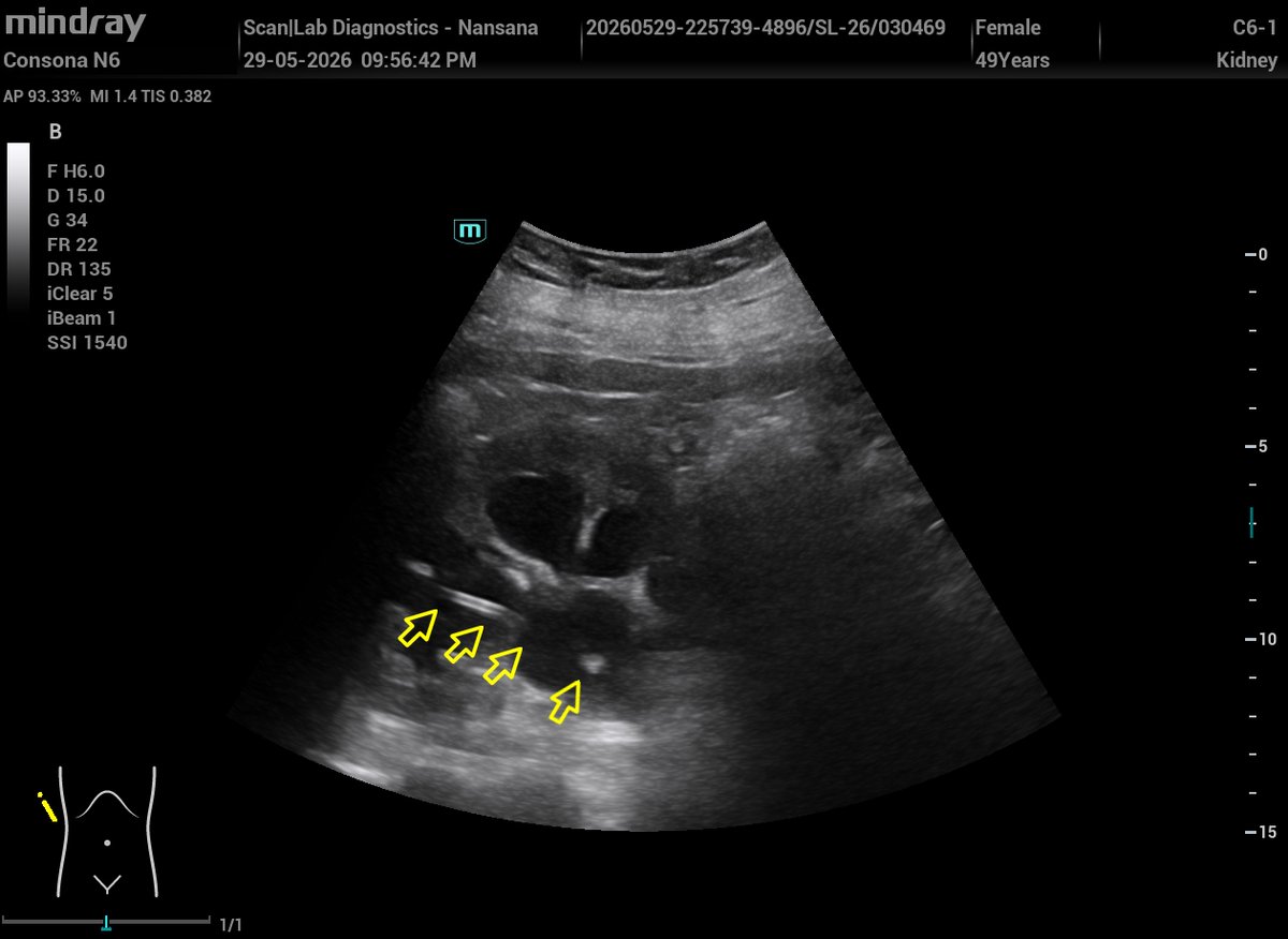
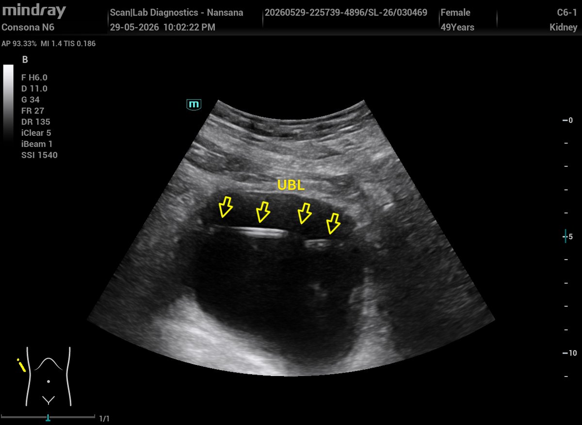
A 49-year-old female with known grade III hydronephrosis. Percutaneous nephrostomy (PCN) done previously; ureteric stent inserted 2 weeks ago.
Today's scan findings.
Right grade III hydronephrosis,
A ureteric stent with its proximal end in the renal pelvis and distal end in the urinary bladder,
Normal left kidney,
Normal left ureteric jet,
Absent right ureteric jet.


3
3
12
574
Scanlab Diagnostics retweeted
Ultrasound speaks. I interpret!
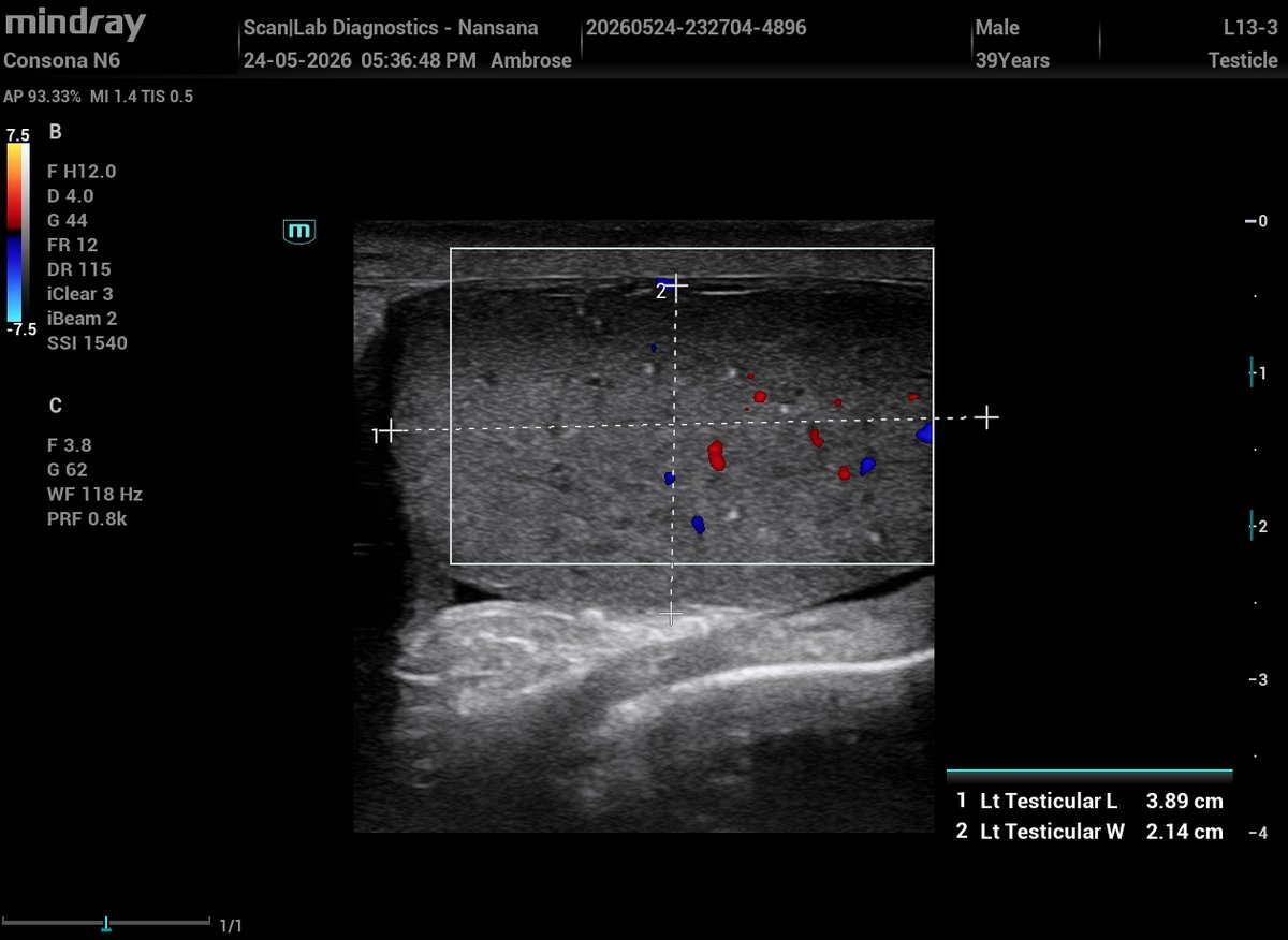
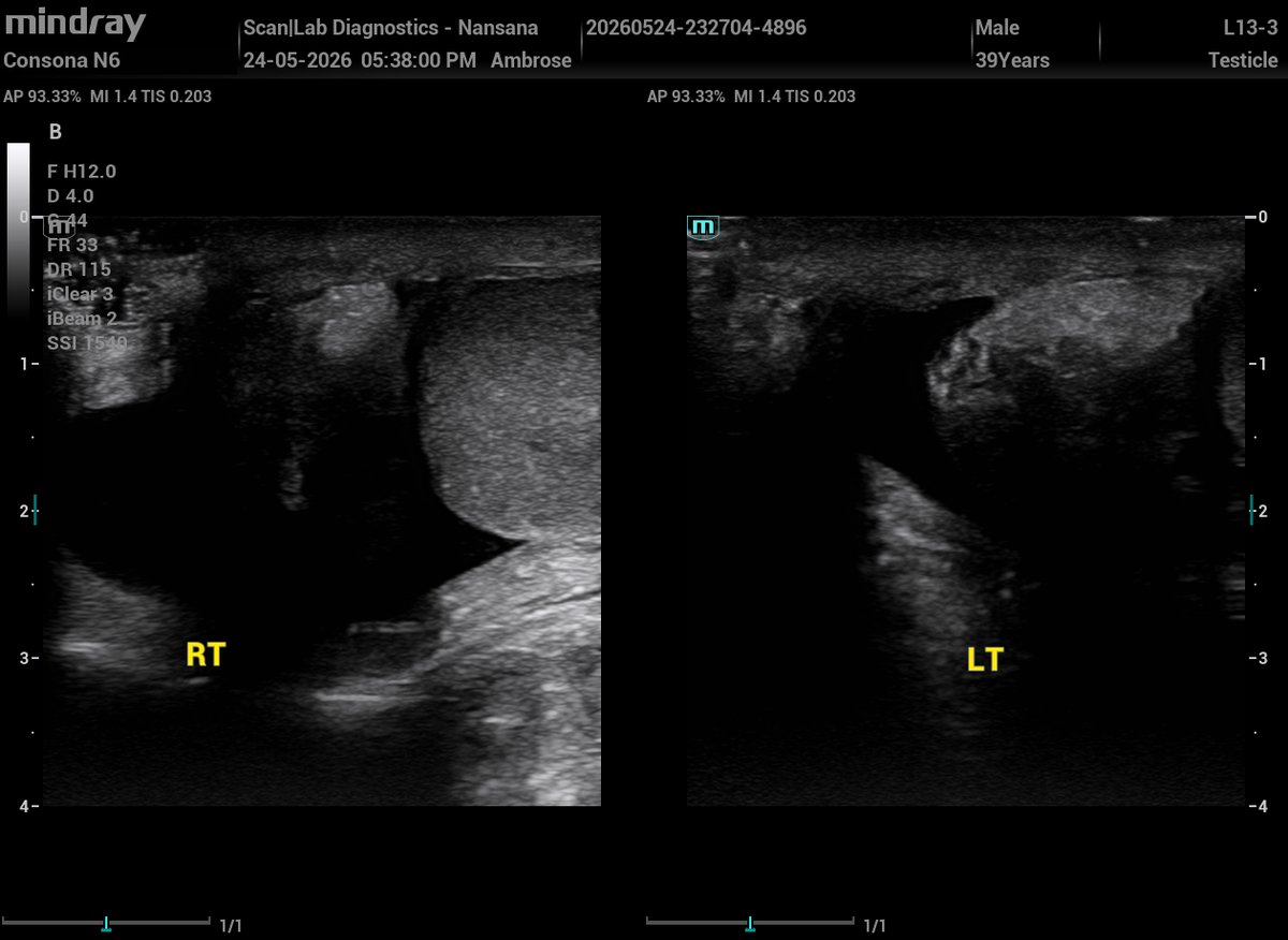
A 39‑year‑old male presenting with painful, palpable scrotal masses.
Sonographic findings:
The testes are normal size, shape, and contour,
Multiple intratesticular tiny echogenic foci in both testes,
The epididymides are thickened and heterogeneous,
Bilateral hydroceles.
Sonograms 👇👇


3
17
955
Scanlab Diagnostics retweeted
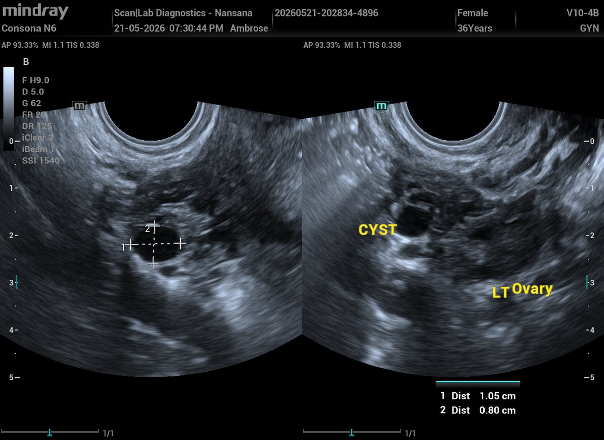
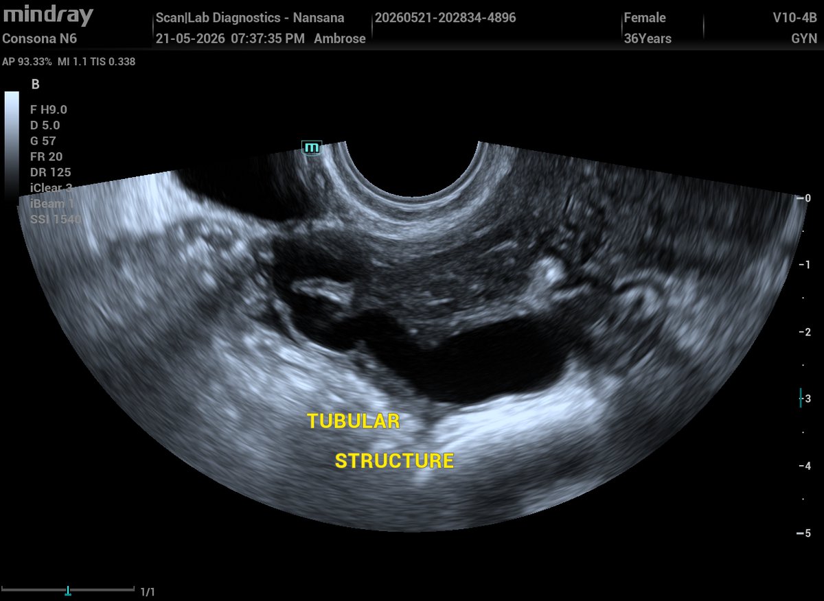
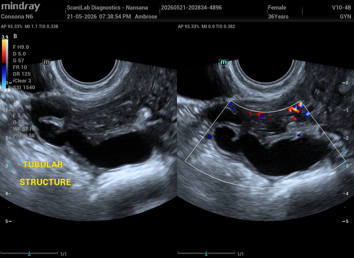
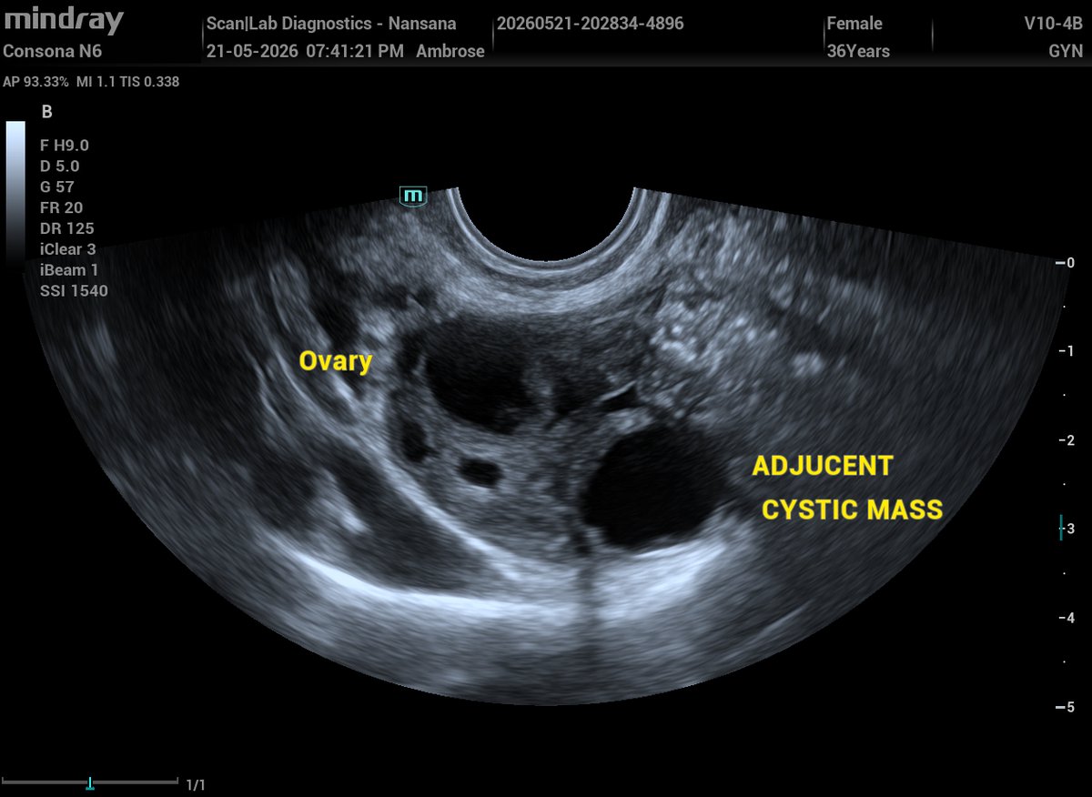
A 36‑yr‑old female with secondary infertility and h/o laparoscopic surgery in January this year, for right hydrosalpinx and left pyosalpinx.
TVS was performed today and here are the findings:
Normal uterus in size, shape and echotexture,
Normal trilaminar endometrium of proliferative phase,
A right adnexa avascular tubular structure adjacent to the ovary,
a small anechoic cystic mass with posterior acoustic enhancement in the left adnexa adjacent to the ovary
Normal ovaries in volume.
So;
What is the likely nature of the right adnexal tubular structure?
What are the differentials for the left anechoic mass in this postoperative context
Sonograms👇👇👇
1
1
17
910
Scanlab Diagnostics retweeted
Arsenal taught me 3 things:
Patience, Persistence and Gumite.
In sonography, those same principles guide every scan you perform;
Patience to find the perfect acoustic window, Persistence to uncover subtle pathology,
Gumite to deliver clarity even in complex cases.
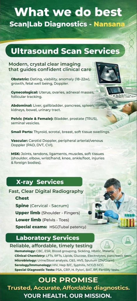
At @ScanlabNansan using these principles we offer: Obstetric scans, Gynecological scans (Transvaginal scans– TVS), Abdominal scans, MSK scans, Small parts (Breast, thyroid, scrotal etc) scans, Prostate scans including Transrectal scan (TRUS), Doppler scans to mention but afew.
We also provide X-ray and laboratory services 💪💪
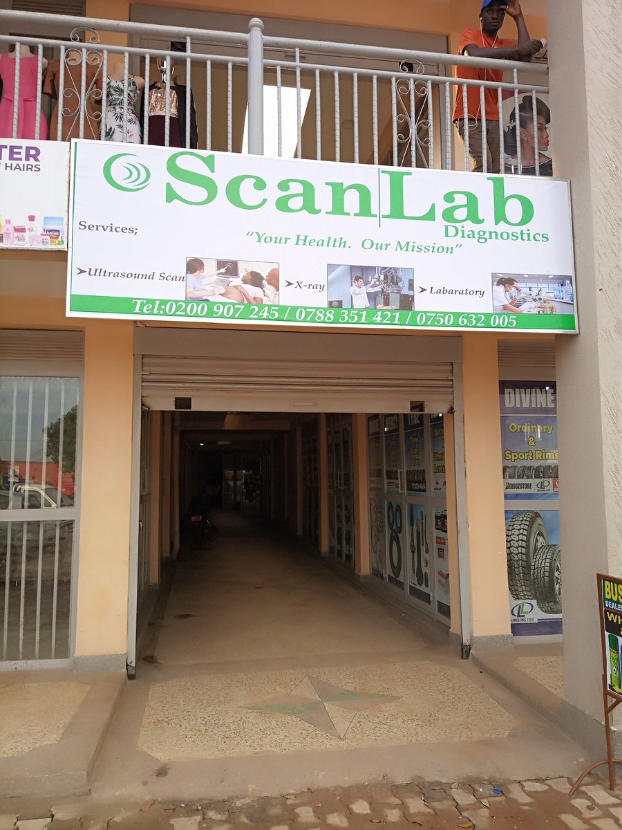
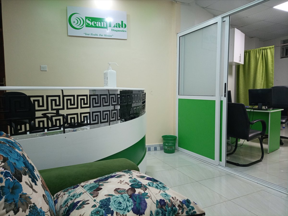
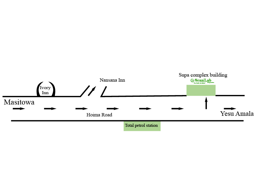
So, if you’re around Nansana and need a quality scan, step into @ScanlabNansana (Scan|Lab Diagnostics) for better medical imaging and laboratory services.
Your health. Our mission.”


1
3
174
Scanlab Diagnostics retweeted
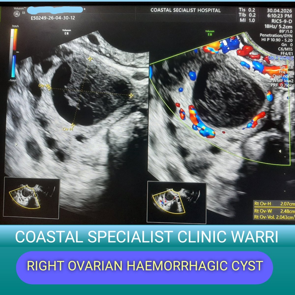
Right hemorrhagic ovarian cyst (HOC) in clot retraction phase. Thank you for this wonderful capture

3
13
480
Scanlab Diagnostics retweeted
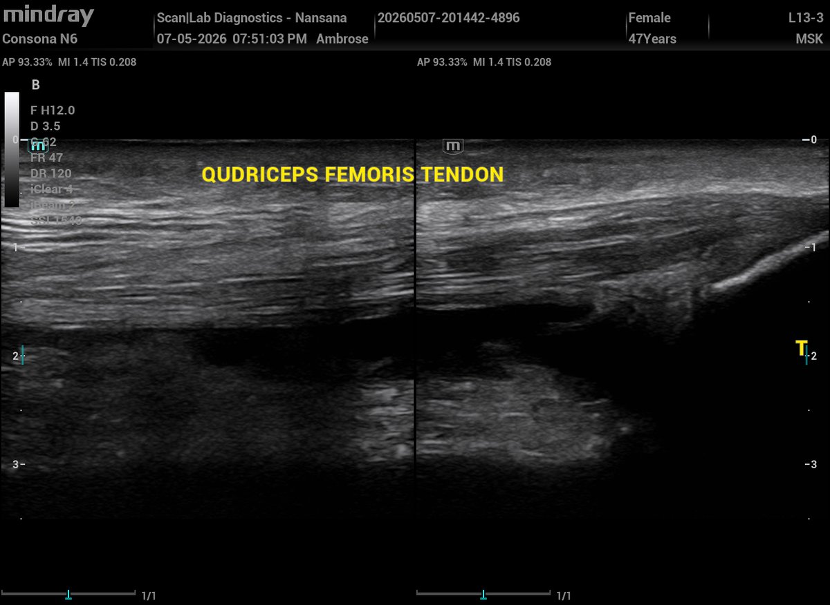
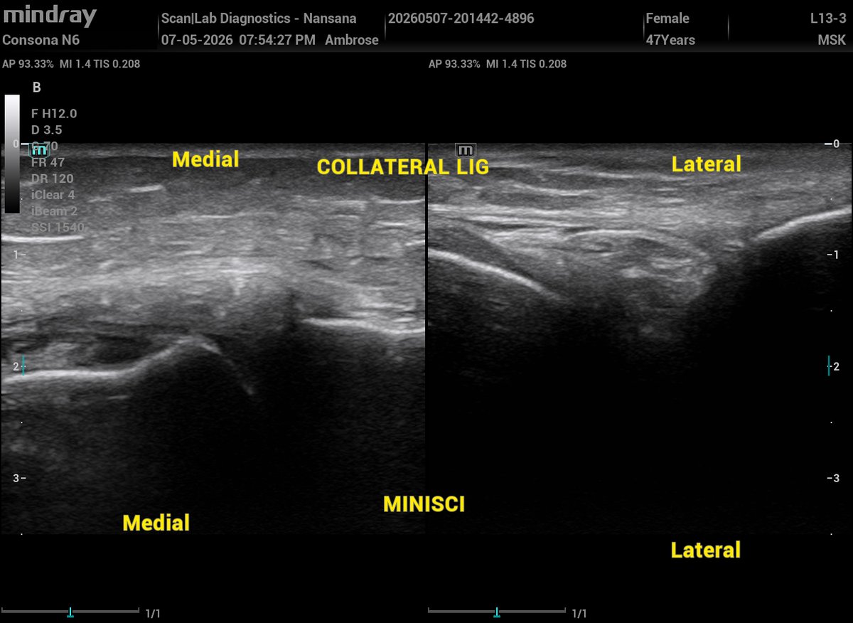
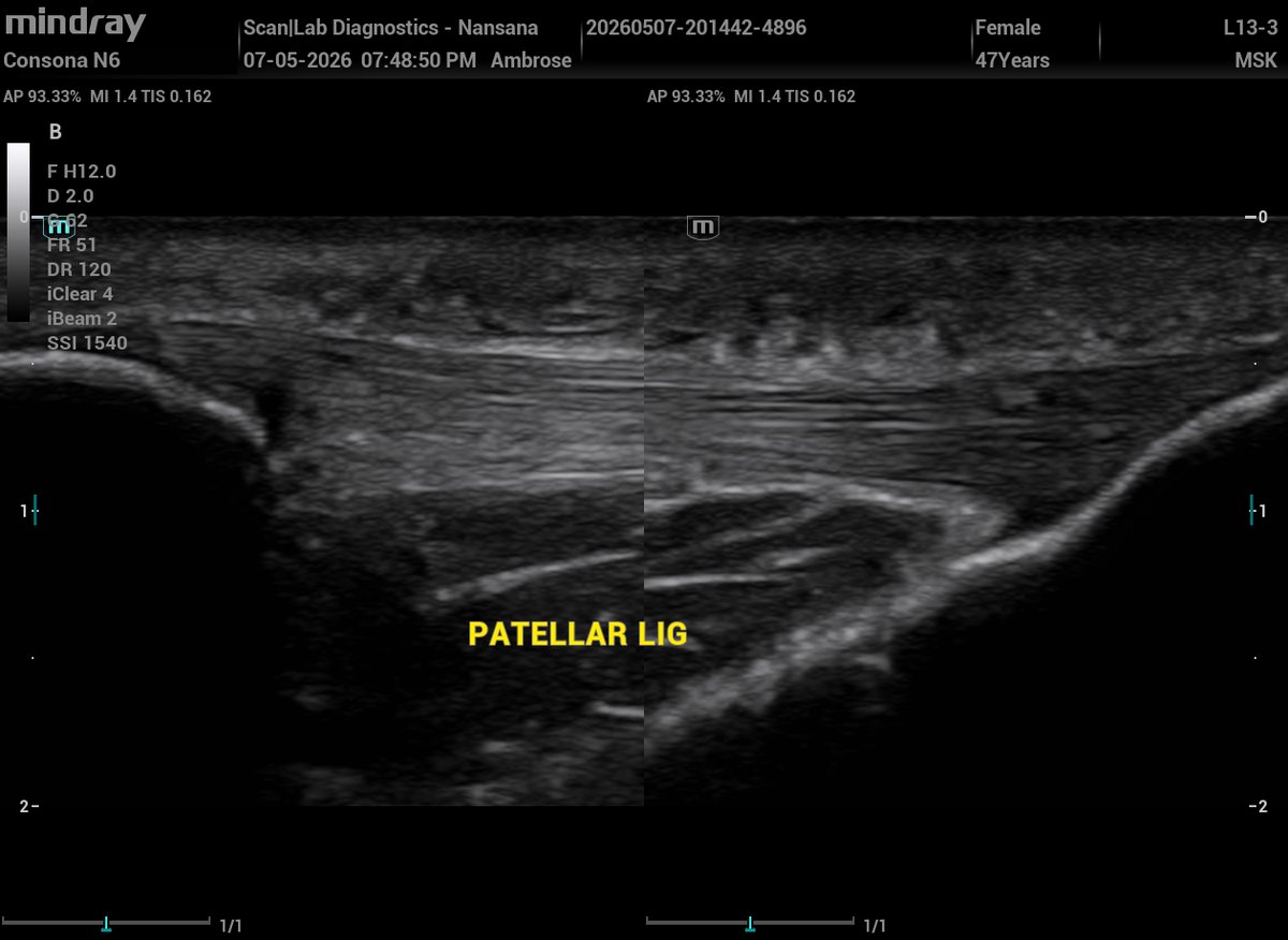
Ultrasound speaks. I interpret!!
Knee Ultrasound (MSK U/S).
A 47‑year‑old female presented with a painful and mildly swollen left knee.
MSK ultrasound was performed.
Findings:
There is a suprapatellar effusion distending the suprapatellar recess and tracking into both the medial and lateral parapatellar recesses.
In the posterior compartment, there is a Baker’s cyst; a well‑defined, fluid‑filled collection located between the medial head of the gastrocnemius and the semimembranosus tendon, with no internal vascularity on Doppler.
Sonographic features:
• Distended suprapatellar recess with free fluid
• Fluid extension into medial & lateral parapatellar recesses
• Intact quadriceps tendon
• Posteromedial fluid collection consistent with a Baker’s cyst
• No internal Doppler flow within the cyst
Here are the sonograms 👇👇

1
4
28
1,210
Scanlab Diagnostics retweeted
Ultrasound speaks. I interpret!!
Hematometra
A hematometra is a uterus distended with blood.
It occurs when there is partial or complete obstruction of the outflow tract, meaning menstrual blood cannot drain from the endometrial cavity.
When the outflow pathway is blocked, menstrual blood accumulates silently for weeks or months. Eventually, the pressure becomes unbearable leading to acute pelvic pain, amenorrhea, and a tense, distended uterus.
Ultrasound features of hematometra include:
Distended endometrial cavity,
Homogeneous fluid with low‑level internal echoes (old blood),
Possible fluid–fluid levels (wasn't present in my case),
No internal Doppler flow at color Doppler,
Cervix appears closed.
Yesterday, I received a 32‑year‑old woman with severe acute lower abdominal pain.
She reported h/o IUFD 1 year ago, and since then she has never resumed her menses.
TVS scan was performed and the findings were consistent with hematometra.
Here are the sonograms 👇👇


4
10
473
Scanlab Diagnostics retweeted
A fluid‑filled large bowel with pendulum movements strongly suggestive of mechanical intestinal obstruction.

2
9
436
Scanlab Diagnostics retweeted
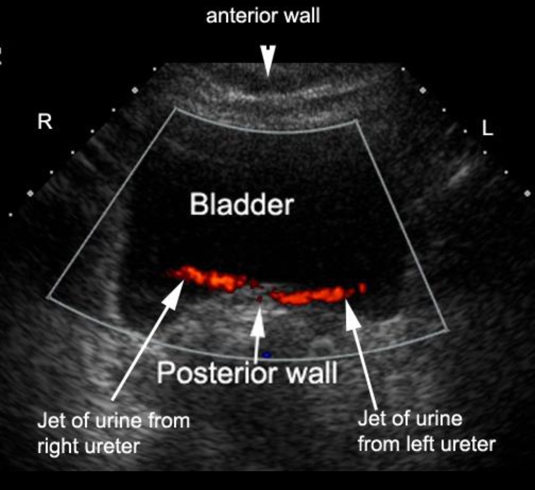
Trigone region

During a pelvic ultrasound, color Doppler demonstrates bilateral ureteric jets entering the posterior inferior aspect of the urinary bladder, forming a triangular region between the ureteric orifices and the internal urethral opening.
What is this anatomical region called?
2
11
288
Scanlab Diagnostics retweeted
Diastasis Recti
A 32 year old multigravida in her third trimester presents with a midline abdominal bulge that becomes more prominent on coughing.
Abdominal ultrasound scan shows widening of the linea alba with separation of the rectus abdominis muscles.
What is the most likely diagnosis?

5
18
564
Scanlab Diagnostics retweeted
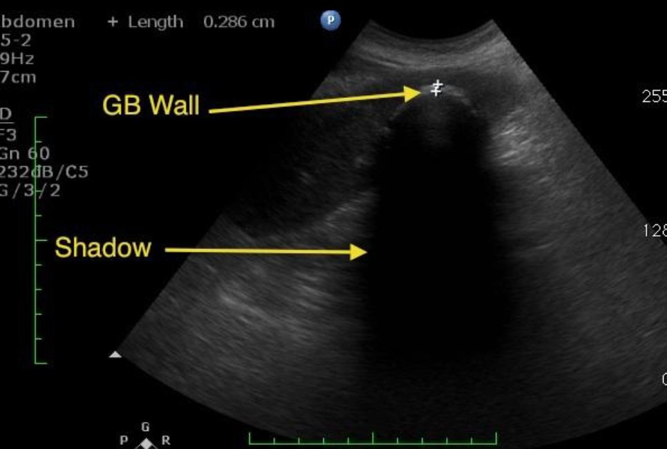
'WES' sign
A 42 year old patient presents with recurrent right upper quadrant pain after meals.
Ultrasound shows posterior acoustic shadowing from the gall bladder.
What classic ultrasound sign is demonstrated?
2
8
350
Scanlab Diagnostics retweeted
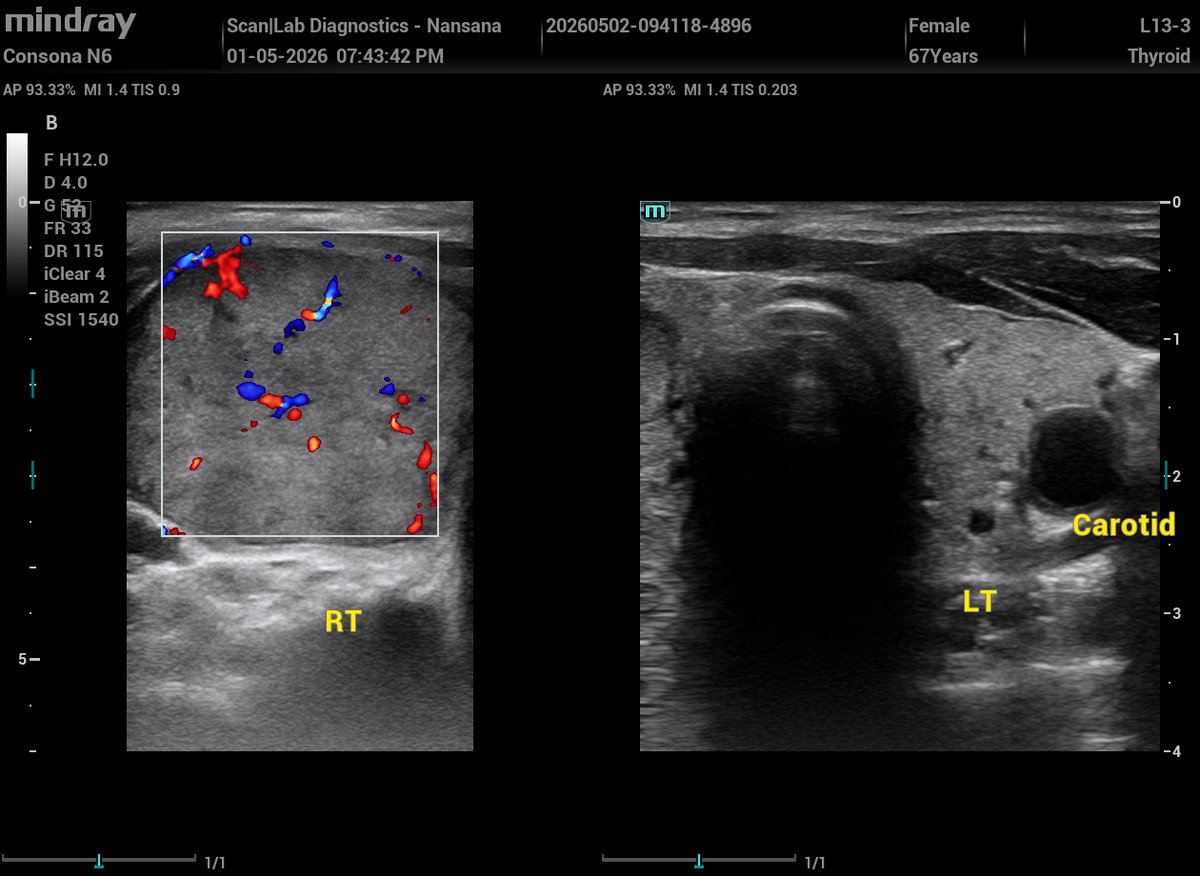
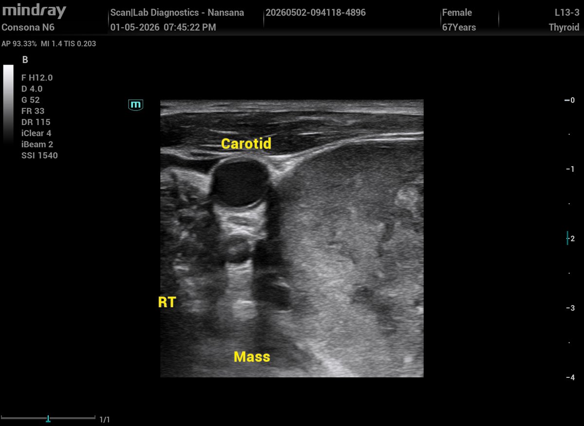
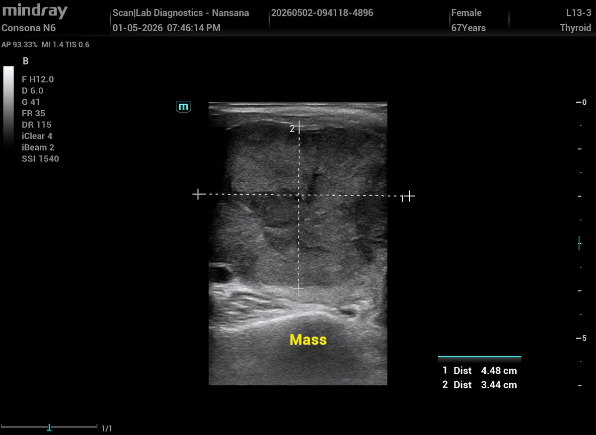
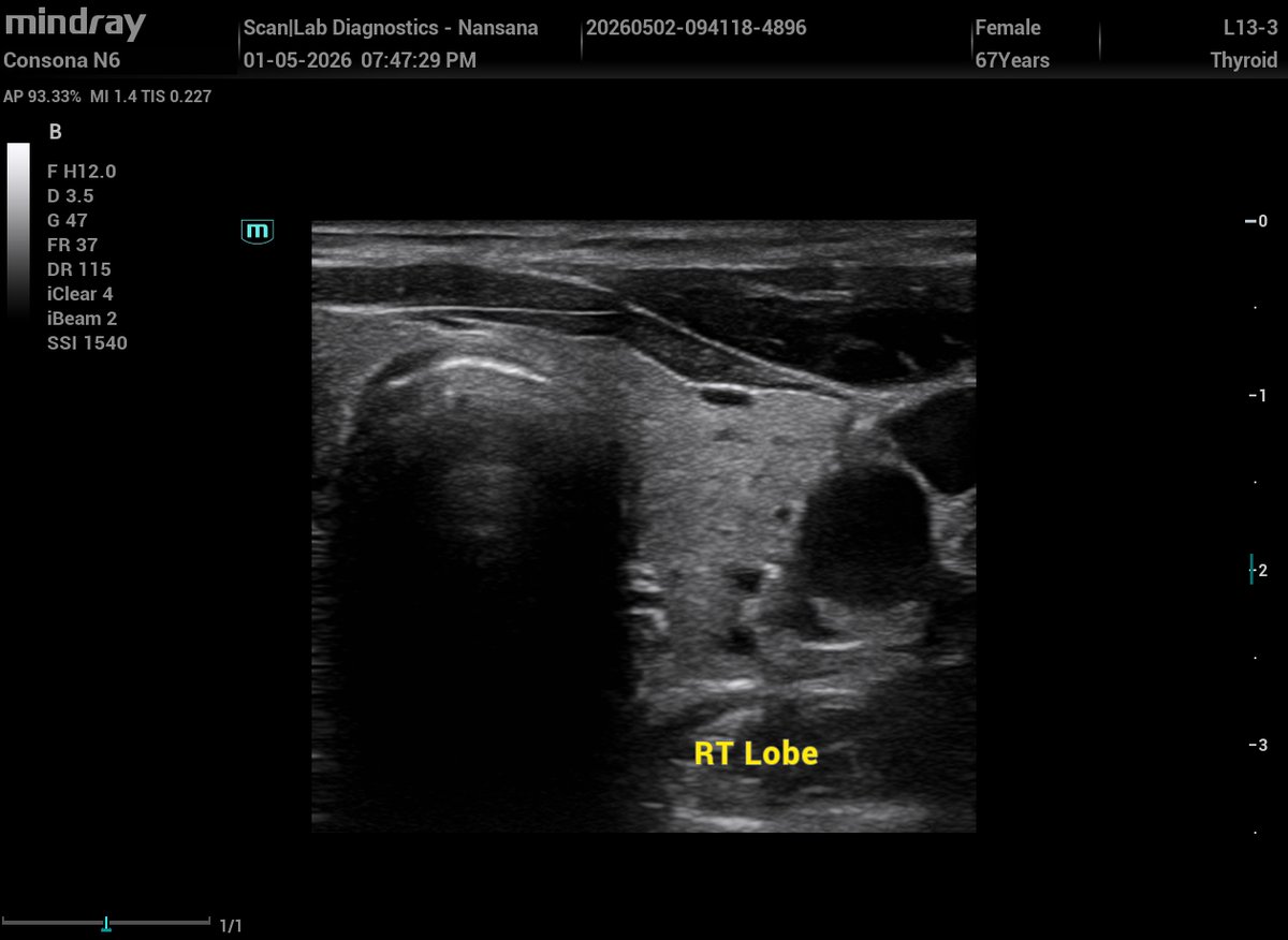
A 67-year-old lady c/o painless anterior neck swelling more to the right side.
Here are sonographic findings!
The right lobe is enlarged due to a dominant heterogeneous hyperechoic nodule occupying most of the lobe. The nodule measures (4.5x3.5) cm.
It shows increased vascularity at color Doppler imaging. There is minimal residual parenchyma thyroid visualized.
The left lobe is normal size; however, it contains tiny cystic nodules and small hyperechoic nodules. No suspicious masses seen.
The isthmus is normal in size and echotexture.
Sonograms 👇👇
Your thoughts
3
2
25
1,094
Scanlab Diagnostics retweeted
Are we not dealing with blighted ovum?
1
3
9
834
Scanlab Diagnostics retweeted
Amniotic Sheet (Synechia) vs Amniotic Band
Amniotic Sheet
Echogenic thick septa just like a separating septum in twin pregnancy,
A broad base attached to uterine wall, free edge but not free‑floating
No attachment to fetal parts,
Often shows Doppler flow along the membrane
Benign and incidental finding, but can influence fetal lie,
Always a single sheet.
Amniotic Band (ABS)
Thin, irregular, free‑floating strands
Attaches to fetal parts restricting movement
Can cause constriction rings, amputations, craniofacial defects
No Doppler flow
It is associated with fetal abnormalities
Case example:
At 34 weeks, a scan shows a thick septum arising from the placental edge and extending across the uterine cavity to the anterior wall.
This is a classic amniotic sheet; It is formed when the amnion drapes over a uterine synechia.
An amniotic sheet is a normal variant with no fetal risk whereas an amniotic band is pathological and can cause serious fetal deformities.
Amniotic sheet 👇👇

2
11
41
1,790