- Tweets 25
- Following 39
- Followers 257
- Likes 42


ALT DICER1 syndrome and thyroid disease. Michael Canfarotta et al. Journal of Pediatric Surgery Case Reports 11(C). June 2016. DOI:10.1016/j.epsc.2016.05.014

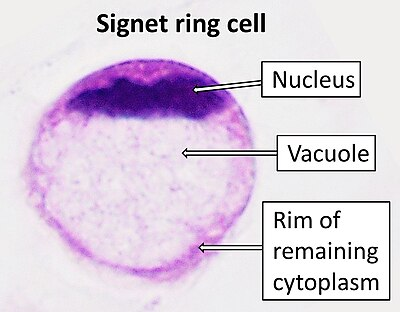
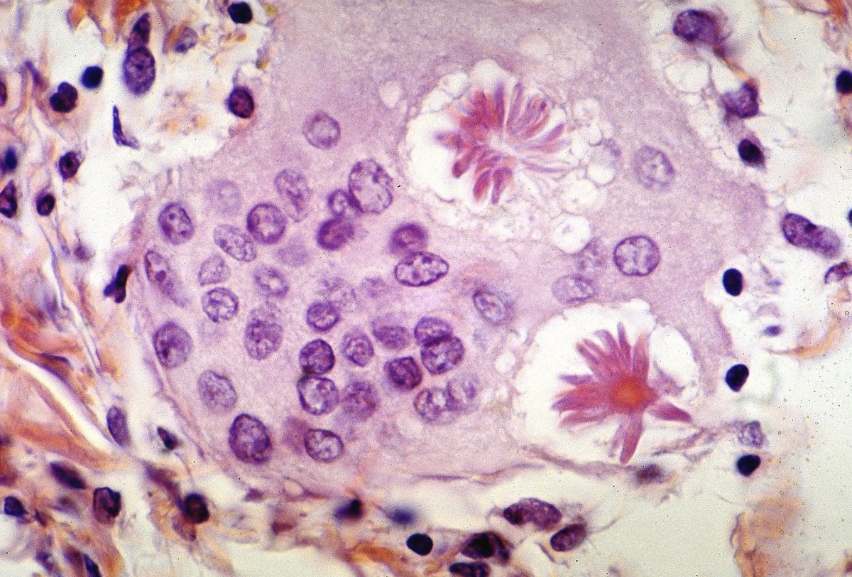
ALT Photo Credits: Bella Lingjia Liu, M.D. (Pathology Outlines)
ALT Photo Credit: Mikael Häggström, M.D. (Wiki)

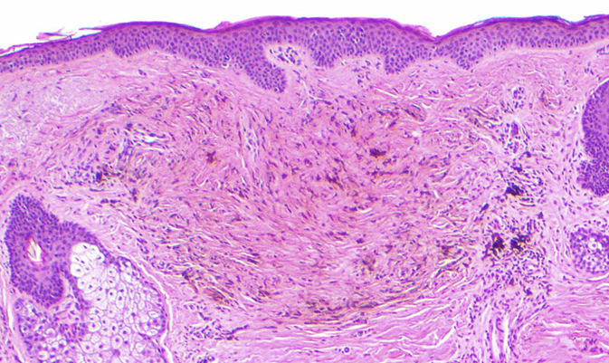
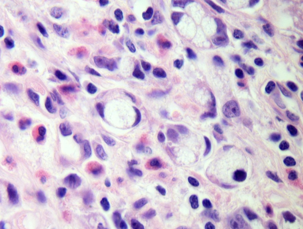
ALT Photo credits: Eric W. Hossler, M.D. (Pathology Outlines)
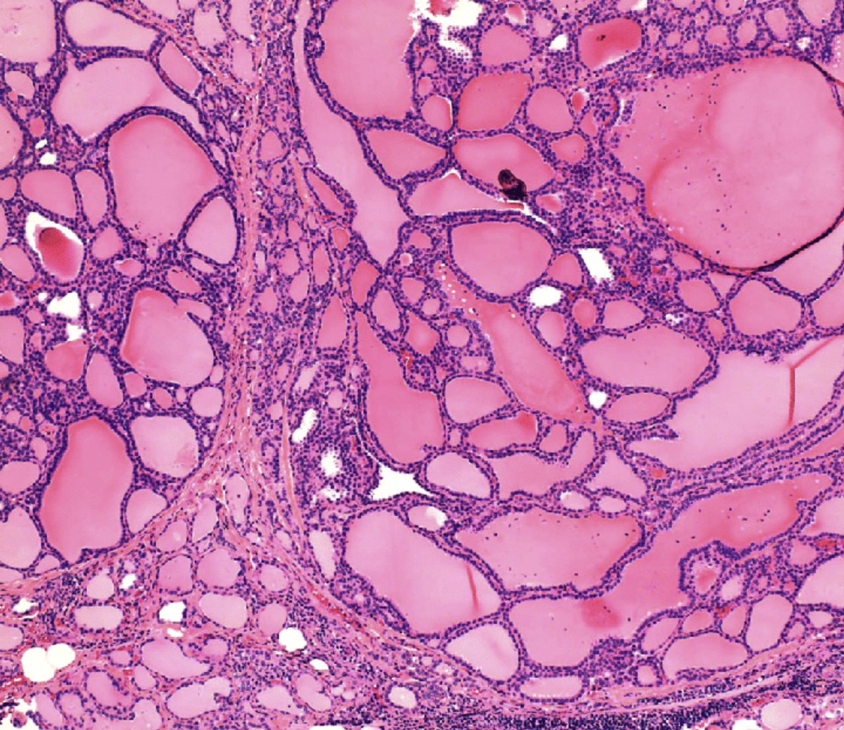
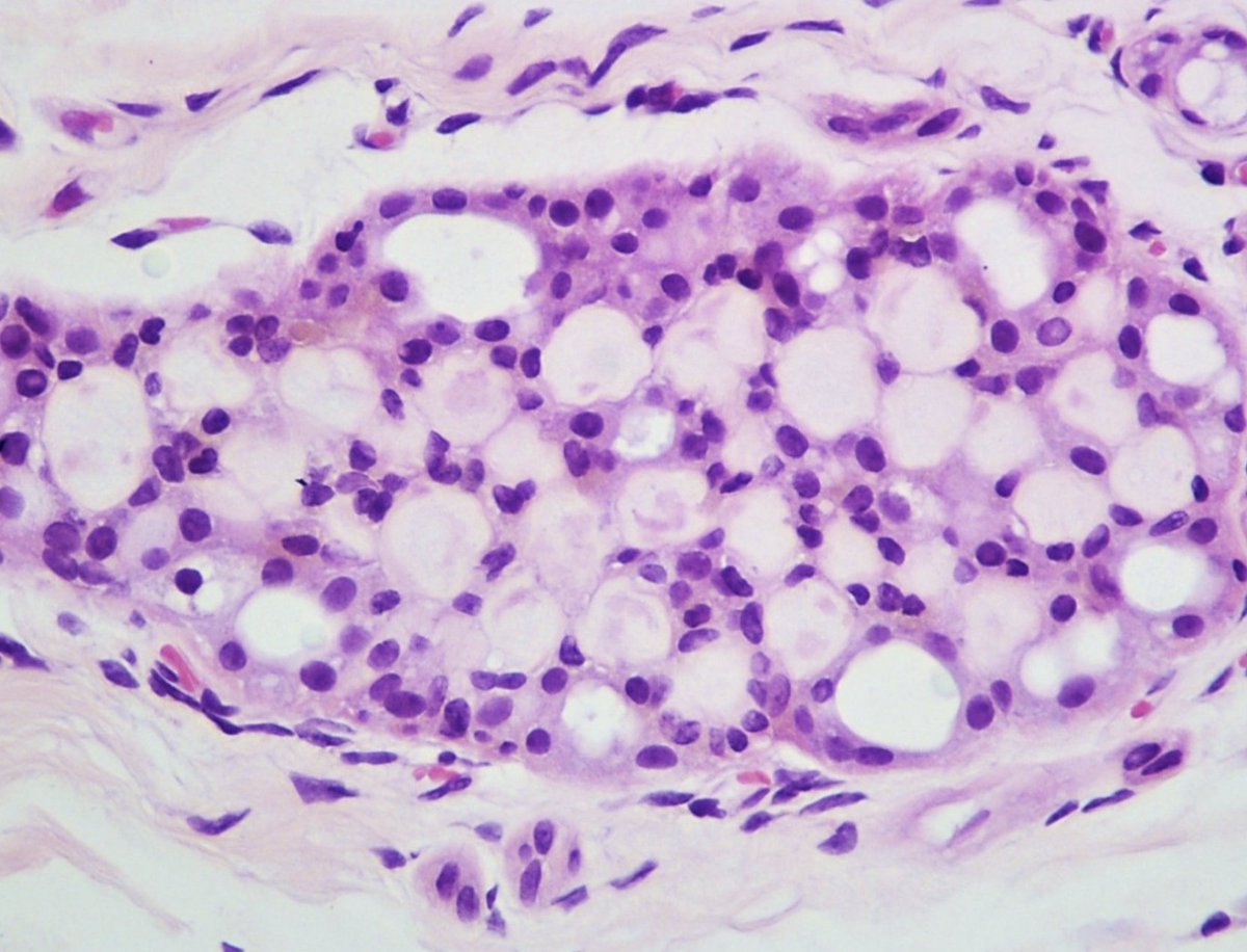
ALT Credit: Iskender Sinan Genco, M.D. and Sabina Hajiyeva, M.D. (Pathology Outlines)
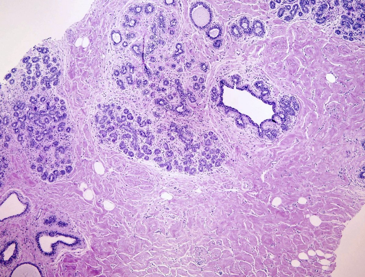
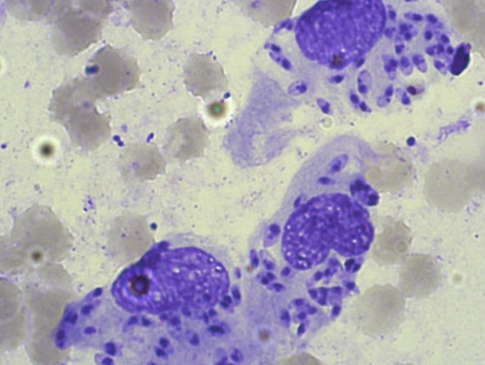
ALT Credit: Pathology Outlines and Puri S, Mohindroo S, Gulati A. Collagenous spherulosis: An interesting cytological finding in breast lesion. Cytojournal. 2015;12:25. Published 2015 Nov 30.

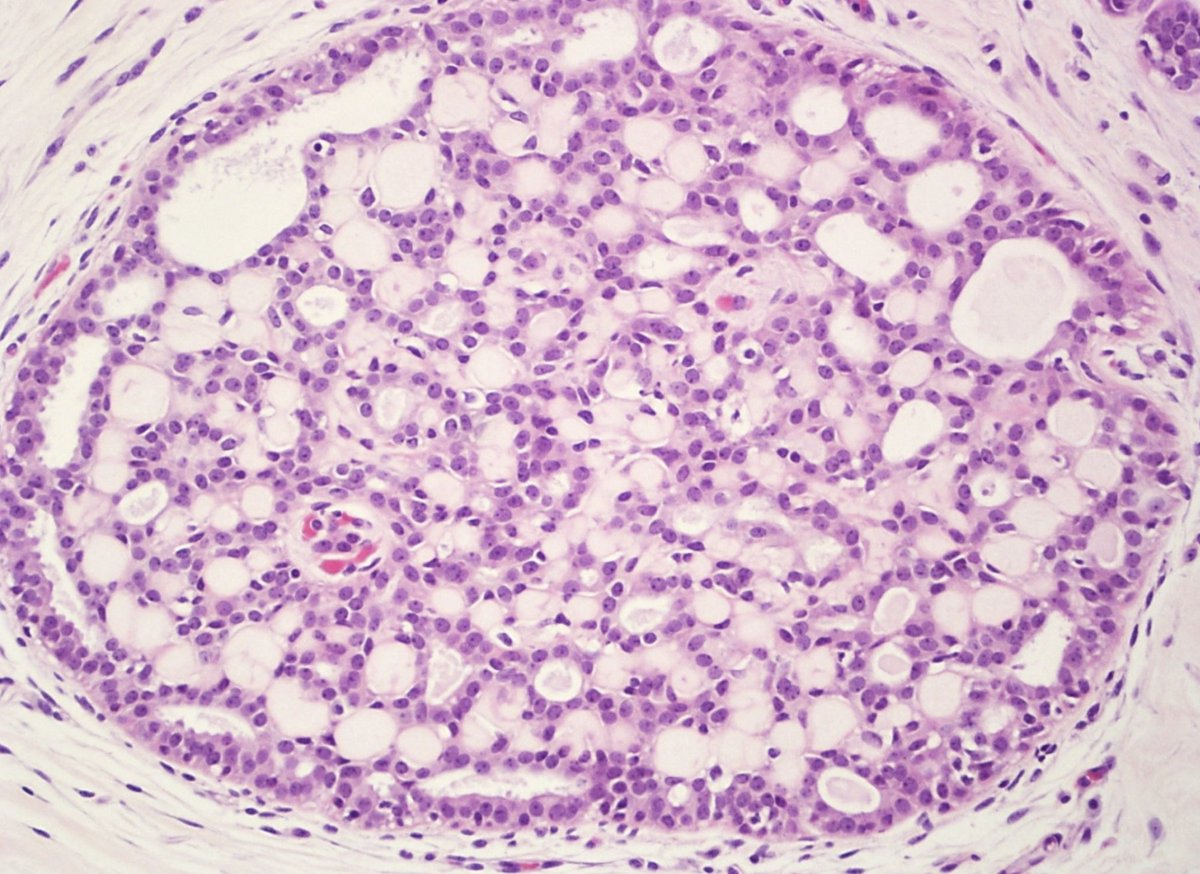
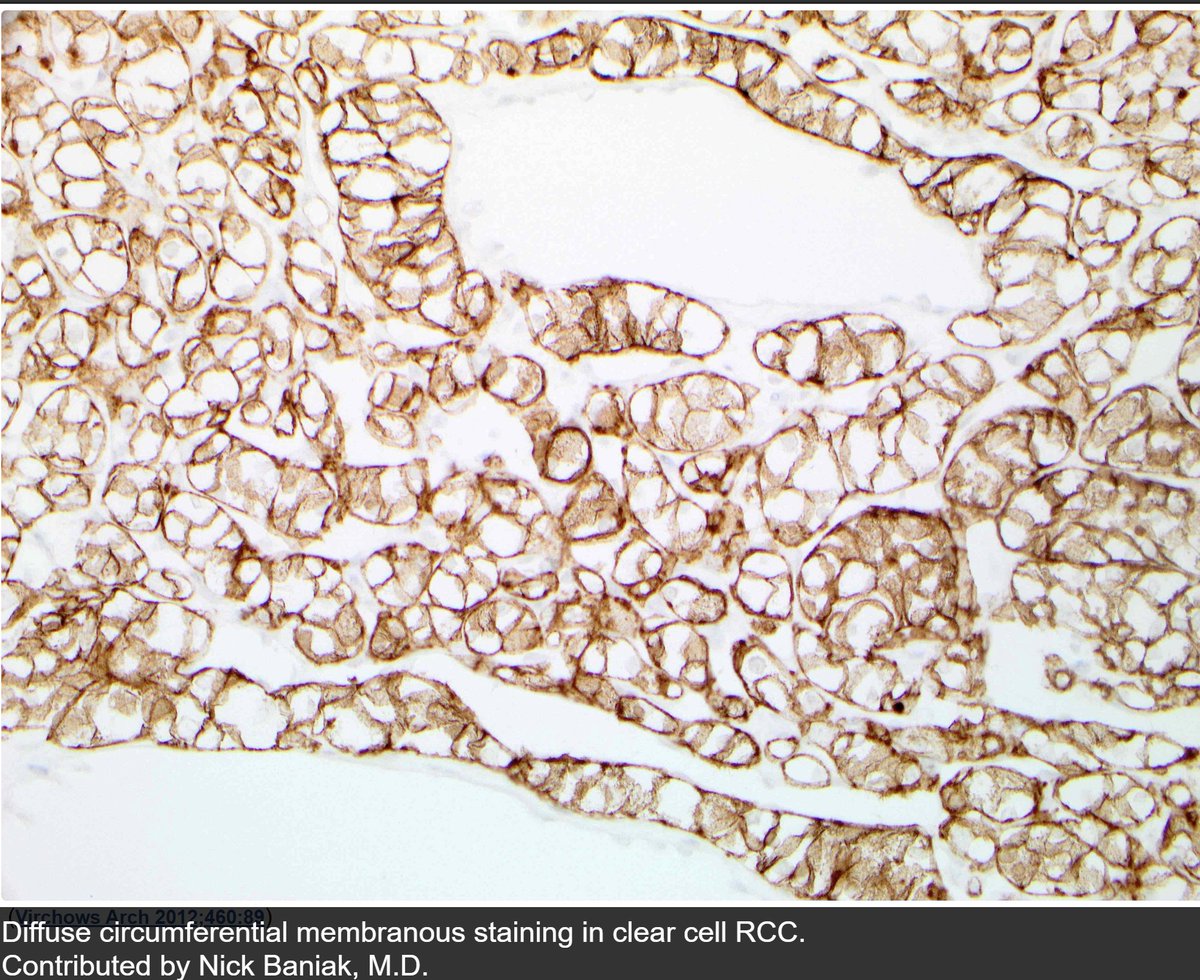
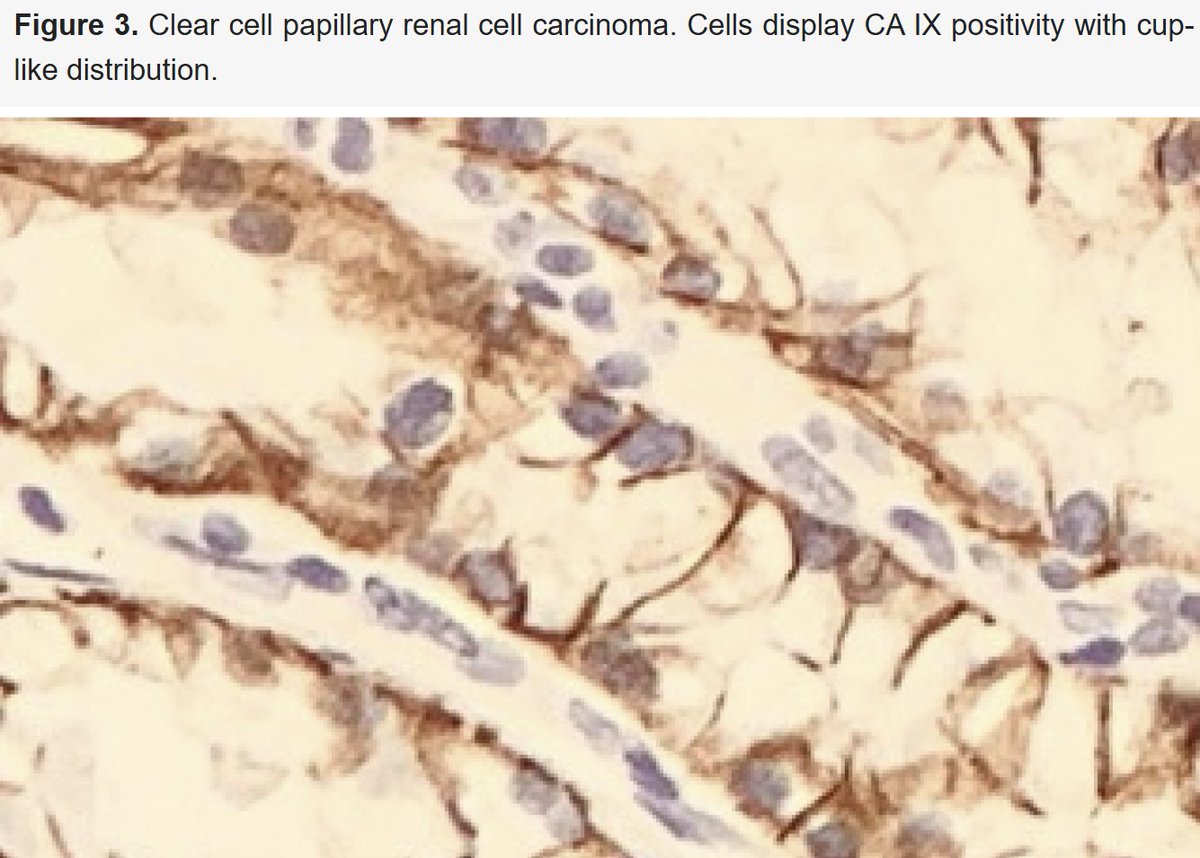
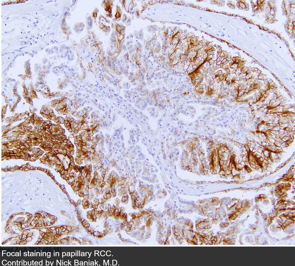
ALT Figures 1 and 3: Pathology Outlines Figure 2: Courcier et al. Carbonic Anhydrase IX in Renal Cell Carcinoma, Implications for Disease Management. International Journal of Molecular Sciences. 2020; 21(19):7146.


ALT Dr. Yale Rosen Atlas of Pulmonary Pathology (Flickr)
ALT 1) Indian J Radiol Imaging. 2016 Apr-Jun;26(2):216–225. doi: 10.4103/0971-3026.184409 2) Julie M. Jorns, M.D. (Pathology Outlines)
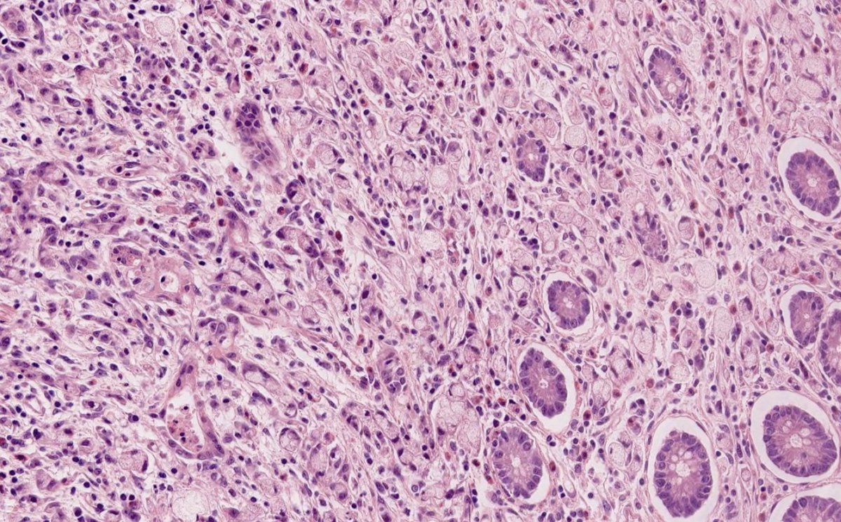
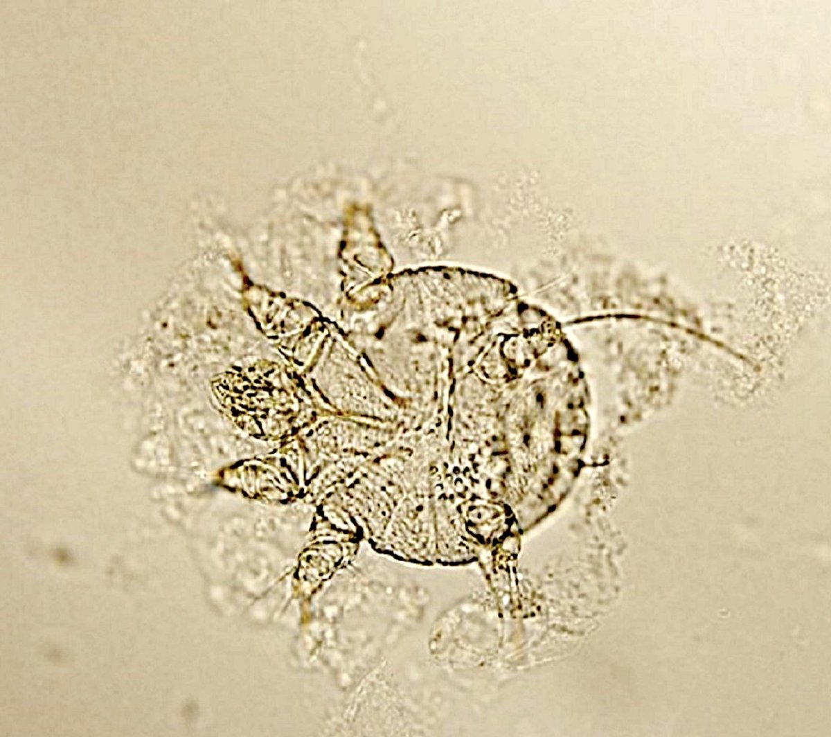
ALT Credit: Indu Agarwal, MD (Pathology Outlines)
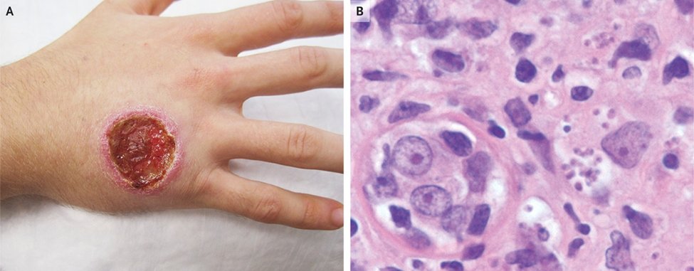
ALT Cutaneous Leishmaniasis, Alexia Knapp, MD, Jonathan Alpern, MD https://www.nejm.org/doi/10.1056/NEJMicm1908092
ALT Joachim Richter https://www.researchgate.net/figure/Leishmania-amastigotes-Giemsa-stain-in-a-smear-of-the-buccal-mucosa-patient-1_fig3_51057580
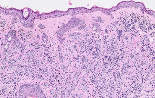
ALT Credit: pathology outlines, Yuri Tachibana, MD and Carlos Torres-Cabala, MD