The ALMF is a part of the Center for Open Research Resources and Equipment at UConn. We provide microscopy expertise and access to cutting edge instruments.
- Tweets 46
- Following 13
- Followers 79
- Likes 20

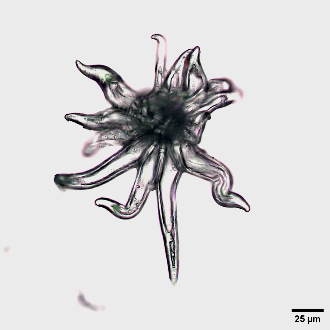


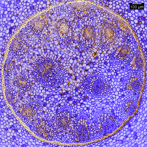
ALT Image of a fern rhizome acquired with a 20X objective and K5C True Color Camera on a Leica Thunder.
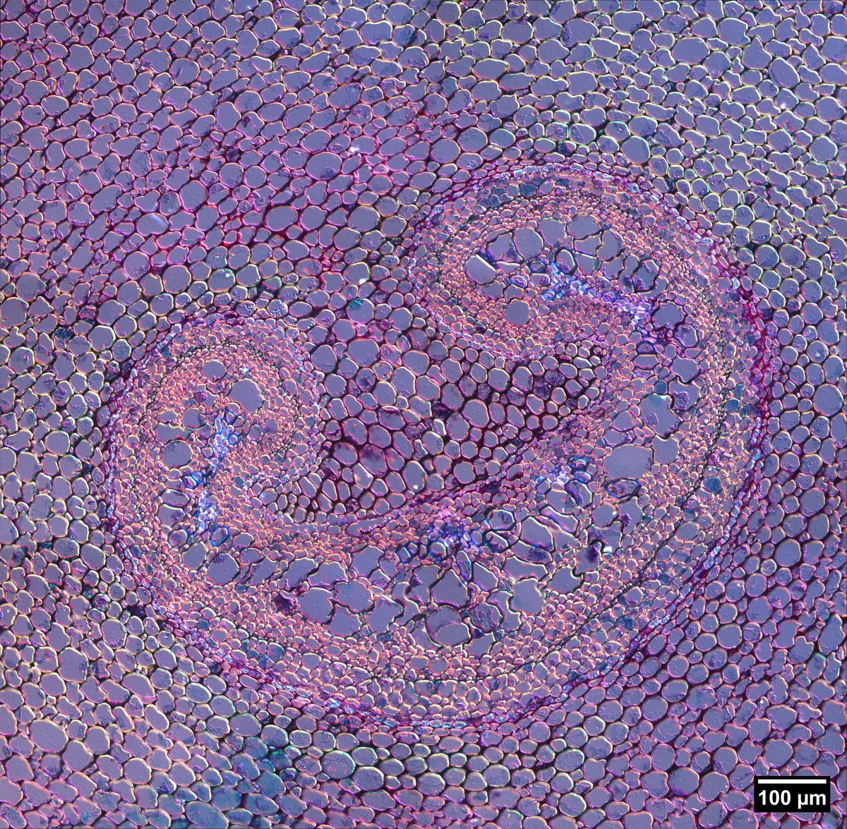
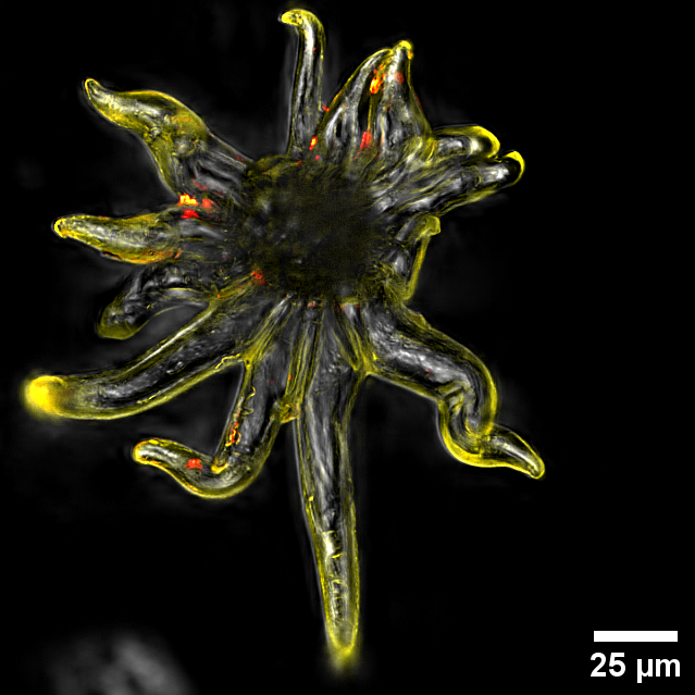
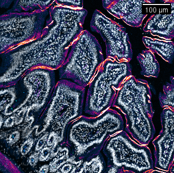
ALT Image of a fern rhizome acquired with a 20X objective and K5C True Color Camera on a Leica Thunder with a DIC prism in the lightpath.
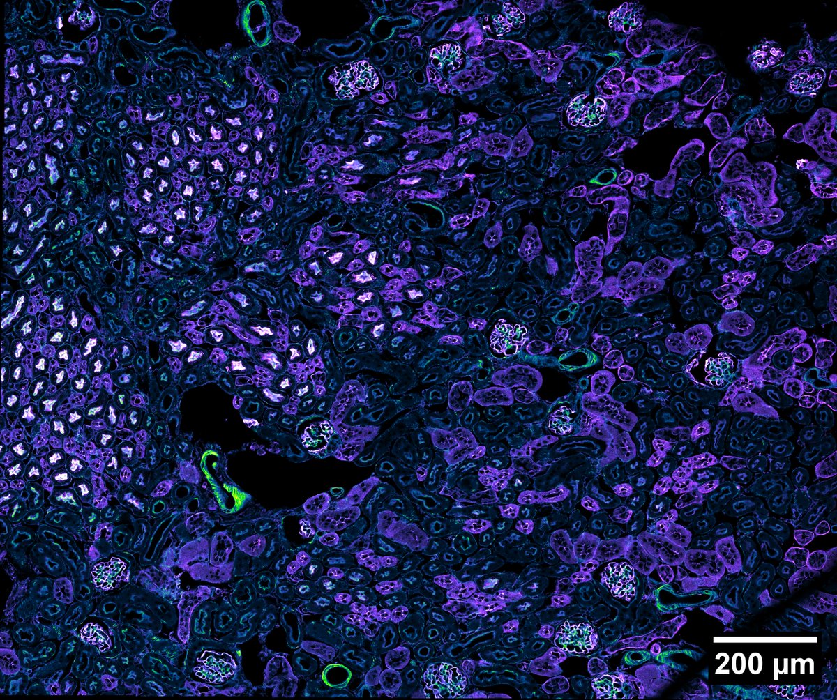
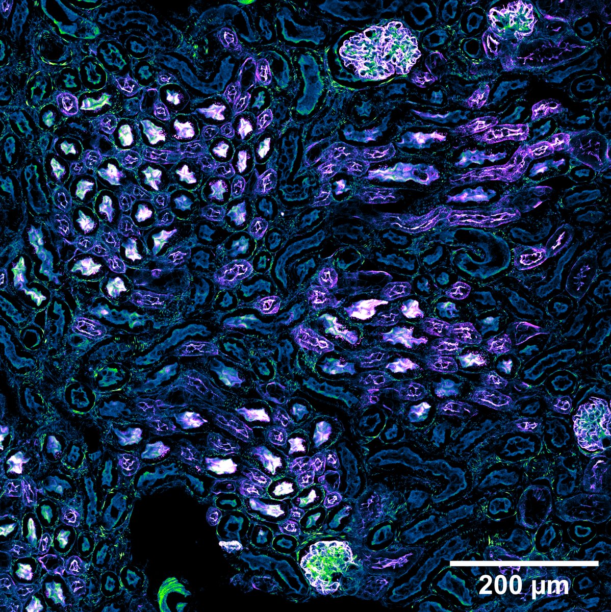
ALT Molecular probes kidney section 2D large image overview with a 40X oil objective on a Nikon AXR confocal. Phalloidin is shown in the Green Fire Blue LUT and wheat germ agglutinin (WGA) is shown in the Magenta Hot LUT on FIJI.
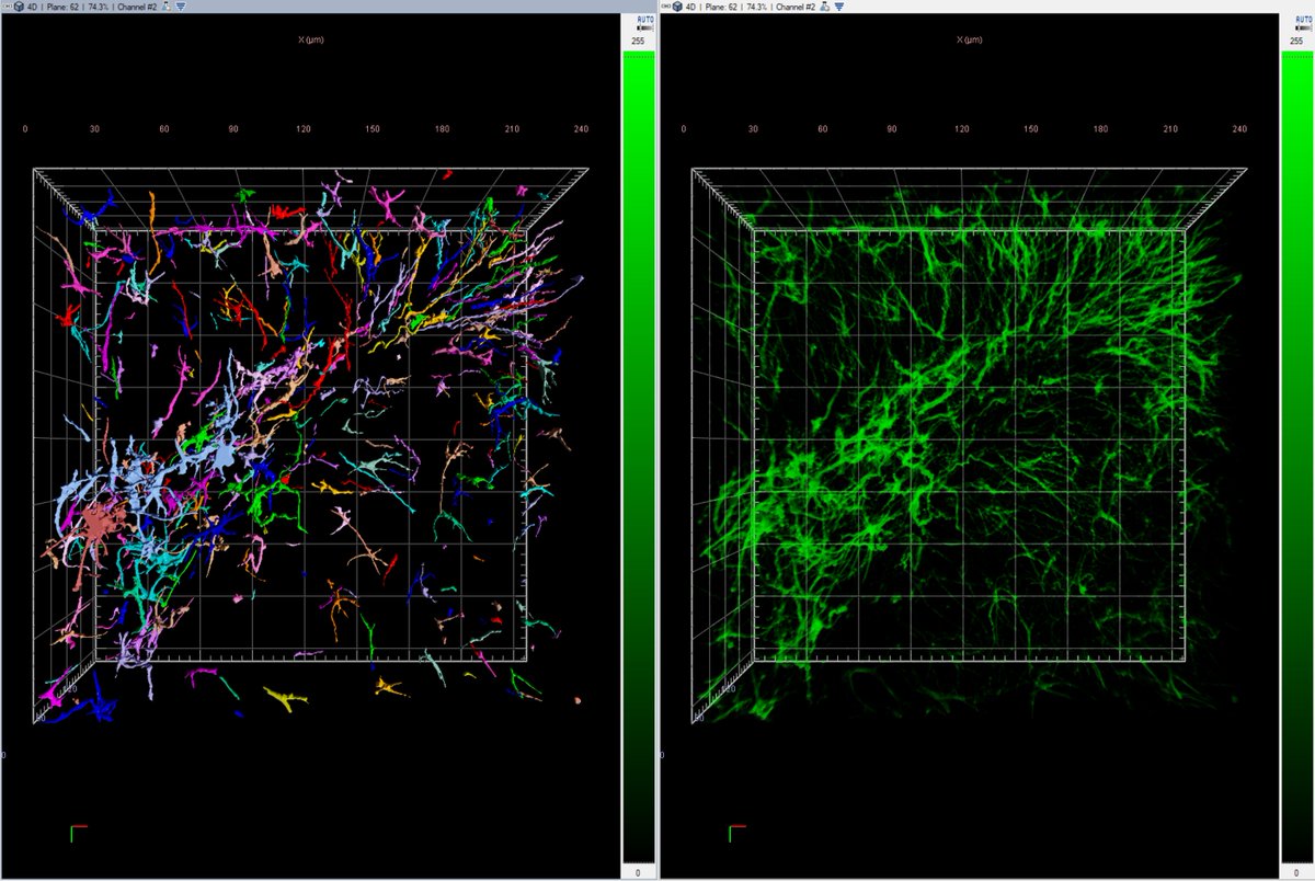
ALT Max intensity projection of stitched tiles from a square ROI from the previous overview. Phalloidin = Green Fire Blue; WGA = Magenta Hot.
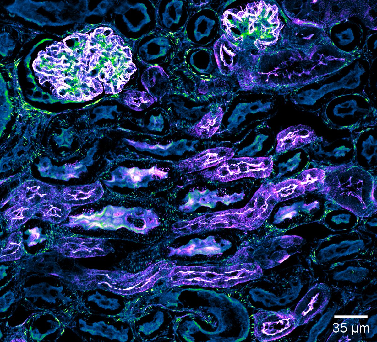
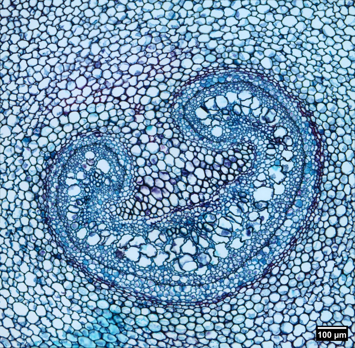
ALT Cropped region from the previous max intensity projection image; Phalloidin = Green Fire Blue; WGA = Magenta Hot.
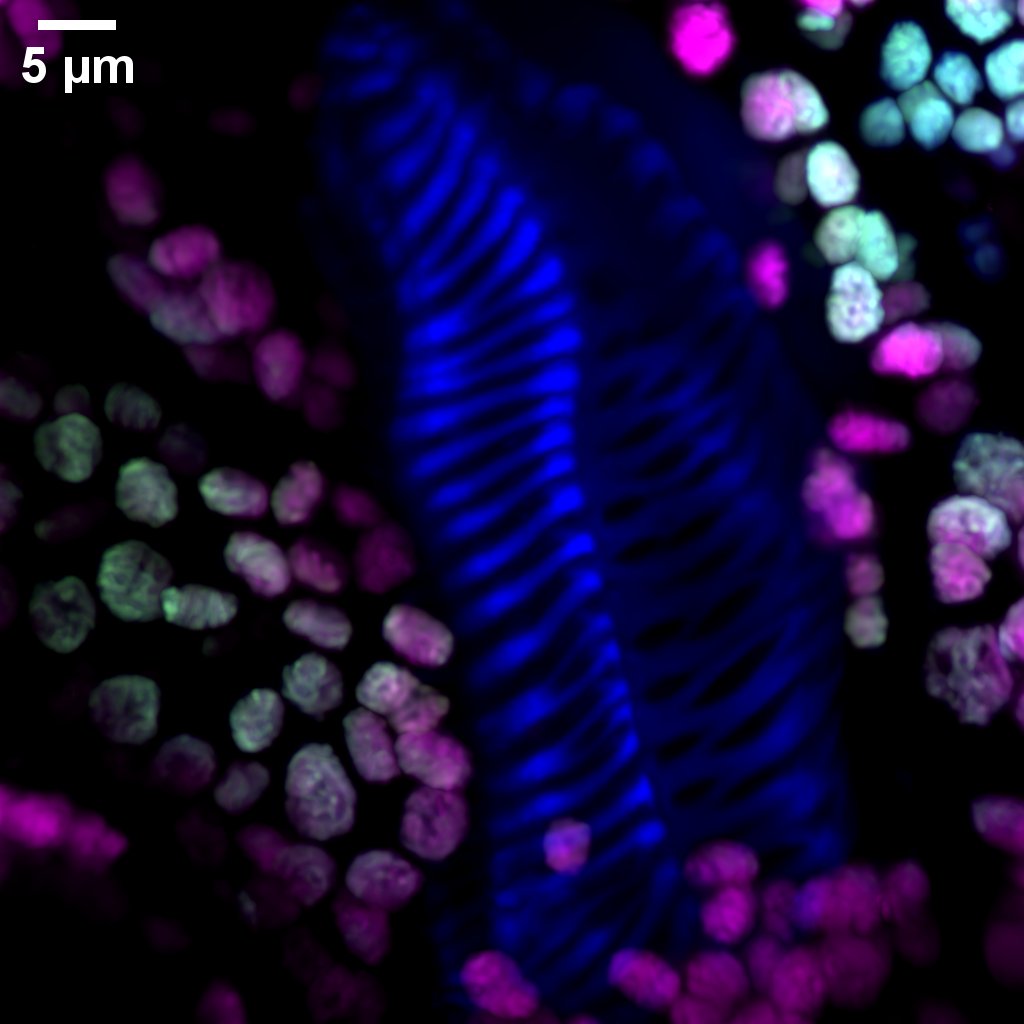
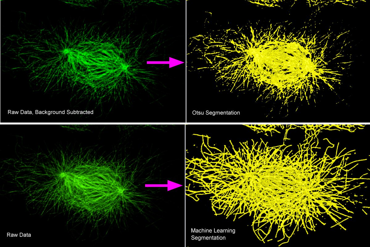
ALT 3D views of tubulin distribution in a prometaphase BPAE cell. For Otsu segmentation, background was first subtracted. For machine learning segmentation, the algorithm was trained manually on the raw data by defining 2 classes, background and objects

ALT Page 1 of UConn COR²E Summer 2023 Newsletter