
microCT scanning Core Lab, located at the University of Michigan Museum of Zoology
Joined April 2024
- Tweets 7
- Following 10
- Followers 10
- Likes 1
5 Photos and videos


UMich Zoology CT Lab retweeted

18 Jun 2024
My first chapter is out! This was an enormously challenging project but I learned a lot and am very excited to share with you the hidden world of ultraviolet coloration in snakes.
nature.com/articles/s41467-0…
9
35
115
10,375

31 May 2024
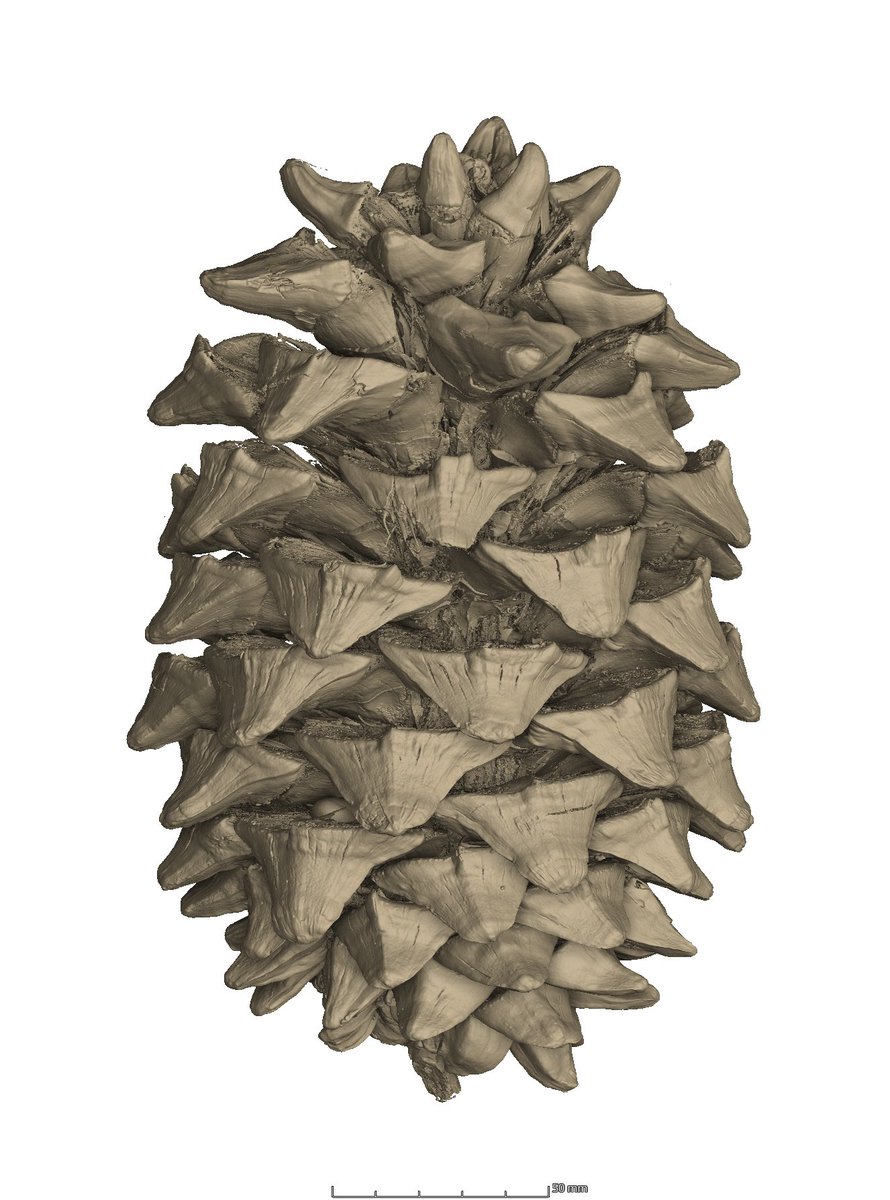
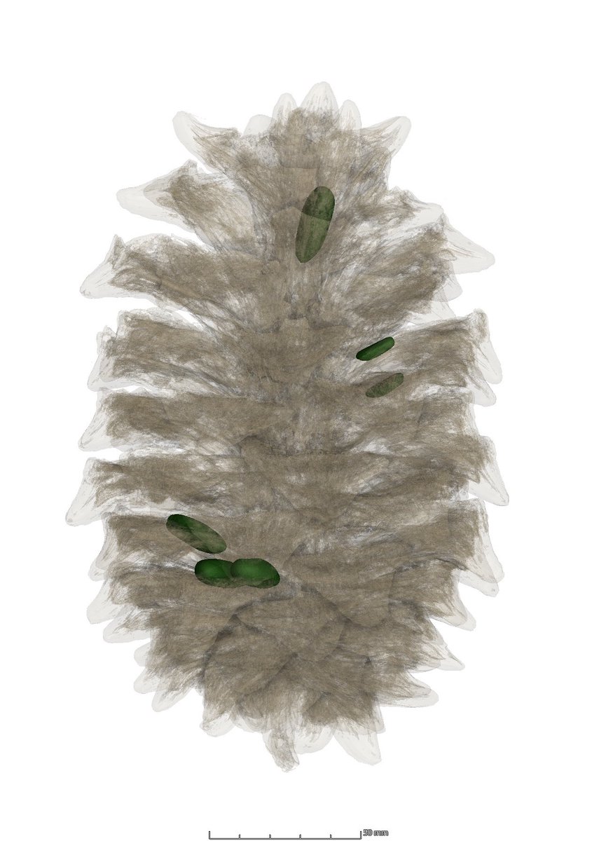
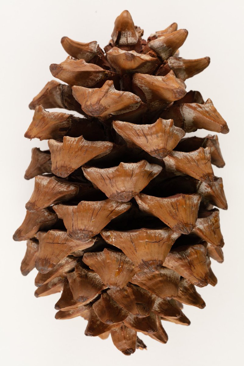
Adding to our herbarium scanning collection, we have a pinecone from a Martinez pinyon (Pinus maximartinezii). This large pinecone scanned in less than 15 minutes, allowing us to see all of the internal structures including the seeds!

1
3
408
17 May 2024
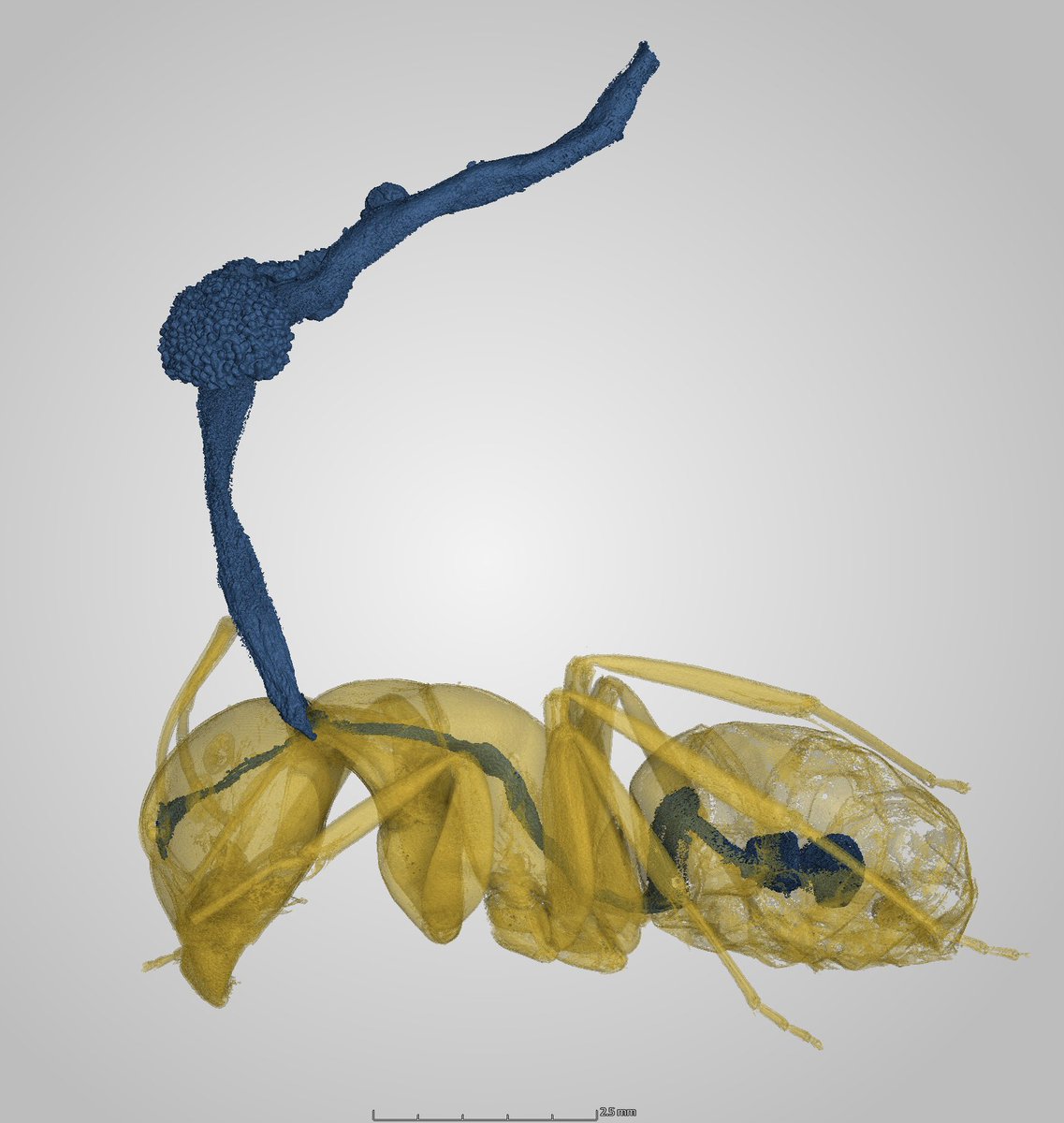
Taking our CT lab in a new direction, scanning fungus! This ant has been infected by a species of cordyceps fungus (that’s same kind of fungus that creates zombies in the Last of Us!)
#fungifriday #ctscan #thelastofus #cordyceps @NIMactual

1
6
19
784

8 May 2024
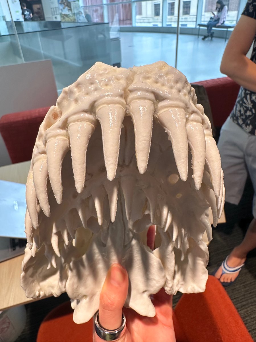
3D mesh of a caecilian skull! If you’re not familiar with them, caecilians are a type of amphibian with a snake-like body form. They live underground and are often referred to as the sharks of the soil due to their large, sharp teeth!
sketchfab.com/3d-models/caec…
23
1 May 2024
AI-generated animation of a twin-spotted rattlesnake (Crotalus pricei) from our skeletal and diceCT scans! Here you can see that scanning specimens as-is gives us a clear visual of the bone, while staining specimens in iodine (dice-staining) allows us to see soft tissue.
31
29 Apr 2024
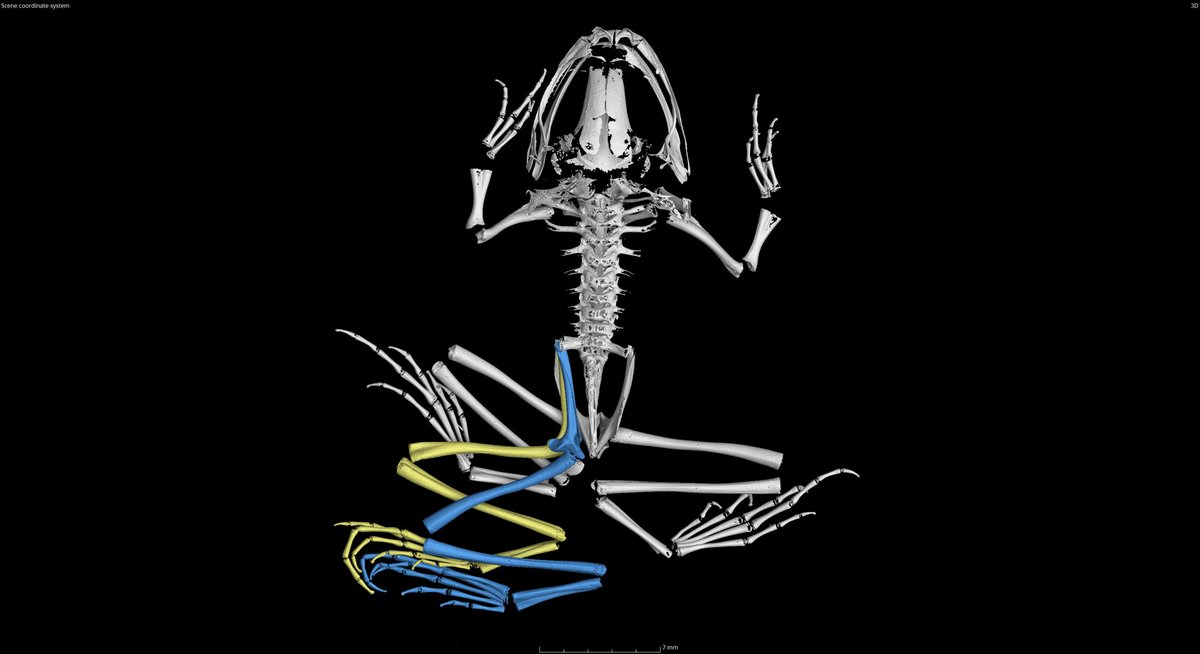
Welcome to the home of all things microCT at the University of Michigan Museum of Zoology! Kicking things off with this skeletal scan of a green frog (Lithobates clamitans) that has an unusual number of legs. This specimen was collected right here in Washtenaw county!
43