
Official X Account of the Molecular Medicine Lab | Virus Cell Biology @ ETH Zürich (Swiss Federal Institute of Technology Zürich)🇨🇭
Joined July 2019
- Tweets 138
- Following 127
- Followers 577
- Likes 66
23 Photos and videos


One of the most-viewed PNAS articles in the last week is “Enhanced visualization of influenza A virus entry into living cells using virus-view atomic force microscopy.” Explore the article here: ow.ly/UiEG50XHyci
For more trending articles, visit ow.ly/e8nb50XHycg.
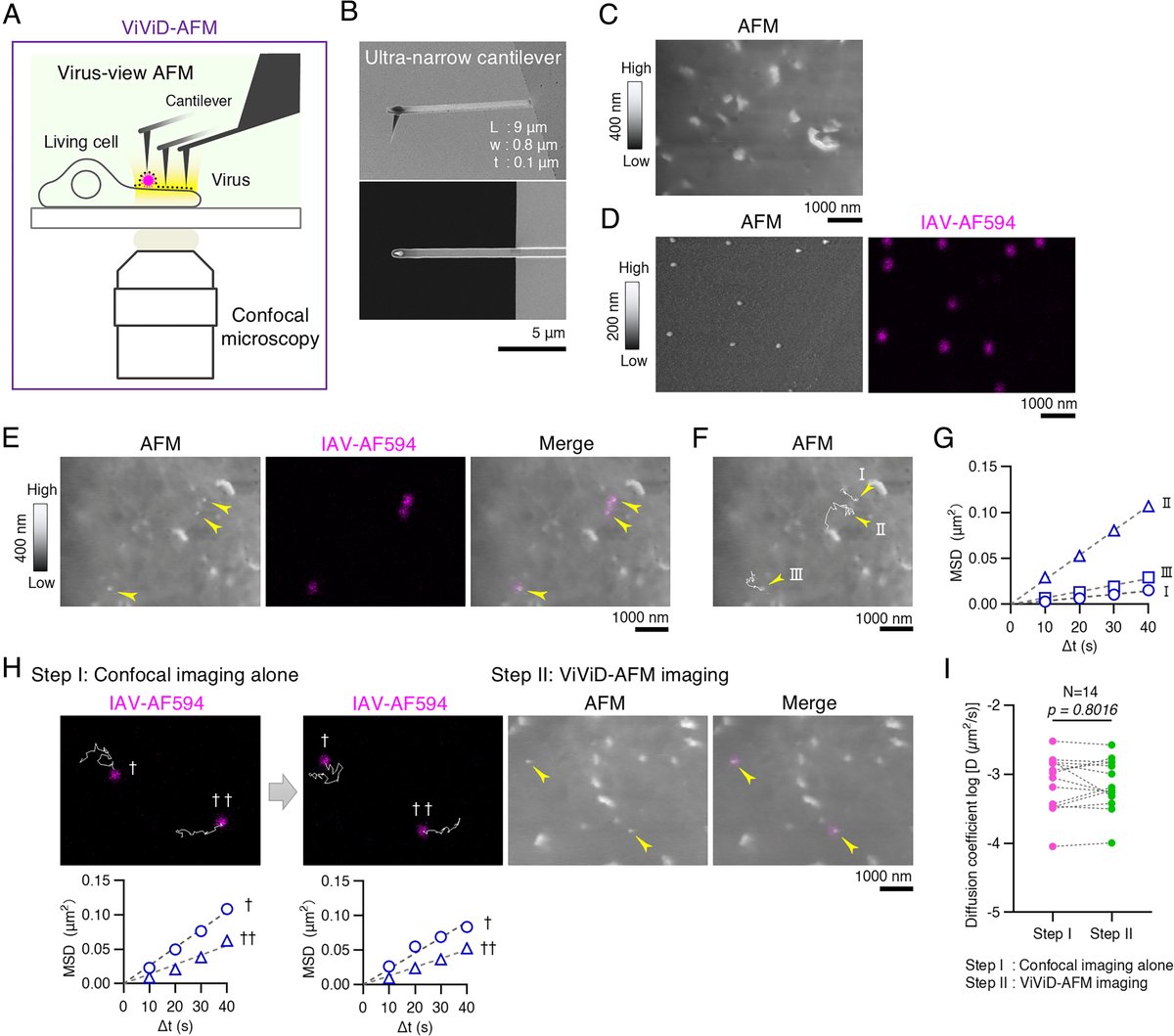
ALT Establishment of virus-view AFM for IAV cell entry studies in MDCK cells.
1
4
9
4,374

8 Oct 2025
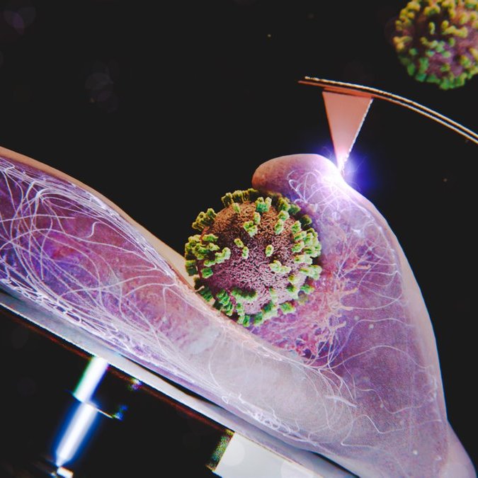
Our ViViD-AFM paper is out in PNAS!
pnas.org/doi/10.1073/pnas.25…
Enhanced visualization of influenza A virus entry into living cells using virus-view atomic force microscopy
ALT ViViD-AFM is a hybrid live imaging technology that captures cell surface nanometric morphological features and fluorescent signals. It uses atomic force microscopy (AFM) specifically tailored for soft surface biological imaging. It is delicate enough to preserves weak virus-host interactions at the live cell surface. The image - showing an influenza A virus taken up into the cell by clathrin-mediated endocytosis - was created by @ScienceBrush Design.
6
215
Yohei Yamauchi retweeted

27 Oct 2023
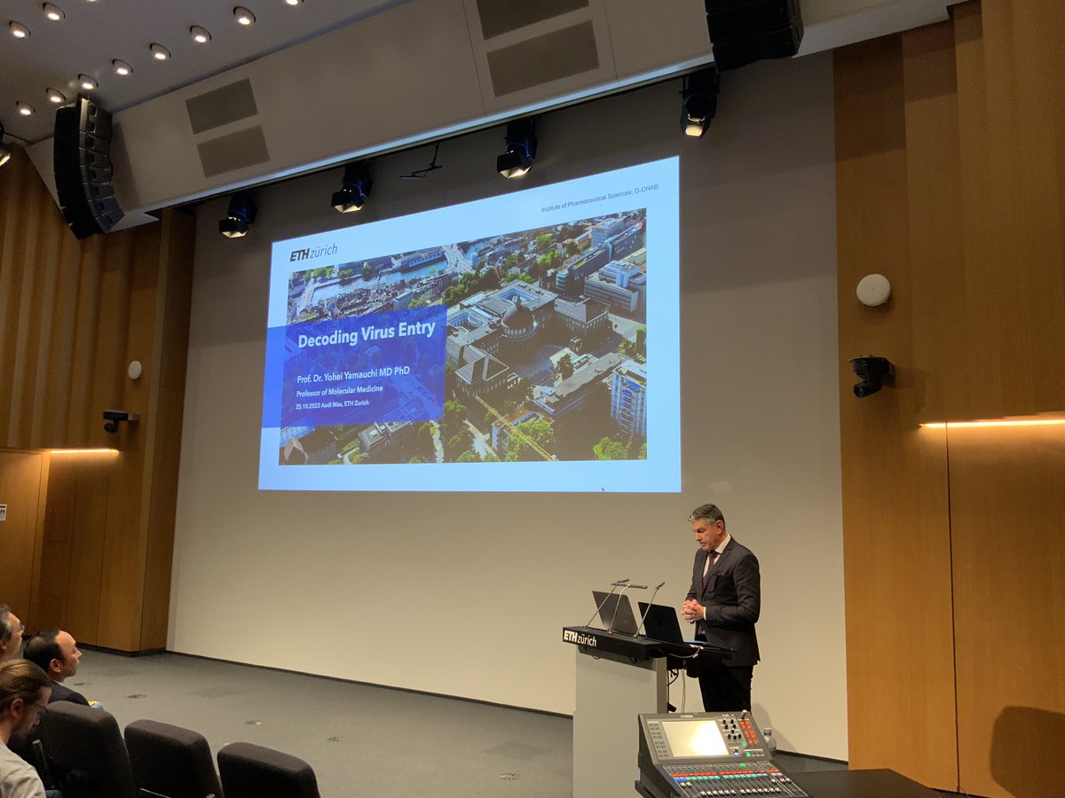
On Oct 25, Kjell Jorner's @kjelljorner @DCL_ETHZ & Yohei Yamauchi's @YamauchiLab inaugural lectures took place in the Auditorium Maximum @ETH_en . In case you missed the event, check out the video recording 👇
video.ethz.ch/speakers/lectu…
1
11
1,506
Yohei Yamauchi retweeted
25 Oct 2023
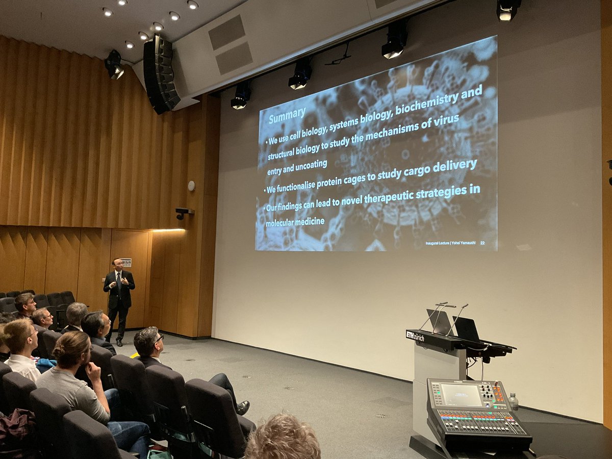
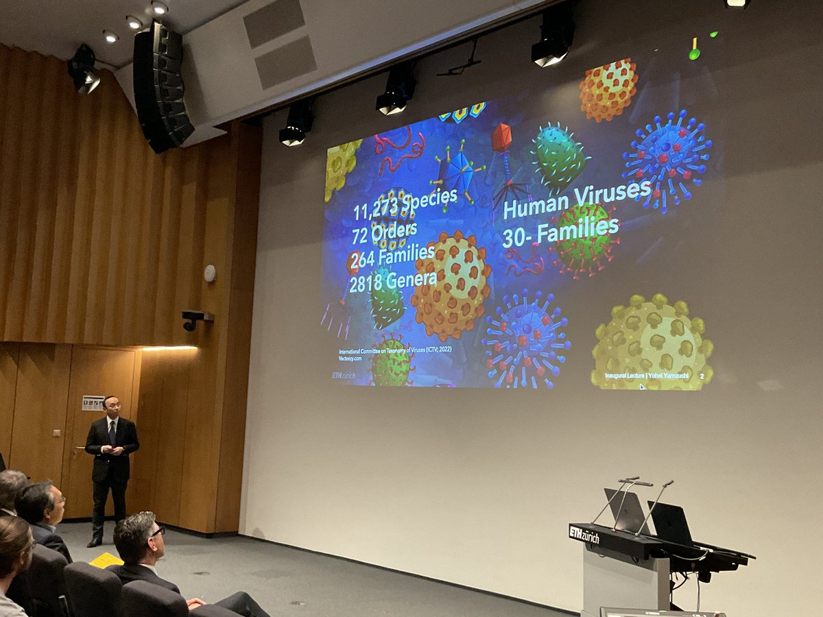
Yamauchi @YamauchiLab presenting his #SARS -CoV-2 co-receptor study which was EU project of the month in Autumn 2020 and his more recent research on virus-inspired protein cages for cellular uptake to facilitate payload delivery in cells.
4
9
1,770
Yohei Yamauchi retweeted
25 Oct 2023
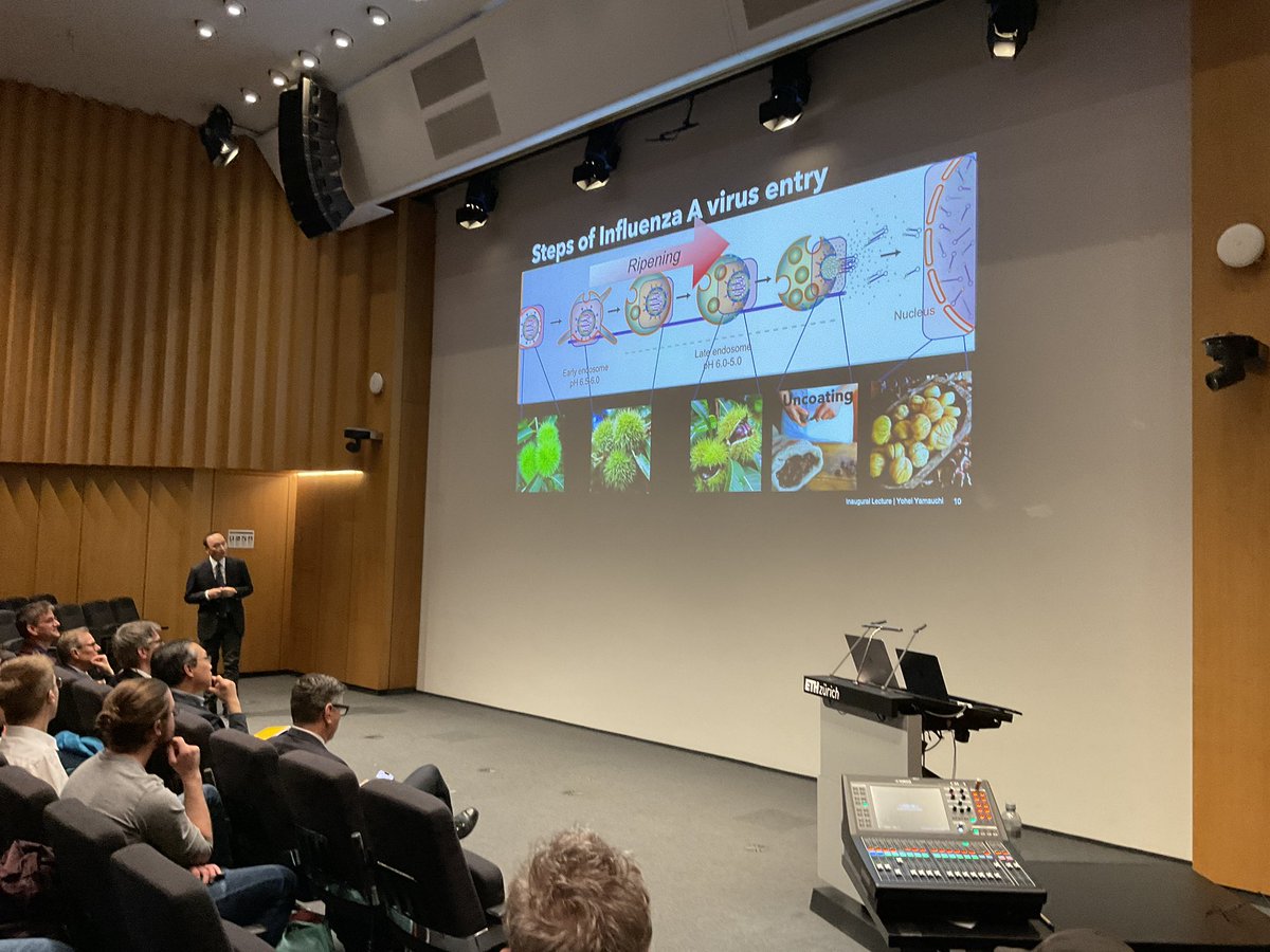
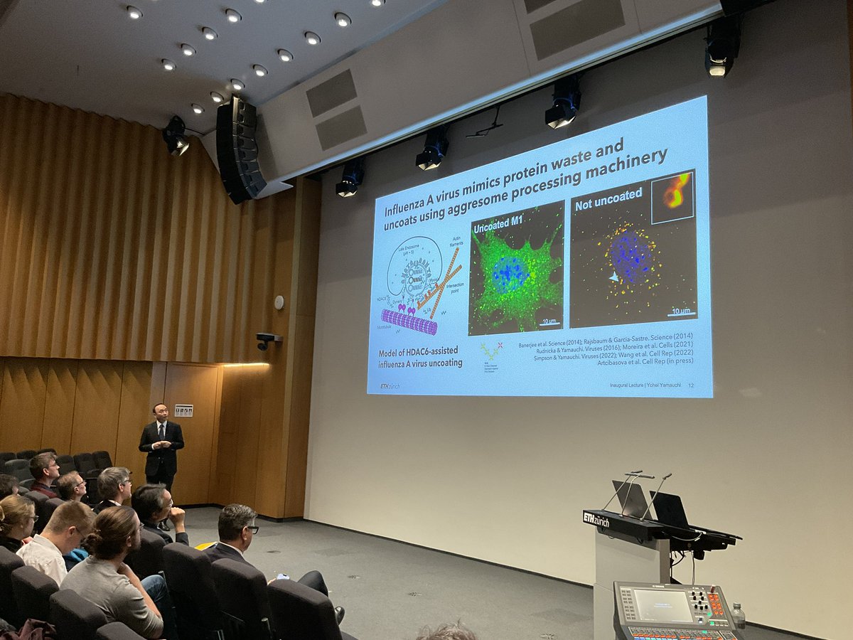
And here we go - this is how the entry of the influenza virus looks like. But the virus has even more stunning tricks - it uses, for instance, the host cell's waste disposal system to break down its capsid as Yamauchi @YamauchiLab explains.
1
2
840
Yohei Yamauchi retweeted
25 Oct 2023
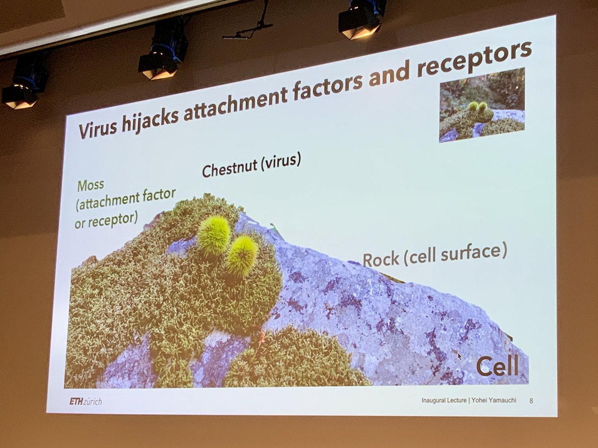
a nice analogy on how viruses can enter the human body by @YamauchiLab, discovered during a hike in southern Switzerland
1
5
876
Yohei Yamauchi retweeted
25 Oct 2023
Now we are off to a new universe - the universe of viruses. Yohei Yamauchi @YamauchiLab giving his inaugural lecture on “Decoding Virus Entry” enlightening the cell biology of virus entry.
1
2
598
Yohei Yamauchi retweeted
25 Oct 2023
now it is time for the next Inaugural Lecture, by Yohei Yamauchi, @YamauchiLab, Roger Schibli gives the introduction
1
3
577
Yohei Yamauchi retweeted

21 Jul 2023
This works comes from a fantastic collaboration with @YamauchiLab at @ETH_DCHAB and Patrick Matthias at @FMIscience on understanding the role of branched Ub chains in viral infections. For this we need homogenous Ub chains to reconstitute the machinery that uncoats viral capsids.
1
1
7
1,954
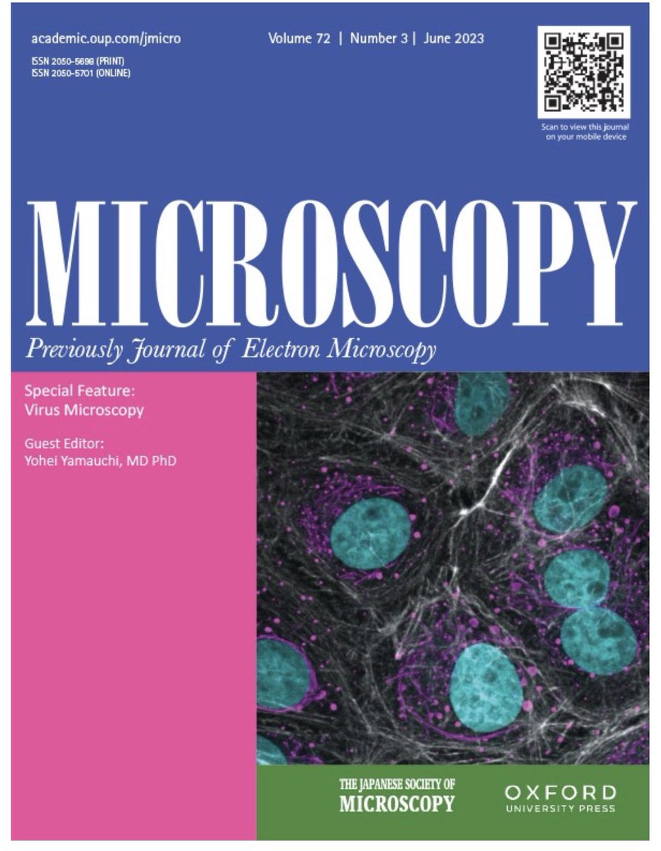
12 Jun 2023
I was lucky to guest edit a Special Feature: Virus Microscopy for the journal Microscopy. Review by S. Padilla-Parra (HIV-1 transmission); T. Noda (filovirus structure); @Chr_Sieben orthohantavirus imaging); U. Greber (label-free microscopy). See issue 👉
academic.oup.com/jmicro/issu…
1
12
1,007
1 Jun 2023
Life beyond the Pixels was a fantastic talk given by computational biologist Peter Horvath @hpke1980 @BiochemBrc for the IPW seminar series hosted by Yohei Y. #AI #singlecell #MolecularMedicineLab @ETH_DCHAB Thanks Peter for your visit to ETH!!
3
20
1,651
25 May 2023
We are recruiting a permanent Technician in Cellular and Molecular Biology #MolecularMedicineLab at ETH Zürich
jobs.ethz.ch/job/view/JOPG_e…
yamauchilab.com/
10
9
2,062
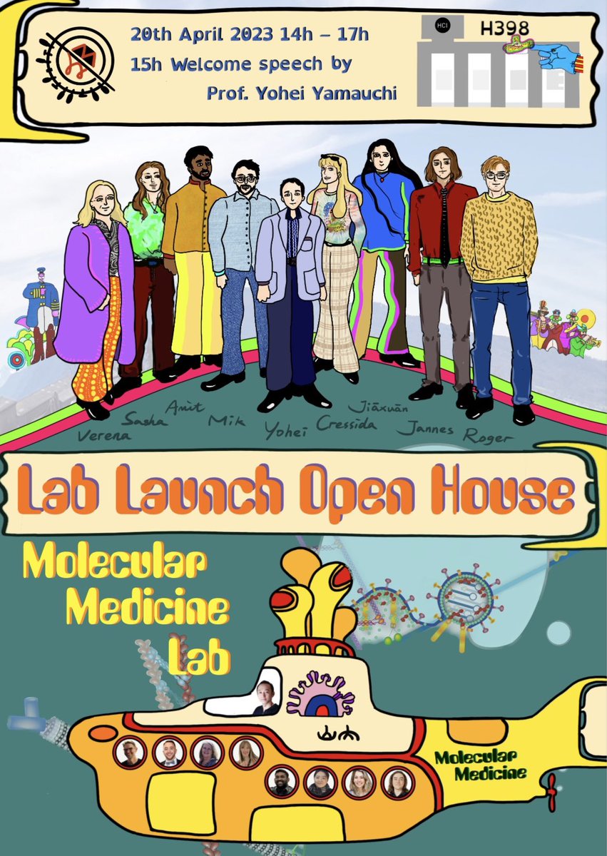
18 Apr 2023
Visit the Molecular Medicine Lab Launch event on Thursday.
Artwork by @fjx0326 @ETH_DCHAB
2
2
17
1,697
13 Apr 2023
I highly recommend taking a look at "Catching the (viral) burglar", an engaging interview article written by Julia Ecker that effectively summarizes our research. chab.ethz.ch/en/news-and-eve… @ETH_DCHAB
2
17
1,938
Yohei Yamauchi retweeted
4 Apr 2023
Where are the cellular entry points of viruses that could be targets for future #therapies ? Yohei Yamauchi @YamauchiLab , new Professor of Molecular Medicine @ETH_DCHAB is hot on the heels of #influenza - & #coronaviruses. Check out the video & article!
chab.ethz.ch/en/news-and-eve…
4
17
4,690
17 Mar 2023
Over the last several months the Molecular Medicine laboratory recruited PhD students Jiaxuan Fan @fjx0326 Cressida Harvey @cressidaharvey Amit Santhu @AmitSanthu and Master student Jannes Brender. Welcome aboard!! 👏@ETH_DCHAB @LifeScienceZH
3
19
1,164
Yohei Yamauchi retweeted
10 Jan 2023
Single #cancer cells separate from their #tumour and dissipate via the bloodstream. Early #detection can help physicians to intervene in time. Check out this article and learn more about Stavros Stavrakis' (@deMelloGroup ) promising #diagnostic method:
ethz.ch/en/industry/industry…
3
9
1,607