
Attended an enriching 3D Echocardiography Workshop and excellent academic discussions with @DrHaniMahmoud at #SHS2026. A valuable step towards enhancing cardiac imaging expertise. #3DEcho #Cardiology #Echofirst
5

📄 Is LV diameter enough in mitral regurgitation… or are we missing the full picture?
🔗 doi.org/10.1093/ehjimp/qyag0…
🫀 In patients with significant mitral regurgitation (MR),
👉 timing of intervention depends heavily on LV remodelling
But here’s the problem:
❌ LV end-systolic diameter (LVESD) is simple…
❗ but often inconsistent and incomplete
✨ This study asks a key question:
👉 Can 3D LV end-systolic volume (3D LVESV) improve risk stratification?
🔬 Study design (3D-PRIME):
👥 227 patients with moderate or greater MR
✔ Primary MR (PMR)
✔ Secondary MR (SMR)
📅 Follow-up: ~2 years
🎯 Outcomes:
➡️ MR progression → intervention
➡️ Death
➡️ HF hospitalization
📊 Key findings:
🔥 Increased 3D LVESV = higher risk
👉 In primary MR:
✔ ~2x higher risk of adverse events
👉 In secondary MR:
✔ ~4x higher risk of death/HF hospitalization
⚠️ Discordance matters:
📉 27% of PMR patients had mismatch between:
✔ LVESD
✔ 3D LVESV
👉 And here’s the key:
💥 Patients with increased 3D LVESV (even with normal diameter)
➡️ Still had high risk
📈 The graphical abstract (page 2) highlights:
✔ 3D LVESV identifies high-risk patients missed by diameter
✔ Best risk prediction when combining both measures
🧠 Why this matters:
👉 LV remodelling is 3D, not linear
❗ Diameter = one dimension
✅ Volume = full geometry
➡️ 3D echo avoids:
✔ Geometric assumptions
✔ LV foreshortening
✔ Underestimation of true dilatation
🚀 Clinical implications:
👉 Don’t rely on LVESD alone
✔ Add 3D LVESV for:
➡️ Better risk stratification
➡️ Earlier identification of high-risk patients
➡️ Improved timing of intervention
💡 Especially in borderline or asymptomatic MR
⚠️ Take-home message:
🧩 The ventricle doesn’t remodel in 1D… so why measure it that way?
👉 3D LVESV adds critical prognostic value beyond diameter.
#Cardiology #Echocardiography #MitralRegurgitation #3DEcho #CardiacImaging #HeartFailure #ValvularHeartDisease #PrecisionMedicine 🫀📊

1
10
29
1,162
📄 Are we misclassifying mitral regurgitation in MVP?
🔗 doi.org/10.1093/ehjimp/qyag0…
🫀 Mitral valve prolapse (MVP) is the leading cause of primary MR…
👉 but accurately quantifying MR remains a major challenge
Why?
❗ Eccentric jets
❗ Multiple jets
❗ Non-holosystolic regurgitation
➡️ Traditional methods (PISA, 2D echo) often fall short
✨ This study explores a smarter approach:
👉 Three-dimensional continuity equation (3D-CE)
🔬 Study design:
👥 72 MVP patients (65% female, mean age ~60)
📊 Compared:
✔ 3D-CE
✔ Conventional diameter-based CE (2D-CE)
✔ CMR (gold standard)
📊 Key findings:
⚠️ 2D-CE overestimates MR severity
➡️ 19 mL bias vs 3D-CE
🔥 3D-CE vs CMR:
✔ Excellent agreement (r ≈ 0.93–0.94)
✔ Minimal bias ( 2.1 mL)
❌ PISA performance limited
➡️ Especially in patients with multiple jets (r = 0.40)
📈 The graphical abstract (page 2) clearly shows:
✔ Strong concordance between 3D-CE and CMR
✔ Systematic overestimation with 2D methods
🧠 Why this matters:
👉 MVP anatomy is complex:
✔ Non-circular annulus
✔ Bileaflet prolapse (86%)
✔ High prevalence of MAD (96%)
❗ Assuming a circular annulus (as in 2D methods) → major source of error
👉 3D imaging captures true anatomy
➡️ More accurate regurgitant volume
🚀 Clinical implications:
👉 In patients with moderate or borderline MR:
✔ Better classification
✔ Better timing of intervention
✔ Reduced risk of under/over-treatment
💡 Supports a multimodality approach (Echo CMR)
⚠️ Take-home message:
🧩 In MVP, geometry matters.
👉 If you measure MR with the wrong shape… you get the wrong answer.
➡️ 3D-CE brings us closer to the truth.
#Cardiology #Echocardiography #MitralRegurgitation #MVP #CardiacImaging #3DEcho #CMR #ValvularHeartDisease #Innovation 🫀📊

2
12
33
1,658

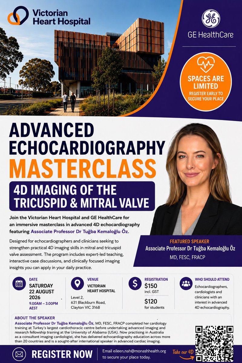
I am delighted to be visiting Melbourne this August to present at the Advanced Echo Masterclass, hosted by the Victorian Heart Hospital and @GEHealthCare
Looking forward to connecting with cardiologists, echocardiographers, and clinicians! #3decho
5
9
410

May 25
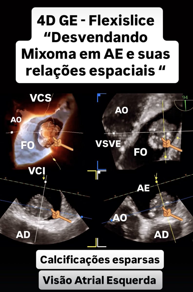
This Is NOT a CT Scan. It’s a 4D Echocardiographic View of a Left Atrial Myxoma
#Myxoma
#3DEcho
#echocardiography
#cardiology
#EchoFirst
#TEE
@ASE360
1
2
8
462
May 24
The leaflet fractured.
4D exposed it.
#Mitral
#3DEcho
#echocardiography
#cardiology
#EchoFirst
#TEE
@ASE360

1
6
197

May 16
At the #3DEcho conference at UN Mehta Institute in Ahmedabad, India @purviparwani @ASE360 @EACVIPresident @EchoSoliman


3
3
10
1,080
May 15
Three planes. One valve. One truth.
3D MPR FlexiSlice redefining mitral valve area assessment in rheumatic stenosis.
#Mitral
#3DEcho
#echocardiography
#cardiology
#EchoFirst
#TEE
@ASE360
8
211
May 15
Late Failure of Rheumatic Mitral Annuloplasty — 4D TEE Dual View
#Mitral
#3DEcho
#echocardiography
#cardiology
#EchoFirst
#TEE
@ASE360
2
242
May 12
Surgeons Love This View 👀 4D Barlow Anatomy
#3DEcho
#echocardiography
#cardiology
#EchoFirst
#TEE
@ASE360
1
10
421
May 12
Inside the Mitral Valve in True 4D
A2 prolapse through the surgeon’s eyes.
#3DEcho
#echocardiography
#cardiology
#EchoFirst
#TEE
@ASE360
1
8
28
1,827

May 11
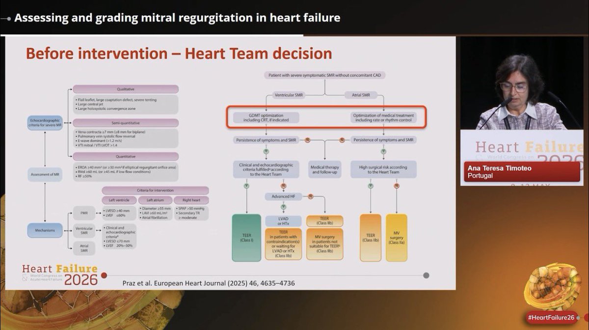
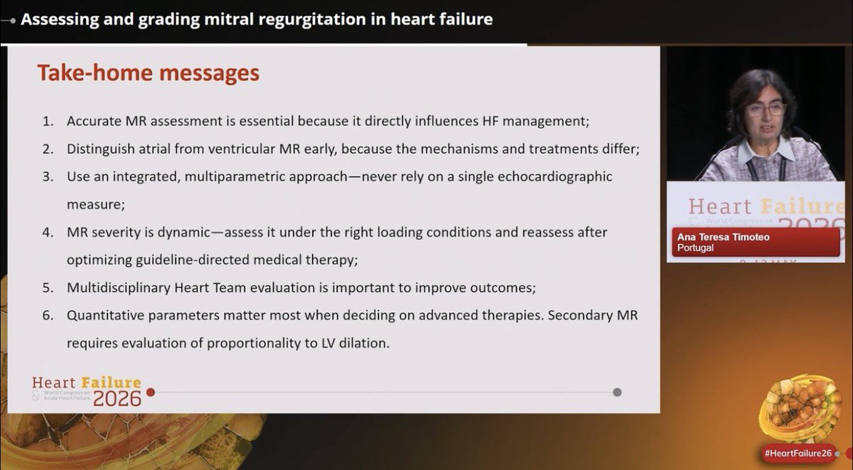
🫀#MR in #HF is far more than “mild, moderate, severe”.
⚠️Accurate MR assessment directly influences HF management & outcomes.
Excellent overview by @anatimoteo46040 at #HeartFailure26 on how we should assess secondary MR in HF:
📍Mechanism matters: Ventricular vs atrial MR, differentiate early
📍MR grading must be multiparametric,
Never rely on a single number alone.
📍Secondary MR is dynamic,
Severity may change after: GDMT optimization, CRT, rhythm/rate control, decongestion. ⚠️Reassess before intervention decisions.
📍Multimodality imaging matters, each provide complementary information on anatomy, mechanism, ventricular remodeling, procedural suitability
📍Heart Team discussion is essential.
📍Quantitative parameters matter most when deciding advanced therapies
@escardio @EACVIPresident @VictoriaDe32503 #EchoFirst #whyCMR #YesCCT #3DEcho #HeartFailure


2
36
91
13,122

May 9
📣 𝐄𝐂𝐇𝐎-𝐒𝐔𝐑𝐆𝐄𝐑𝐘 𝐋𝐀𝐁 𝐕𝐈𝐈 𝐄𝐝𝐢𝐭𝐢𝐨𝐧
The course where Echo and Surgery meet each other!
💡 Not only frontal lessons, but true interactive sessions and workshops with imagers, cardiac surgeons, interventionalists and anatomopathologist.
👉 TEE simulator, 3Decho workstations, Wet Lab on pig's heart and inspection of anatomic specimen.
🔥 Let's take a look to this video in order to compare 3D echo findings of rheumatic mitral stenosis and a real rheumatic valve after cardiac surgery.
✅commissures fusion
✅fish mouth opening
✅AML doming
✅thickening and retraction of subvalvular apparatus
📅 𝟐𝟐-𝟐𝟑 𝐌𝐀𝐘 𝟐𝟎𝟐𝟔
📍 𝐒𝐚𝐧𝐭𝐚 𝐌𝐚𝐫𝐢𝐚 𝐇𝐨𝐬𝐩𝐢𝐭𝐚𝐥 - 𝐆𝐕𝐌 , 𝐁𝐚𝐫𝐢
Program: mics.mitralacademy.it/it-it/…
𝑊𝑒 𝑎𝑟𝑒 𝑟𝑒𝑎𝑑𝑦 𝑓𝑜𝑟 𝑎 𝑤𝑜𝑛𝑑𝑒𝑟𝑓𝑢𝑙 𝑒𝑑𝑢𝑐𝑎𝑡𝑖𝑜𝑛𝑎𝑙 𝑒𝑥𝑝𝑒𝑟𝑖𝑒𝑛𝑐𝑒
1
1
6
355
Apr 28
From P1 to P3: Mapping the Alfieri Stitch in 3D with Multi-Planar TEE
#3DEcho
#echocardiography
#cardiology
#EchoFirst
#TEE
@ASE360
1
3
7
761
Apr 28
Double-Orifice Mitral Valve One Year After Alfieri: When 3D TEE Tells the Whole Story
#3DEcho
#echocardiography
#cardiology
#EchoFirst
#TEE
@ASE360

1
1
4
118

#3DEcho for #LAAC
The best way to guide this procedure (planning / implant / follow up).
#watchmanflx
@bostonsci @BostonSciLatam @BSCCardiology @SONECOM_AC @SOME_IC @ImagenCardiaca @iamritu @NMerke @MigueldeBoedo @MAecocardio @GEHealthCare @GE_IanMc @HugoMartinezCMR @ASE360
2
7
260

Thanks @alexsfelixecho for your excellent talk and continued support to our #Master! #3DEcho at its best 🪄
Hope to see you in person next time 🩷
Apr 18

🌟It was a great honor to participate as faculty in II Livelo Master of Echocardiography in Universita Cattolica del Sacro Cuore 2025/2026 invited by my friends Prof. Francesca Graziani @FGraziani_Grace , Prof. Antonella Lombardo @Anto_Lombard e Prof. Gabriella Locorotondo @GabriellaLocor1 🌟
A great opportunity to discuss the global burden of rheumatic heart disease, even in Europe and how #echofirst can be an essential tool from diagnosis to intervention, with advanced techniques as 3D echocardiography 😀
@Unicatt
@dicsbc @RosaLillo14 @meucci_chiara

1
2
9
675
“TUSI Valve: A Striking Case of Drug-Induced Mitral Disease on Advanced Imaging”
#3DEcho
#echocardiography
#cardiology
#EchoFirst
#TEE
@ASE360
2
4
18
2,561

11
14
51
8,809

ASE experts connect with industry partners to drive advances in the field like #Strain #3DEcho & #ArtificialIntelligence while focusing on disease diagnosis in #HCM #Amyloidosis #ValveDisease & more! bit.ly/4247EQ4
#ASEIRT #PartnersInInnovation #HeartofASE #ASEMemberDay


2
3
6
551