🫀 4D Dual-Source Photon-Counting CT in GUCH Patients
From Anatomy to True Morpho-Functional Assessment
In grown-up congenital heart (GUCH) patients, imaging is rarely simple.
Complex anatomy, prior surgeries, devices, and altered hemodynamics require more than static snapshots.
Conventional CT gives excellent morphology—but limited function.
Other modalities add function—but often at the cost of resolution or accessibility.
4D dual-source Photon-Counting CT (PCCT) brings both together.
🧠 Where conventional imaging struggles
- Complex post-surgical anatomy difficult to fully map
- Limited temporal resolution for dynamic assessment
- Fragmented evaluation across multiple modalities
👉 Result: partial understanding of a complex system
⚡ What 4D dual-source PCCT enables
- Ultra-high spatial resolution → precise anatomy, even in complex repairs
- High temporal resolution (dual-source) → true cardiac dynamics
- 4D datasets → valve motion, ventricular function, flow-related changes
- High contrast resolution (with strong iodine signal) → clear chamber and vessel opacification
- Spectral capability → added tissue and flow characterization
🎯 The shift
From: Static anatomy
To: Integrated morph-functional imaging
In GUCH patients, understanding structure is not enough.
You need to see how it works—beat by beat.
That’s where 4D dual-source PCCT redefines cardiac imaging. ⚡🫀
#PhotonCountingCT #PCCT #CardiacCT #GUCH #CongenitalHeartDisease #4DCT #DualSourceCT #CardiacImaging #RadiologyInnovation #yesCCT
1
9
656
Seconda Edizione 2026
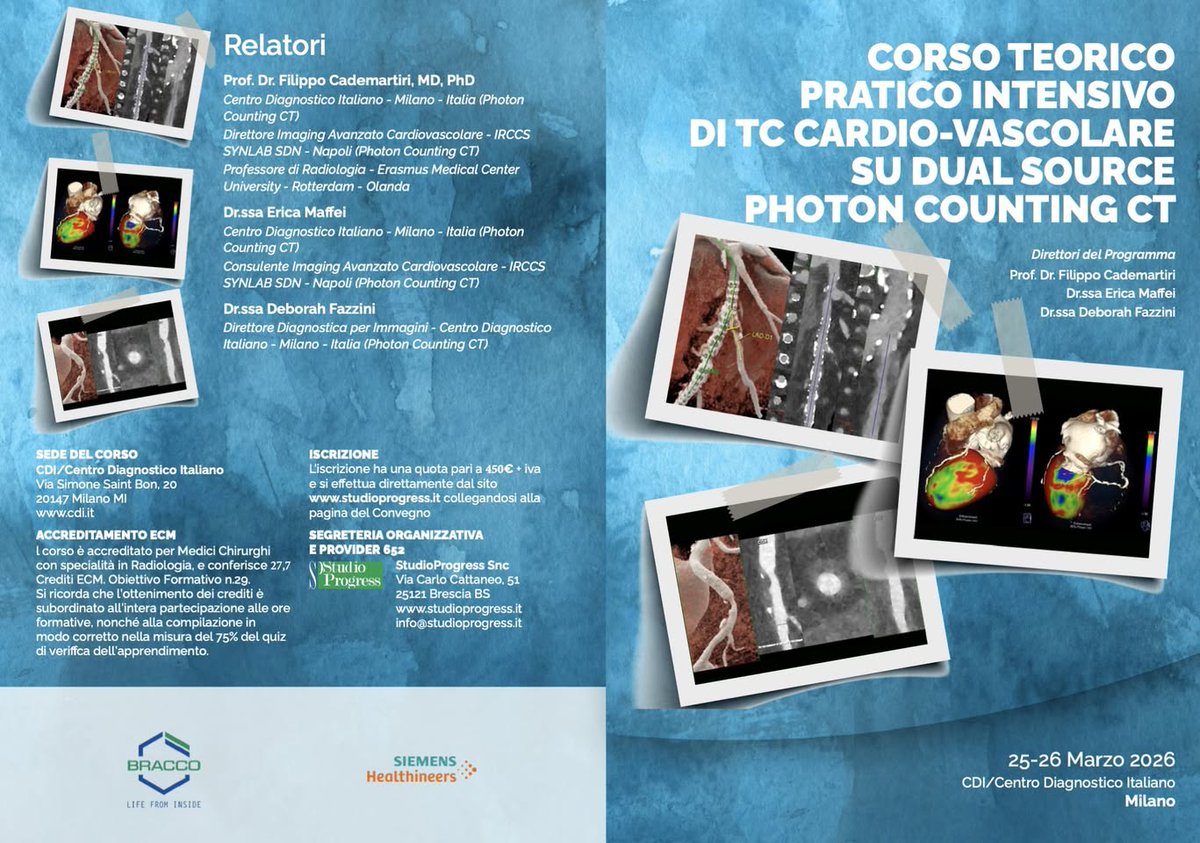
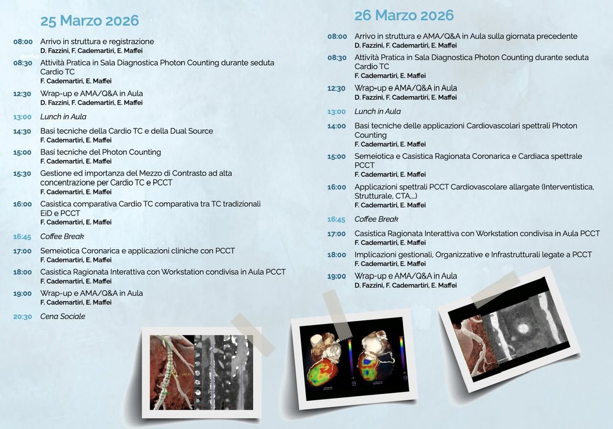
🚀 CORSO TEORICO PRATICO INTENSIVO DI TC CARDIO-VASCOLARE SU DUAL SOURCE PHOTON COUNTING CT 🫀⚡
📍 Milano | CDI Centro Diagnostico Italiano
📅 20-21 Maggio 2026
La prima edizione è già completamente prenotata, quindi abbiamo deciso di proporne immediatamente una seconda.
La Cardio TC sta entrando in una nuova era ed il Photon Counting porta questa diagnostica ad un livello eccezionale.
E questa volta non si parla di teoria astratta, ma di pratica reale sul campo.
👉 Corso teorico-pratico intensivo di TC Cardio-Vascolare su Photon Counting CT
👉 Il primo corso hands-on in Italia interamente dedicato alla Cardio PCCT
💡 Cosa rende questo corso unico
- Attività pratica in sala
diagnostica su Photon Counting CT reale
- Deep-dive negli aspetti tecnici della Cardio TC
e del Photon Counting
- Gestione avanzata del Mezzo di contrasto ad alta
concentrazione
- Valutazione alla Workstation, casi ragionati e
interattivi
- Approccio clinico, tecnico e
organizzativo completo
- Confronto diretto PCCT vs TC tradizionale
(EID)
- Focus su coronarie, imaging tessutale
spettrale, mezzo di contrasto, workflow
Ampio spazio a AMA / Q&A senza filtri
👨🏫 Faculty
Dr. Filippo Cademartiri
Dr. Erica Maffei
Dr.ssa Deborah Fazzini
🎓 27,7 Crediti ECM – Radiologia
💶 Quota: 450 € IVA
🔴 Se fai Cardio TC oggi e vuoi aggiornarti
🔴 Se stai valutando o già lavorando con PCCT
🔴 Se vuoi capire davvero cosa cambia nella pratica quotidiana
👉 Questo corso è per te.
📌 Iscrizioni e info: studioprogress.it
📌 Programma completo e dettagli: studioprogress.it/convegno/c…
#PhotonCountingCT #CardioCT #PCCT #DualSourceCT #CardiacImaging #Radiologia #ImagingAvanzato #HandsOn #ECM #Milano
2
907
🚀 I CORSO CORSO TEORICO PRATICO INTENSIVO DI TC CARDIO-VASCOLARE SU DUAL SOURCE PHOTON COUNTING CT 🫀⚡
📍 Milano | CDI – Centro Diagnostico Italiano
📅 25–26 Marzo 2026
La Cardio TC sta entrando in una nuova era ed il Photon Counting porta questa diagnostica ad un livello eccezionale.
E questa volta non si parla di teoria astratta, ma di pratica reale sul campo.
👉 Corso teorico-pratico intensivo di TC Cardio-Vascolare su Photon Counting CT
👉 Il primo corso hands-on in Italia interamente dedicato alla Cardio PCCT
💡 Cosa rende questo corso unico
- Attività pratica in sala diagnostica su Photon Counting CT reale
- Deep-dive negli aspetti tecnici della Cardio TC e del Photon Counting
- Gestione avanzata del Mezzo di contrasto ad alta concentrazione
- Valutazione alla Workstation, casi ragionati e interattivi
- Approccio clinico, tecnico e organizzativo completo
- Confronto diretto PCCT vs TC tradizionale (EID)
- Focus su coronarie, imaging tessutale spettrale, mezzo di contrasto, workflow
Ampio spazio a AMA / Q&A senza filtri
👨🏫 Faculty
Prof. Dr. Filippo Cademartiri
Dr.ssa Dr. Erica Maffei
Dr.ssa Deborah Fazzini
🎓 27,7 Crediti ECM – Radiologia
💶 Quota: 450 € IVA
🔴 Se fai Cardio TC oggi e vuoi aggiornarti
🔴 Se stai valutando o già lavorando con PCCT
🔴 Se vuoi capire davvero cosa cambia nella pratica quotidiana
👉 Questo corso è per te.
📌 Iscrizioni e info: studioprogress.it
📌 Programma completo e dettagli: studioprogress.it/convegno/c…
#PhotonCountingCT #CardioCT #PCCT #DualSourceCT #CardiacImaging #Radiologia #ImagingAvanzato #HandsOn #ECM #Milano

1
3
352

28 Dec 2025
🚀 I CORSO CORSO TEORICO PRATICO INTENSIVO DI TC CARDIO-VASCOLARE SU DUAL SOURCE PHOTON COUNTING CT 🫀⚡
📍 Milano | CDI – Centro Diagnostico Italiano
📅 25–26 Marzo 2026
La Cardio TC sta entrando in una nuova era ed il Photon Counting porta questa diagnostica ad un livello eccezionale.
E questa volta non si parla di teoria astratta, ma di pratica reale sul campo.
👉 Corso teorico-pratico intensivo di TC Cardio-Vascolare su Photon Counting CT
👉 Il primo corso hands-on in Italia interamente dedicato alla Cardio PCCT
💡 Cosa rende questo corso unico
- Attività pratica in sala diagnostica su Photon Counting CT reale
- Deep-dive negli aspetti tecnici della Cardio TC e del Photon Counting
- Gestione avanzata del Mezzo di contrasto ad alta concentrazione
- Valutazione alla Workstation, casi ragionati e interattivi
- Approccio clinico, tecnico e organizzativo completo
- Confronto diretto PCCT vs TC tradizionale (EID)
- Focus su coronarie, imaging tessutale spettrale, mezzo di contrasto, workflow
Ampio spazio a AMA / Q&A senza filtri
👨🏫 Faculty
Prof. Dr. Filippo Cademartiri
Dr.ssa Dr. Erica Maffei
Dr.ssa Deborah Fazzini
🎓 27,7 Crediti ECM – Radiologia
💶 Quota: 450 € IVA
🔴 Se fai Cardio TC oggi e vuoi aggiornarti
🔴 Se stai valutando o già lavorando con PCCT
🔴 Se vuoi capire davvero cosa cambia nella pratica quotidiana
👉 Questo corso è per te.
📌 Iscrizioni e info: studioprogress.it
📌 Programma completo e dettagli: studioprogress.it/convegno/c…
#PhotonCountingCT #CardioCT #PCCT #DualSourceCT #CardiacImaging #Radiologia #ImagingAvanzato #HandsOn #ECM #Milano
8
808
19 Dec 2025
🚀 I CORSO CORSO TEORICO PRATICO INTENSIVO DI TC CARDIO-VASCOLARE SU DUAL SOURCE PHOTON COUNTING CT 🫀⚡
📍 Milano | CDI – Centro Diagnostico Italiano
📅 25–26 Marzo 2026
La Cardio TC sta entrando in una nuova era ed il Photon Counting porta questa diagnostica ad un livello eccezionale.
E questa volta non si parla di teoria astratta, ma di pratica reale sul campo.
👉 Corso teorico-pratico intensivo di TC Cardio-Vascolare su Photon Counting CT
👉 Il primo corso hands-on in Italia interamente dedicato alla Cardio PCCT
💡 Cosa rende questo corso unico
- Attività pratica in sala diagnostica su Photon Counting CT reale
- Deep-dive negli aspetti tecnici della Cardio TC e del Photon Counting
- Gestione avanzata del Mezzo di contrasto ad alta concentrazione
- Valutazione alla Workstation, casi ragionati e interattivi
- Approccio clinico, tecnico e organizzativo completo
- Confronto diretto PCCT vs TC tradizionale (EID)
- Focus su coronarie, imaging tessutale spettrale, mezzo di contrasto, workflow
Ampio spazio a AMA / Q&A senza filtri
👨🏫 Faculty
Prof. Dr. Filippo Cademartiri
Dr.ssa Dr. Erica Maffei
Dr.ssa Deborah Fazzini
🎓 27,7 Crediti ECM – Radiologia
💶 Quota: 450 € IVA
🔴 Se fai Cardio TC oggi e vuoi aggiornarti
🔴 Se stai valutando o già lavorando con PCCT
🔴 Se vuoi capire davvero cosa cambia nella pratica quotidiana
👉 Questo corso è per te.
📌 Iscrizioni e info: studioprogress.it
📌 Programma completo e dettagli: studioprogress.it/convegno/c…
#PhotonCountingCT #CardioCT #PCCT #DualSourceCT #CardiacImaging #Radiologia #ImagingAvanzato #HandsOn #ECM #Milano


4
377
10 Nov 2025
DUAL SOURCE PHOTON COUNTING CT AND CARDIAC IMAGING
Spatial Temporal Spectral Resolution at their best
The temporal resolution of Dual Source scanners is the highest achievable in clinical practice (66ms) and it does not rely on magic tricks and algorithms to show you static Cardiac images. When more than 25 years I started doing Cardiac CT we need very low heart rates (native or induced by negative chronotropic drugs, mostly beta-blockers). Then rotation times of the gantry/detector systems increased and we have better results even at slightly higher heart rates.
The real step forward, however, happened when Dual Source CT was introduced drastically cutting in half the time required to acquire one single image (slabs of images). There have been 3 major generations of Dual Source CT and the current benchmark for temporal resolution is 66ms with the 3rd generation of Dual Source CT (FORCE) that is already available since 2014 (!!! 12 years ago). With this technology I personally stopped using IV beta-blockers which was a very common practice for non dual source CT (and still is).
I could scan all patients coming in every day regardless the heart rate (high, irregular, and so forth). Arrhythmia is never a contraindication to Cardiac CT in my clinical practice on Dual Source CTs.
With Photon Counting that is the starting point.
In addition you put on top the outstanding performance of the new detector in terms of noise reduction, spatial resolution, and spectral capabilities; all massively increased.
When I say PCCT it's a new imaging modality, I mean it. It generates a new semeiology for Coronary Atherosclerosis and Cardiovascular imaging in general, both because it massively improves the power of your electronic microscope and because it gives you many more imaging tools for tissue imaging making of CT a multiparametric imaging modality.
We are entering a phase of great modifications around the Cardiovascular field because of Photon Counting CT. And as it happened with previous leaps all this innovation and improvements will fallout into all the other applications.
#PhotonCountingCT #PCCT #DualSourceCT #CoronaryStents #BifurcationImaging #InStentRestenosis #SpectralCT #CoronaryCTA #CardiacImaging #PrecisionCardiology #InnovationInImaging
3
15
2,142
8 Nov 2025
💥 Photon-Counting CT in Cardiac Amyloidosis: Dual insight into myocardium coronaries! 🫀⚡️
📚 Study: “Evaluation of Extracellular Volume and Coronary Artery Disease in Cardiac Amyloidosis Using Photon-Counting CT” (Popp et al., 2025, Investigative Radiology)
🎯 Purpose:
Compare extracellular volume (ECV) measurements from photon-counting CT (PCCT) with cardiac MRI (CMR)🧩, and assess coronary artery disease (CAD) in patients with ATTR cardiac amyloidosis (CA).
👥 Cohort:
30 CA patients (77 ± 8 yrs) scanned with NAEOTOM Alpha; nearly half had AF (47%), and 70% were on tafamidis.
🧮 Methods:
Late-phase PCCT after CCTA to compute ECV via:
1️⃣ Single-energy (SE) subtraction method
2️⃣ Dual-energy (DE) iodine-map method
Compared to CMR-derived ECV 🧠
Also analyzed coronary calcium, stenosis (CAD-RADS 2.0), and plaque burden.
📊 Results:
Mean ECV values (%): CMR 42.9 | SE 42.5 | DE 40.7
🔗 Excellent correlations:
DE vs SE r = 0.98 💪
CMR vs DE r = 0.89
CMR vs SE r = 0.88
Minor DE underestimation (≈ −2%) vs CMR → clinically irrelevant ✅
Segmental agreement strongest in septal regions (main CA site).
Acquisition mode (systole vs diastole) had no major effect on ECV precision.
🫀 Coronary Findings:
CAD present in 97% (!)
Mean Agatston score ≈ 1,086 (range 0–6,849) 🧱
58% non-obstructive CAD (CAD-RADS 1-2)
Only 2 patients (6.7%) had obstructive CAD ≥ 70%
Image quality preserved in 97% — even with AF ⚙️
🔬 Take-home messages:
✅ PCCT provides accurate ECV quantification vs CMR 🧠
✅ Enables simultaneous evaluation of myocardial structure coronary plaque in a single exam 💡
✅ Even in elderly patients with calcified plaques and AF, diagnostic quality remains excellent 👏
⚠️ Radiation ≈ 10 mSv (total CCTA late phase) should be optimized for younger patients.
🚀 Conclusion:
Photon-Counting CT is a game-changer for comprehensive cardiac amyloidosis imaging — capturing fibrosis (ECV) coronary disease in one shot.
🩶 Precision | Efficiency | Dual diagnostic value
DOI: 10.1097/RLI.0000000000001198
#PhotonCountingCT #PCCT #DualSourceCT #CoronaryStents #BifurcationImaging #InStentRestenosis #SpectralCT #CoronaryCTA #CardiacImaging #PrecisionCardiology #InnovationInImaging

1
1
8
1,688
8 Nov 2025
📄 New study: Cost-effectiveness of Photon-Counting CT for CAD diagnosis (Finnish healthcare) 🇫🇮💶
💡 Goal: Compare photon-counting CT (PCD-CT) vs energy-integrating CT (EID-CT) for diagnosing coronary artery disease (CAD).
🧠 Why it matters: PCD-CT offers sharper images 🩻, fewer artifacts, and better accuracy in detecting coronary stenosis — but costs more upfront 💰.
📊 Methods:
Monte Carlo simulation 🧮 of 10,000 virtual patients over 10 years.
Two diagnostic pathways tested (literature-based vs local Finnish practice).
Included downstream test costs and scanner price difference (€ 1.5 M).
📈 Results:
🔻 Downstream costs cut by 27–34%.
💰 Savings per patient: €190–€310.
⚙️ Break-even point:
Scenario 1 ➡️ after ~7,880 patients
Scenario 2 ➡️ after ~4,950 patients
❤️ Fewer unnecessary stress tests & invasive angiographies → fewer complications and safer care 👏
🏥 Takeaway:
✅ PCD-CT is cost-effective in high-volume centers and improves diagnostic accuracy.
✅ It supports smarter, safer, and leaner CAD diagnostic pathways.
✅ Open-source code lets hospitals test local cost models 🧩
🚀 Conclusion:
Photon-Counting CT isn’t just a tech leap — it’s an economic win for precision cardiac imaging and patient safety. ❤️💡📉
DOI: 10.1016/j.ejrad.2025.112245
#PhotonCountingCT #PCCT #DualSourceCT #CoronaryStents #BifurcationImaging #InStentRestenosis #SpectralCT #CoronaryCTA #CardiacImaging #PrecisionCardiology #InnovationInImaging

3
2
15
1,455
7 Nov 2025
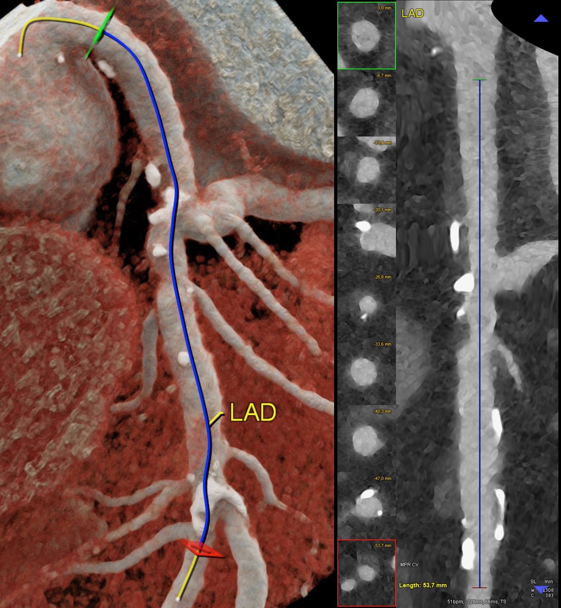
🧠 Dual-Source Photon-Counting CT for Coronary Plaque Visualization, Quantification, and Characterization
Photon-Counting CT (PCCT) represents a major leap forward in coronary atherosclerosis imaging, offering the ability not only to visualize but also to quantify and characterize plaque composition with unprecedented precision.
Leveraging dual-source architecture, PCCT delivers:
🔍 Ultra-high spatial resolution (Voxel 0.1 mm), enabling confident delineation of the vessel wall, plaque boundaries, and microcalcifications.
⚡ High temporal resolution (~66 ms) for motion-free coronary imaging, even at higher heart rates.
🌈 Spectral and quantitative capabilities through energy-resolved detection, allowing precise iodine quantification, calcium differentiation, and non-calcified plaque composition analysis.
📊 True density-based quantification (not influenced by electronic noise or partial volume effects), enabling reliable assessment of low-attenuation plaque, fibrofatty components, and spotty calcifications.
The combination of spatial, temporal, and spectral fidelity allows PCCT to transcend traditional CT angiography — evolving into a morpho-functional, quantitative tool for plaque vulnerability assessment and patient-specific risk stratification.
Example: very large and long atherosclerotic non calcified plaque on LM and LAD non obstructive plaque atherosclerosis.
#PhotonCountingCT #PCCT #DualSourceCT #CoronaryStents #BifurcationImaging #InStentRestenosis #SpectralCT #CoronaryCTA #CardiacImaging #PrecisionCardiology #InnovationInImaging
1
11
57
4,149
4 Nov 2025
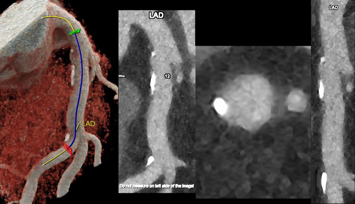
🧠 Dual-Source Photon-Counting CT for Coronary Plaque Visualization, Quantification, and Characterization
Photon-Counting CT (PCCT) represents a major leap forward in coronary atherosclerosis imaging, offering the ability not only to visualize but also to quantify and characterize plaque composition with unprecedented precision.
Leveraging dual-source architecture, PCCT delivers:
🔍 Ultra-high spatial resolution (Voxel 0.1 mm), enabling confident delineation of the vessel wall, plaque boundaries, and microcalcifications.
⚡ High temporal resolution (~66 ms) for motion-free coronary imaging, even at higher heart rates.
🌈 Spectral and quantitative capabilities through energy-resolved detection, allowing precise iodine quantification, calcium differentiation, and non-calcified plaque composition analysis.
📊 True density-based quantification (not influenced by electronic noise or partial volume effects), enabling reliable assessment of low-attenuation plaque, fibrofatty components, and spotty calcifications.
The combination of spatial, temporal, and spectral fidelity allows PCCT to transcend traditional CT angiography — evolving into a morpho-functional, quantitative tool for plaque vulnerability assessment and patient-specific risk stratification.
Example: distal LM/ostial LAD non calcified low density non obstructive plaque atherosclerosis.
🚨 If you want to learn, understand, discover the clinical potential of Photon Counting CT register and attend my online course 🚨
17 - 19 Dec 2025
⏳ Limited spots—secure yours NOW!
🔬 What’s in it for you?
✅ Hands-on training with the latest PCCT tech
✅ CME credits for continued professional growth
✅ Real clinical cases to immediately apply to your practice
✅ Expert-led guidance from industry leaders
✅ Interactive virtual labs to sharpen your skills
Why Join Now?
Master PCCT and elevate your practice with cutting-edge techniques that boost diagnostic accuracy and reduce radiation—before your peers do.
🔗 academy.unilabs.com/radiolog…
📧 Questions? Reach out at academy.radiology@unilabs.com
#PhotonCountingCT #PCCT #DualSourceCT #CoronaryStents #BifurcationImaging #InStentRestenosis #SpectralCT #CoronaryCTA #CardiacImaging #PrecisionCardiology #InnovationInImaging

5
27
3,614
4 Nov 2025
🧠 Dual-Source Photon-Counting CT for Coronary Plaque Visualization, Quantification, and Characterization
Photon-Counting CT (PCCT) represents a major leap forward in coronary atherosclerosis imaging, offering the ability not only to visualize but also to quantify and characterize plaque composition with unprecedented precision.
Leveraging dual-source architecture, PCCT delivers:
🔍 Ultra-high spatial resolution (Voxel 0.1 mm), enabling confident delineation of the vessel wall, plaque boundaries, and microcalcifications.
⚡ High temporal resolution (~66 ms) for motion-free coronary imaging, even at higher heart rates.
🌈 Spectral and quantitative capabilities through energy-resolved detection, allowing precise iodine quantification, calcium differentiation, and non-calcified plaque composition analysis.
📊 True density-based quantification (not influenced by electronic noise or partial volume effects), enabling reliable assessment of low-attenuation plaque, fibrofatty components, and spotty calcifications.
The combination of spatial, temporal, and spectral fidelity allows PCCT to transcend traditional CT angiography — evolving into a morpho-functional, quantitative tool for plaque vulnerability assessment and patient-specific risk stratification.
Example: LM-LAD with diffuse mild mixed non obstructive atherosclerosis.
🚨 If you want to learn, understand, discover the clinical potential of Photon Counting CT register and attend my online course 🚨
17 - 19 Dec 2025
⏳ Limited spots—secure yours NOW!
🔬 What’s in it for you?
✅ Hands-on training with the latest PCCT tech
✅ CME credits for continued professional growth
✅ Real clinical cases to immediately apply to your practice
✅ Expert-led guidance from industry leaders
✅ Interactive virtual labs to sharpen your skills
Why Join Now?
Master PCCT and elevate your practice with cutting-edge techniques that boost diagnostic accuracy and reduce radiation—before your peers do.
🔗 academy.unilabs.com/radiolog…
📧 Questions? Reach out at academy.radiology@unilabs.com
#PhotonCountingCT #PCCT #DualSourceCT #CoronaryStents #BifurcationImaging #InStentRestenosis #SpectralCT #CoronaryCTA #CardiacImaging #PrecisionCardiology #InnovationInImaging
1
8
36
3,628
2 Nov 2025
HOW PASSION MAKES YOUR PROFESSIONAL LIFE EXCITING
I have always been passionate about technology since I was little kid.
Technology is what makes me feel like a little kid even today.
Understanding the inner details of the "Horse" I ride every day is part of my being.
I guess this is also part of what makes me better than average in what I do for a living. I hope so.
The best thing about feeling ignorant is that you can correct it (with a lot humility and effort).
If you want to learn and share my passion CT and Photon Counting CT continue read.
🚨 If you want to learn, understand, discover the clinical potential of Photon Counting CT register and attend my online course 🚨
17 - 19 Dec 2025
⏳ Limited spots—secure yours NOW!
🔬 What’s in it for you?
✅ Hands-on training with the latest PCCT tech
✅ CME credits for continued professional growth
✅ Real clinical cases to immediately apply to your practice
✅ Expert-led guidance from industry leaders
✅ Interactive virtual labs to sharpen your skills
Why Join Now?
Master PCCT and elevate your practice with cutting-edge techniques that boost diagnostic accuracy and reduce radiation—before your peers do.
🔗 academy.unilabs.com/radiolog…
📧 Questions? Reach out at academy.radiology@unilabs.com
#PhotonCountingCT #PCCT #DualSourceCT #CoronaryStents #BifurcationImaging #InStentRestenosis #SpectralCT #CoronaryCTA #CardiacImaging #PrecisionCardiology #InnovationInImaging

8
769
30 Oct 2025
Photon-Counting CT for Cardiovascular imaging: the new imaging modality 🚀
For years we’ve been pushing energy-integrating detector (EiD) CT to its limits. Even the best-in-class system — the 3rd-generation Dual Source CT (Siemens SOMATOM Force) — has shown how far you can go with high temporal resolution, low kV, Tin filtration, and smart IR/AI recon. It’s an extraordinary scanner.
But Photon-Counting CT (PCCT) changes the game because it changes the detector.
Here’s why it’s a quantum leap ⬇️
1. Detector physics, not just better software
EiD CT sums all incoming X-ray energy into one signal. PCCT counts and bins individual photons according to their energy.
2. Coronary arteries finally at their native size
Coronary imaging is all about small, moving, high-contrast structures. PCCT brings:
- ultra-high spatial resolution
- minimized blooming
- perfect lumen assessment
3. Spectral always-on = better plaque characterization
With PCCT you don’t need to choose “dual-energy mode” — it’s intrinsic.
4. Dose–IQ decoupling
5. Beyond what Force can do
SOMATOM Force is still the benchmark for EiD CT
…but Force still integrates energy.
So the message is simple 👇
Force showed us the ceiling of EiD CT. Photon-Counting CT removes that ceiling.
📌 Take-home for coronary imaging
Better diagnostic confidence in calcified/stented segments
More consistent image quality across patient sizes
Native spectral information for smarter, physiology-oriented CT reports
A platform ready for AI that works on richer, cleaner data
🚨 If you want to learn, understand, discover the clinical potential of Photon Counting CT register and attend my online course 🚨
17 - 19 Dec 2025
⏳ Limited spots—secure yours NOW!
🔬 What’s in it for you?
✅ Hands-on training with the latest PCCT tech
✅ CME credits for continued professional growth
✅ Real clinical cases to immediately apply to your practice
✅ Expert-led guidance from industry leaders
✅ Interactive virtual labs to sharpen your skills
Why Join Now?
Master PCCT and elevate your practice with cutting-edge techniques that boost diagnostic accuracy and reduce radiation—before your peers do.
🔗 academy.unilabs.com/radiolog…
📧 Questions? Reach out at academy.radiology@unilabs.com
#PhotonCountingCT #PCCT #DualSourceCT #CoronaryStents #BifurcationImaging #InStentRestenosis #SpectralCT #CoronaryCTA #CardiacImaging #PrecisionCardiology #InnovationInImaging
5
3
40
4,392
29 Oct 2025
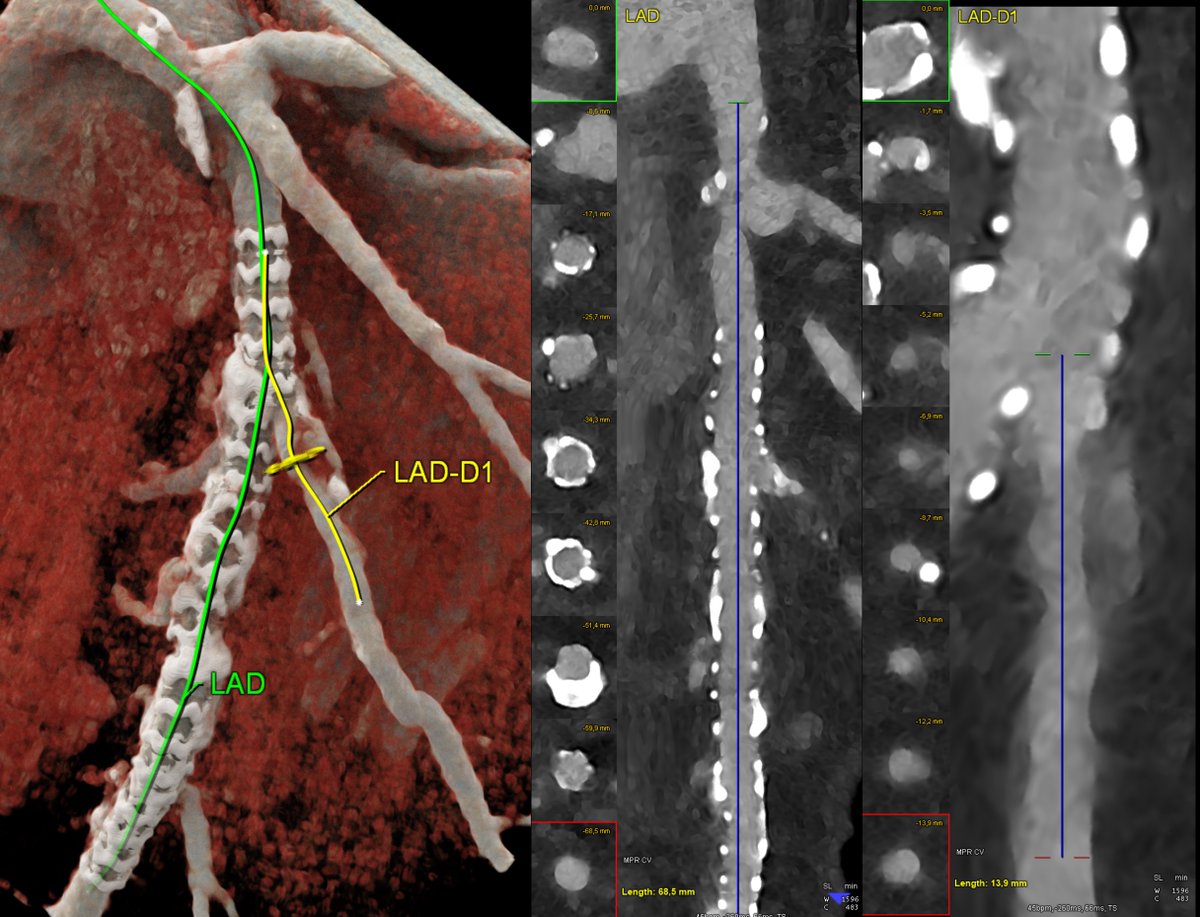
🩻 Dual-Source Photon-Counting CT for In-Stent and Bifurcation Imaging: Bridging the Gap with Intravascular Modalities
Assessing coronary stents and adjacent bifurcations has traditionally required invasive intravascular imaging such as IVUS or OCT to overcome the limitations of conventional CT in spatial resolution, blooming, and metal artifact suppression.
With dual-source Photon-Counting CT (PCCT), this gap is narrowing rapidly.
PCCT enables:
🔬 Ultra-high spatial resolution (voxel 0.1 mm) and minimal blooming artifacts, allowing clear visualization of stent struts, neointimal coverage, and in-stent lumen patency.
⚡ High temporal resolution (~66 ms) with dual-source acquisition, minimizing motion artifacts even in distal or tortuous segments.
🌈 Spectral and quantitative imaging that differentiates iodine from metal, enhancing lumen visualization and supporting iodine-density mapping for neointimal or peristent tissue characterization.
🌿 Accurate assessment of bifurcation anatomy and flow-limiting geometries, including side-branch ostiaemerging from stented segments — a scenario previously accessible only to invasive techniques.
By integrating morphological, spectral, and temporal precision, PCCT approaches intravascular-level insight non-invasively, redefining how we evaluate stent integrity, restenosis, and bifurcation hemodynamics.
Image: LAD stent (patent with no intimal hyperplasia) and large D1 with postal mixed plaque determining intermediate (45% diameter) stenosis.
🚨 If you want to learn, understand, discover the clinical potential of Photon Counting CT register and attend my online course 🚨
17 - 19 Dec 2025
⏳ Limited spots—secure yours NOW!
🔬 What’s in it for you?
✅ Hands-on training with the latest PCCT tech
✅ CME credits for continued professional growth
✅ Real clinical cases to immediately apply to your practice
✅ Expert-led guidance from industry leaders
✅ Interactive virtual labs to sharpen your skills
Why Join Now?
Master PCCT and elevate your practice with cutting-edge techniques that boost diagnostic accuracy and reduce radiation—before your peers do.
🔗 academy.unilabs.com/radiolog…
📧 Questions? Reach out at academy.radiology@unilabs.com
#PhotonCountingCT #PCCT #DualSourceCT #CoronaryStents #BifurcationImaging #InStentRestenosis #SpectralCT #CoronaryCTA #CardiacImaging #PrecisionCardiology #InnovationInImaging
1
8
46
3,343
28 Oct 2025
🧠 Dual-Source Photon-Counting CT for Coronary Plaque Visualization, Quantification, and Characterization
Photon-Counting CT (PCCT) represents a major leap forward in coronary atherosclerosis imaging, offering the ability not only to visualize but also to quantify and characterize plaque composition with unprecedented precision.
Leveraging dual-source architecture, PCCT delivers:
🔍 Ultra-high spatial resolution (Voxel 0.1 mm), enabling confident delineation of the vessel wall, plaque boundaries, and microcalcifications.
⚡ High temporal resolution (~66 ms) for motion-free coronary imaging, even at higher heart rates.
🌈 Spectral and quantitative capabilities through energy-resolved detection, allowing precise iodine quantification, calcium differentiation, and non-calcified plaque composition analysis.
📊 True density-based quantification (not influenced by electronic noise or partial volume effects), enabling reliable assessment of low-attenuation plaque, fibrofatty components, and spotty calcifications.
The combination of spatial, temporal, and spectral fidelity allows PCCT to transcend traditional CT angiography — evolving into a morpho-functional, quantitative tool for plaque vulnerability assessment and patient-specific risk stratification.
Example: RCA multiple different plaques non obstructive.
#PhotonCountingCT #PCCT #CoronaryCT #PlaqueCharacterization #AtherosclerosisImaging #SpectralCT #CardiacImaging #DualSourceCT #PrecisionCardiology #InnovationInImaging
1
1
8
486
28 Oct 2025
🧠 Dual-Source Photon-Counting CT for Coronary Plaque Visualization, Quantification, and Characterization
Photon-Counting CT (PCCT) represents a major leap forward in coronary atherosclerosis imaging, offering the ability not only to visualize but also to quantify and characterize plaque composition with unprecedented precision.
Leveraging dual-source architecture, PCCT delivers:
🔍 Ultra-high spatial resolution (Voxel 0.1 mm), enabling confident delineation of the vessel wall, plaque boundaries, and microcalcifications.
⚡ High temporal resolution (~66 ms) for motion-free coronary imaging, even at higher heart rates.
🌈 Spectral and quantitative capabilities through energy-resolved detection, allowing precise iodine quantification, calcium differentiation, and non-calcified plaque composition analysis.
📊 True density-based quantification (not influenced by electronic noise or partial volume effects), enabling reliable assessment of low-attenuation plaque, fibrofatty components, and spotty calcifications.
The combination of spatial, temporal, and spectral fidelity allows PCCT to transcend traditional CT angiography — evolving into a morpho-functional, quantitative tool for plaque vulnerability assessment and patient-specific risk stratification.
Example: LM-LAD large mixed plaque non obstructive.
#PhotonCountingCT #PCCT #CoronaryCT #PlaqueCharacterization #AtherosclerosisImaging #SpectralCT #CardiacImaging #DualSourceCT #PrecisionCardiology #InnovationInImaging
5
30
2,039
25 Oct 2025
🔬 Dual-Source Photon-Counting CT for Morphological and Functional Assessment of Myocardial Bridging
The evaluation of myocardial bridging (MB) has traditionally relied on morphological imaging, often underestimating its dynamic systolic behavior and potential hemodynamic impact.
With the advent of dual-source Photon-Counting CT (PCCT), both morphological and functional characterizationof MB can now be approached within a single non-invasive acquisition.
Key enabling features include:
⚙️ Ultra-high spatial resolution (0.1 mm) for accurate delineation of the intramyocardial segment and perivascular tissue interface.
⚡ High temporal resolution (~66 ms) through dual-source acquisition, allowing visualization of systolic compression (“milking”) and diastolic decompression across the cardiac cycle.
🌈 Spectral quantification capabilities, enabling iodine mapping and potentially dynamic perfusion analysis, to correlate luminal narrowing with downstream myocardial enhancement patterns.
This integration of temporal, spatial, and spectral information positions PCCT as a unique tool for quantitative, phase-resolved assessment of myocardial bridging — offering insights previously limited to invasive or functional imaging modalities.
#PhotonCountingCT #PCCT #DualSourceCT #MyocardialBridging #FunctionalCardiacImaging #SpectralImaging #CardiacCT #PrecisionCardiology #AdvancedImaging
2
13
45
6,263
20 Aug 2025
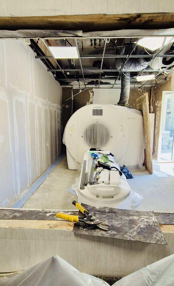
TIME TO MAKE ROOM FOR NEW HIGH PERFORMANCE CT SCANNER IN NAPLES - IRCCS SYNLAB SDN
COMPUTED TOMOGRAPHY is the cornerstone of advanced diagnostic imaging. When something gets relevant/serious or complex you usually need a good CT examination. This technology comes from 50 years of development, research and discoveries.
In Naples where I've been working for many years now, the work has started. In the picture the glorious Siemens SOMATOM Force (one of the first installed in Italy in early 2017) that I've been using in Naples over the past 8 years to build the Cardiovascular CT program in the city of Naples. We had several thousands of patients undergoing Cardiac and Vascular CT on this scanner every year beside the many thousands of CT in every field of diagnostic imaging on this workhorse.
But innovation never stops and we are making room for a newer technology that very soon will push the boundaries of what we can do and offer to our patients further and further.
More info coming soon 😉
#CCT #AdvancedCCT #DualSourceCT #PhotonCountingCT #SpectralImaging #CardiacCT #PlaqueVulnerability #AIinCardiology #CTRevolution #PlaqueImaging #InnovationInImaging #NAEOTOMAlpha #SiemensHealthineers
3
430
20 Aug 2025
📢 Upcoming Radiology Fellowships 2025
With Filippo Cademartiri, M.D., Ph.D.
CT leader • Cardiovascular imaging pioneer • Educator
Organised by Unilabs Academy, an EACCME Trusted Provider
(CME accreditation submitted to UEMS/EBR)
1️⃣ Advanced Comprehensive Cardiac CT Imaging
🗓 1 – 3 September 2025 | Online
➡️ A hands-on expert-led fellowship to elevate your skills in:
Coronary CT angiography
Plaque characterization
Myocardial perfusion
📌 Designed for radiologists and cardiologists aiming to strengthen their expertise in advanced cardiac CT imaging.
2️⃣ Morphological and Spectral Photon Counting Cardiac CT
🗓 17 – 19 December 2025 | Online
➡️ A deep dive into Photon Counting CT (PCCT) applied to cardiac imaging:
High-precision morphological visualization of cardiac structures
Spectral capabilities for improved tissue and plaque analysis
Emerging applications of PCCT in cardiovascular imaging
📌 Perfect for imaging specialists eager to explore the next frontier of CT technology.
✅ Both courses provide interactive online learning, with direct mentorship from Dr. Cademartiri.
✅ Places are limited – early registration is recommended.
🔗 Register now and secure your spot!
academy.unilabs.com/radiolog…
#CCT #AdvancedCCT #DualSourceCT
#PhotonCountingCT #SpectralImaging #CardiacCT #PlaqueVulnerability #AIinCardiology #CTRevolution #PlaqueImaging #InnovationInImaging #NAEOTOMAlpha #SiemensHealthineers


1
11
1,064
13 Jun 2025
📌 CT Protocol for Pre-TAVI Planning using Dual Source CT: A Quick Refresher
By Filippo Cademartiri, MD, PhD – Cardiovascular Imaging
Transcatheter Aortic Valve Implantation (TAVI) has become a cornerstone treatment for aortic stenosis. DSCT CT imaging plays a crucial role in pre-procedural planning, ensuring optimal outcomes and minimizing complications.
🧭 Pre-TAVI CT Protocol
1. Patient Preparation
IV access: 18–20G cannula, preferably antecubital
Heart rate control: Not mandatory, but <75 bpm preferred for motion-free aortic root imaging. DSCT makes independent of Heart Rate.
Breath-hold training: Essential to reduce motion artifacts
2. Scan Acquisition – One/Two Phases
🔹 A. ECG-Gated Aortic Root & Valve Assessment
Type: Prospective ECG-triggered or retrospective ECG-gated
Coverage: From tracheal bifurcation to diaphragm (include the entire aortic root and annulus)
Phases: Typically 30–75% R-R interval (for dynamic analysis)
kVp: 100–120 depending on patient size
Slice thickness: 0.5/0.6 mm, overlapping reconstructions
Contrast: 60–90 mL at 4 mL/s saline flush
Bolus tracking: ROI in ascending aorta, trigger at 120 HU
🔹 B. Non-ECG-Gated Thoracic/Aortoiliofemoral CTA
Purpose: Evaluate access route (calcification, tortuosity, vessel diameter)
Slice thickness: 1 mm
Reconstruction: MPR, MIP, curved MPR for iliofemoral vessels
🧠 Essential Information Extracted
✔️ Annulus diameter, perimeter, area (best measured in systole)
✔️ Sinotubular junction and coronary ostia heights
✔️ Leaflet morphology & calcification burden
✔️ LVOT characteristics
✔️ Aortic angulation
✔️ Access vessel diameters, calcifications, kinks
✔️ Tortuosity score (access route planning)
🚀 Why Dual Source CT Makes a Difference
DSCT systems provide critical advantages in pre-TAVI assessment:
✅ High temporal resolution: Up to 66 ms – ideal for imaging aortic root without motion artifacts, even at higher heart rates
✅ Lower contrast dose: Thanks to faster acquisition and dual-energy capability
✅ Robust image quality: Even in arrhythmic or tachycardic patients
✅ Reduced radiation dose: With high-pitch spiral and prospective ECG triggering
✅ Simultaneous evaluation of calcium, lumen, and access with dual-energy imaging
✅ Reliable systolic phase acquisition for annular sizing (typically 30–40% R-R)
📊 Takeaway
A well-structured pre-TAVI CT not only reduces procedural risks (e.g. coronary obstruction, annular rupture, vascular complications), but also improves device selection, sizing, and patient outcomes.
💬 Do you routinely use DSCT for TAVI planning? Share your protocol tips or any challenges you’ve encountered below ⬇️
#TAVI #CardiacCT #DualSourceCT #Radiology #StructuralHeart #AorticStenosis #CardiovascularImaging #ImagingProtocols #CTPlanning #StructuralIntervention #HeartTeam📌 CT Protocol for Pre-TAVI Planning using Dual Source CT: A Quick Refresher
By Filippo Cademartiri, MD, PhD – Cardiovascular Imaging
Transcatheter Aortic Valve Implantation (TAVI) has become a cornerstone treatment for aortic stenosis. DSCT CT imaging plays a crucial role in pre-procedural planning, ensuring optimal outcomes and minimizing complications.
🧭 Pre-TAVI CT Protocol
1. Patient Preparation
IV access: 18–20G cannula, preferably antecubital
Heart rate control: Not mandatory, but <75 bpm preferred for motion-free aortic root imaging. DSCT makes independent of Heart Rate.
Breath-hold training: Essential to reduce motion artifacts
2. Scan Acquisition – One/Two Phases
🔹 A. ECG-Gated Aortic Root & Valve Assessment
Type: Prospective ECG-triggered or retrospective ECG-gated
Coverage: HEART.
Phases: Typically 30–75% R-R interval (for dynamic analysis)
kVp: 70–90 depending on patient size
Slice thickness: 0.5/0.6 mm, overlapping reconstructions
Contrast: 60–90 mL at 4 mL/s saline flush
Bolus tracking: ROI in ascending aorta, trigger at 120 HU
🔹 B. Non-ECG-Gated Thoracic/Aortoiliofemoral CTA
Purpose: Evaluate access route (calcification, tortuosity, vessel diameter)
Slice thickness: 1 mm
Reconstruction: MPR, MIP, curved MPR for iliofemoral vessels
🧠 Essential Information Extracted
✔️ Significant coronary artery stenosis
✔️ Annulus diameter, perimeter, area (best measured in systole)
✔️ Sinotubular junction and coronary ostia heights
✔️ Leaflet morphology & calcification burden
✔️ LVOT characteristics
✔️ Aortic angulation
✔️ Access vessel diameters, calcifications, kinks
✔️ Tortuosity score (access route planning)
🚀 Why Dual Source CT Makes a Difference
DSCT systems provide critical advantages in pre-TAVI assessment:
✅ High temporal resolution: Up to 66 ms – ideal for imaging aortic root without motion artifacts, even at higher heart rates
✅ Lower contrast dose: Thanks to faster acquisition and dual-energy capability
✅ Robust image quality: Even in arrhythmic or tachycardic patients
✅ Reduced radiation dose: With high-pitch spiral and prospective ECG triggering
✅ Simultaneous evaluation of calcium, lumen, and access with dual-energy imaging
✅ Reliable systolic phase acquisition for annular sizing (typically 30–40% R-R)
📊 Takeaway
A well-structured pre-TAVI CT not only reduces procedural risks (e.g. coronary obstruction, annular rupture, vascular complications), but also improves device selection, sizing, and patient outcomes.
💬 Do you routinely use DSCT for TAVI planning? Share your protocol tips or any challenges you’ve encountered below ⬇️
#TAVI #CardiacCT #DualSourceCT #Radiology #StructuralHeart #AorticStenosis #CardiovascularImaging #ImagingProtocols #CTPlanning #StructuralIntervention #HeartTeam
3
8
60
5,995