
opencell تجربة الشراء من ترينديول Trendyol مع ترندول تصبح أوفر ب كود يمنح خصم باستخدام كوبون 👼🧚♀️🏪.
▬تريندول▬ترنديول▬
ty.gl/p9xstys5fynz8

3

Benzer sorun bende de oldu garanti biter bitmez bozuldu televizyon opencell arızası. Garantiden değiştirdiler, kısa süre sonra yine bozuldu garanti bitmiş birşey yapamayız dediler. Her kontrole gelmeye de fahiş servis ücreti istiyorlar kendi hataları olmasına rağmen.
1
67

Jun 10
opencell عروض اديداس adidas مع تطبيق المطار Almatar و⊴ستايلي⊴⊴ Styli تتحسن عند إدخال كود يمنح خصم باستخدام كوبون 🚣🎞

1
1
4

Jun 10
opencell عروض ⊴ستايلي⊴⊴ Styli الجديدة تتحسن عند إدخال كود يمنح خصم باستخدام كوبون 🙋♂️👭🏮

1

opencell تجربة التسوق من صيدلية النهدي Nahdi تصبح أفضل عند استخدام كود يمنح خصم باستخدام كوبون 🥣😮💨🐲🦇.
cnews كؤد خصم صيدليه النهدي كؤبون

48

opencell رمضان كريم مع ترينديول Trendyol أفضل منصة تسوق أونلاين، استخدم كود يمنحك خصم رائع مع كوبون فعال على عروض ترنديول 🥶🥏👥.\
ty.gl/207iyps3aei67

3

opencell 👧📫👷♀️.\
كؤد خصم تيمو

2
2
8

It’s not fiber it’s a opencell foam - full of air and fuel
1
4
118

1 Dec 2025
p53 mutations are found in 36% of tumors and contribute to the development and progression of cancer. We have tried to develop anti-cancer therapies against p53 mutant tumors before but have been unsuccessful (see x.com/aditharun_/status/1995…). In a recent paper, @Sadagopan_A, @wgibson and team present an elegant approach for targeting tumors that harbor p53 missense mutations.
I've been very interested in the p53 drug space and this is clever. I hope they spin this out and try to bring it to patients. They did a lot of detailed cellular experiments to characterize and validate their findings. And, it is presented in a way that non-basic scientists like me can understand. Great writing. Would highly recommend reading the whole piece and please see William's original post (x.com/wgibson/status/1817571…)
I'll summarize what I believe to be the key findings and their therapeutic strategy here.
p53 missense mutations are dominant negative - a p53 tetramer composed of one mutant subunit is sufficient to stop downstream signaling. However, even when p53 mutations occur, the pathways that sense cell stress are still typically intact and work to activate p53 by increasing its abundance and promoting its nuclear translocation. AND mutant p53 can't drive transcription of MDM2, its destroyer. So, half life of mutant p53 goes from 20 mins to several hours! The only protein whose abundance is significantly increased in p53-mutant cancers is p53 itself (based on CCLE data). The authors translate mutant p53 protein abundance into a cell death signal.
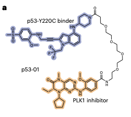
They engineer a bifunctional molecule made of p53 binder, linker, and small molecule toxin. Toxin concentrates in p53 mutant cells to cause death after certain level of accumulation in a cell.
How to design the toxin? Something with high essentiality and low abundance so a small amount of toxin buildup can neutralize the target and render the cell nonviable. DepMap CRISPR gene essentiality scores and OpenCell protein abundances revealed 5 targets of which PLK1 was chosen mostly because it already has an established ligand previously used to synthesize bifunctional small molecules for targeted protein degradation.
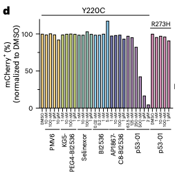
So, they put a p53-Y220C binder PEG4'd to PLK1i. PLK1i from BI and p53 Y220C from PMV Pharma ( $PMVP). This molecule kills cells that express p53 Y220C but doesn't kill cells with p53 R273H. This approach is RIPTAC-esque (x.com/aditharun_/status/1995…) but the target protein is a tumor suppressor (p53) here instead of a oncogene (like AR).
Components alone did not affect viability much. And, drug given to non-Y220C mutant cells did not impact viability.
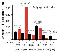
They observed that when treated with drug, PLK1 colocalized with p53Y220C on chromatin and they speculate that "this mislocalization [of PLK1] could disrupt the function of PLK1 as a mitotic kinase beyond simple steric blockade of its active site". Also, the drug induces apoptosis after 1 day of treatment in target cell type (p53-Y220C cancer cells) at much higher rates than toxin alone given at the same concentration.
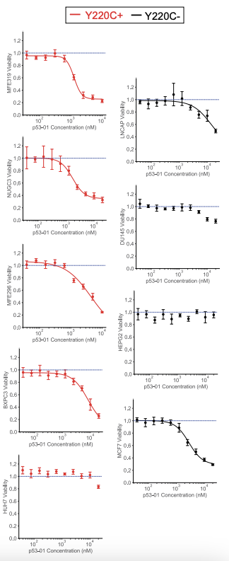
Also, their drug had specificity for Y220C mutant cell lines. Dose-viability curves here
Linker tinkering (peg2, peg4, peg6, c12, c6, c3, etc.) revealed that shorter linkers outperformed longer linkers but PEG2,4,6 all performed similarly. Let's just take the peg4 EC50s which is 850 nM and 2120 nM. Normally, drugs sit at an order of magnitude lower. So, we need to engineer the molecule to be more potent.
The opportunity exists to find the right indication / line of therapy / setting, engineer a good bifunctional compound (maybe not PLK1i, maybe something else; better p53 ligand; optimize design for best properties), and try to bring it to market. Very early days here but exciting.
30 Nov 2025

trials of anti-cancer p53 drugs: basically nothing has worked so far
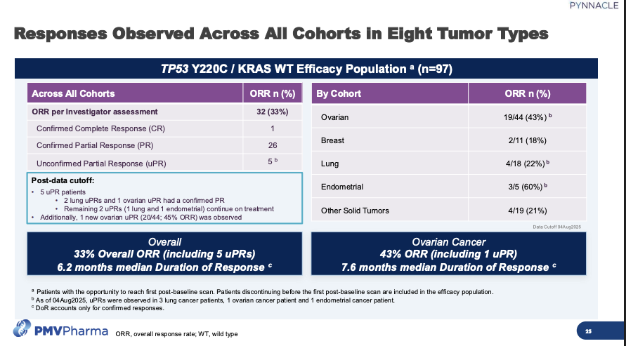
- ph 1/2 PC14586 (p53-Y220C refolder) in advanced solid tumors w/ Y220C mutation. Monotherapy in patients ORR 33% in Phase 2, still ongoing, and 43% ORR in ovarian cancer patients. A lot of partial responses.
- ph 1/2 $PMVP PC14586 (p53-Y220C refolder) in advanced solid tumors w/ Y220C mutation. Monotherapy in patients ORR 33% in Phase 2, still ongoing, and 43% ORR in ovarian cancer patients. A lot of partial responses. (see image)
- ph3 idasanutlin (MDM2i) in AML was stopped for futility based on interim analysis (idasanutlin cytarabine vs cytarabine) NCT02545283 MIRROS $RHHBY. Work on WT p53 and toxic (cytopenias, GI AEs, etc.) and quick aquired resistance
- KT-253 from $KYMR in R/R solid tumors, ALL, and lymphoma completed phase 1 but no mention of it since on their pipeline
- Gendicine, APPROVED in china in 2003 (recombinant human WT p53 packaged in AAV) for tx of HSNCC. mechanism is questionable and valid concerns for immunogenicity.
"Though their studies show that, when combined with traditional chemotherapy regimens, patients treated with rAD-p53 live longer post-diagnosis, the interpretation of these results must be put in the context of the finding that p53 mutation status did not significantly influence efficacy outcomes and long-term survival rate for Ad-p53-treated patients" (Zhang et al 2018 The first approved gene therapy product for cancer ad-p53 (gendicine): 12 years in the clinic. Hum. Gene. Ther.)
- Ph3 advexin (adenoviral vector that carries wt p53) with no real efficacy in recurrent HSCC. FDA issues "not sufficiently complete to file" in response to BLA and program died shortly thereafter. Pulled from EMA too.
- Arsenic trioxide (induces E3 ligase ubiquinitation of mutant p53 resuce mutant p53) in phase 2 and 3 in p53 mt MDS but status unknown (NCT03377725, NCT03381781)
- ph 1/2 p53 vaccine no effect in vaccine vs control in SCLC response rate (Chiappori et al. Cancer Immunol. Immunother 2019)
12
79
405
161,780

20 Oct 2025
TCL al dostum. Bizzat ürettim. Adamların malına verdiği değeri de gördüm. Hem opencell ( TV paneli ) olarak, hem şase olarak, hemde malzeme kalitesi olarak şu an LG ve Samsung’a kafa tutar. İddaa ediyorum. TCL yakında tüm TV pazarını eline alacak.
10
27
9,836

14 Oct 2025
Bridging Protein Sequences and Microscopy Images with Unified Diffusion Models
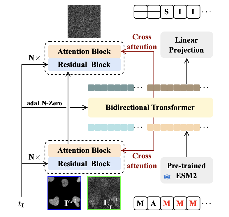
1. A novel study introduces CELL-Diff, a unified diffusion model that enables bidirectional transformations between protein sequences and their corresponding microscopy images. This model leverages both continuous and discrete diffusion models within a single framework, significantly enhancing the resolution and accuracy of generated protein images compared to previous methods.
2. CELL-Diff integrates cell morphology images, such as those of the nucleus, endoplasmic reticulum, and microtubules, as conditional inputs. This allows the model to generate detailed protein images from sequences and vice versa, providing a powerful tool for investigating subcellular protein localization and function.
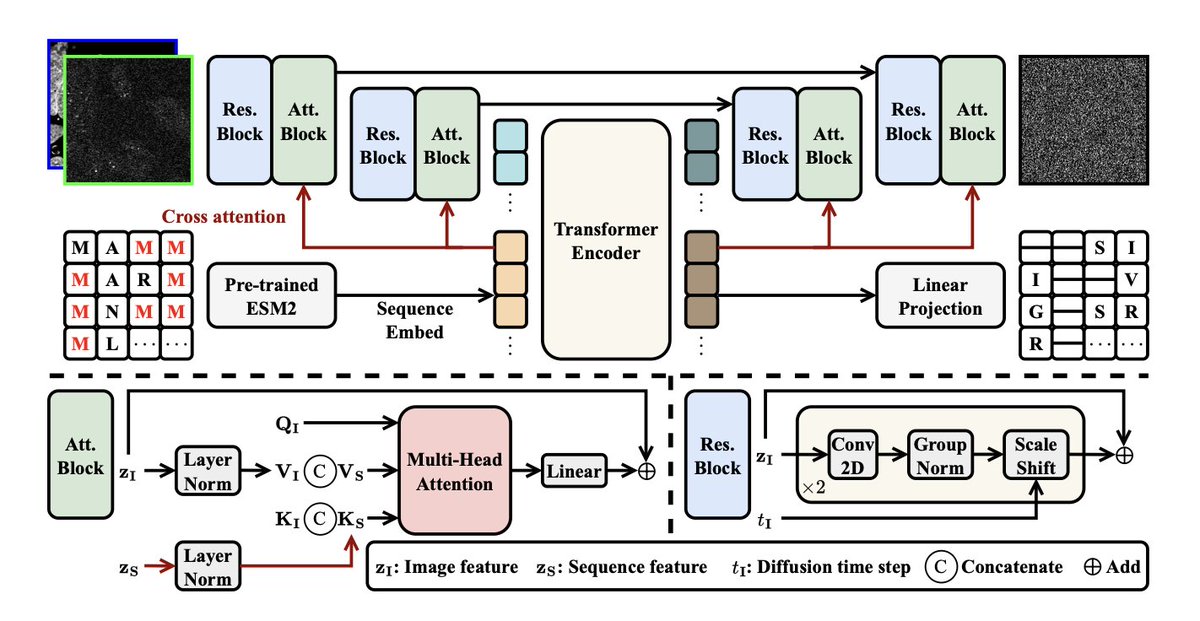
3. The model employs a transformer-based U-Net architecture enhanced with cross-attention mechanisms. This design effectively integrates information from both images and sequences, leading to superior performance in both image-to-sequence and sequence-to-image transformations.
4. CELL-Diff was trained on the Human Protein Atlas (HPA) dataset and fine-tuned on the OpenCell dataset. The results demonstrate that CELL-Diff outperforms existing methods in terms of image fidelity and the ability to resolve fine subcellular structures, making it a practical tool for virtual screening of protein localization signals and virtual staining.
5. Potential applications of CELL-Diff include virtual screening of protein localization signals, such as Nuclear Localization Signals (NLS) and Nuclear Export Signals (NES), as well as generating novel protein localization signals. This opens new avenues for studying intracellular protein dynamics and interactions.
6. The study highlights the potential of CELL-Diff to accelerate advancements in therapeutic target identification, drug discovery, and the investigation of biochemical pathways by bridging the gap between protein sequences and their cellular functions as visualized through microscopy.
📜Paper: raw.githubusercontent.com/ml…
1
1
4
935

8 Apr 2025
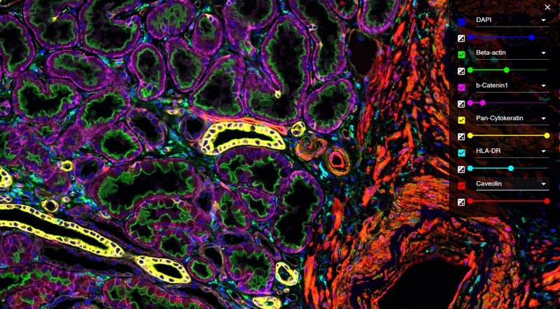
Nature's tech feature explores spatial proteomics, moving beyond the "one gene, one protein, one function" paradigm. Thanks for highlighting our OpenCell network and DVP platform revealing disease mechanisms at single-cell resolution. #SpatialProteomics nature.com/articles/d41586-0…
9
55
3,556

1 Mar 2025
3 year ince @Semicon_India and no movement on OpenCell /Display Fab investment. All initial proposals are in limbo. @AshwiniVaishnaw @RajeevRC_X @GoI_MeitY
We are ending up with a display module-related upstream investment. Adds some value but is still dependent on imports.
1 Mar 2025

Dixon expects to begin manufacturing display modules by October, followed by the launch of a camera module plant.
#MakeInIndia
business-standard.com/specia…
5
8
61
2,167

The @ProteinAtlas dataset was just the beginning! SubCell is being evaluated on OpenCell AllenCell datasets (s/o to our colleagues at @AllenInstitute), the breast cancer cell dataset from Bridge2AI’s Cell Maps 4 AI project & the JUMP dataset, with promising results
1
1
341

9 Dec 2024
4/ The model generalizes well: To cell lines not included in the training, drug-perturbed cells, different image resolutions, and varying reference marker channels. See applications including MoA prediction on OpenCell, Bridge2AI and JUMP datasets.
1
4
373
9 Dec 2024
SubCell: Vision foundation models for microscopy capture single-cell biology
1. SubCell is a suite of self-supervised deep learning models that analyze fluorescence microscopy images, capturing cellular morphology, protein localization, and biological organization at a single-cell level.
2. Trained on the extensive Human Protein Atlas (HPA) dataset, SubCell outperforms state-of-the-art methods in tasks like protein localization prediction, drug perturbation analysis, and mechanism of action identification.
3. Demonstrates exceptional generalizability across datasets without fine-tuning, effectively analyzing diverse datasets such as OpenCell, JUMP Cell Painting, and drug-treated breast cancer cells.
4. Introduces innovative “cell-specific” and “protein-specific” objectives in its multi-task framework, enhancing model performance for both cell morphology and subcellular localization tasks.
5. Constructs the first proteome-wide hierarchical map directly learned from imaging data, revealing subcellular compartments, protein complexes, and dynamic cellular behaviors with unprecedented resolution.
6. Achieves robust results in drug perturbation prediction, distinguishing treatment effects and mechanisms of action with high accuracy, surpassing models trained explicitly on drug-treated datasets.
7. Offers a practical, open-source solution for researchers, with ready-to-use tools, detailed tutorials, and interactive visualization of proteome-wide cellular maps.
8. Lays the foundation for future applications in phenotypic screening, drug discovery, and integrating imaging data with other biological modalities like transcriptomics and proteomics.
@Prof_Lundberg @Tkaraletsos @ulrikaaxelsson @AnthonyCesnik @wdleineweber @K_Kahnert @agupta
💻Code: github.com/czi-ai/SubCellPor…
📜Paper: biorxiv.org/content/10.1101/…
#MicroscopyAI #SingleCellBiology #Bioinformatics #DeepLearning #ProteinLocalization #DrugDiscovery

5
23
2,366
3 Nov 2024
Deep generative model for protein subcellular localization prediction
1. deepGPS introduces a pioneering deep generative model that not only predicts protein subcellular localization but also generates synthetic fluorescence images of predicted localizations. This dual-output capability addresses the need for visual insights alongside textual labels in protein localization prediction.
2. By training on both protein sequences and fluorescence images from the OpenCell database, deepGPS accurately predicts nuclear and cytoplasmic localization, outperforming traditional models that rely solely on textual outputs.
3. Unlike existing image-based models that require fluorescence images as inputs, deepGPS functions as a “text-to-image” predictor, making it highly versatile for applications where only sequence data is available.
4. The model leverages ESM-2 and U-Net architectures, integrating advanced encoding of protein sequences and nuclear images to produce high-quality localization predictions with enhanced image realism and spatial accuracy.
5. Further extensions of the model, including deepGPS-single-4 and deepGPS-all, expand its predictive capabilities to multiple organelles and multi-localized proteins, achieving high performance in complex cellular environments.
6. The online openGPS platform provides a user-friendly interface, allowing researchers to submit protein sequences and obtain both localization predictions and visual representations. This interactive feature broadens accessibility and fosters collaborative data expansion within the research community.
7. Comprehensive evaluation demonstrates that deepGPS maintains robustness across cell types and independent datasets, highlighting its generalizability and potential for broader biological and medical applications.
💻Code: github.com/royal-dargon/deep…
📜Paper: biorxiv.org/content/10.1101/…
#deepGPS #ProteinLocalization #DeepLearning #SubcellularLocalization #Bioinformatics #GenerativeAI #ProteinResearch

8
31
2,154

30 Oct 2024
🚨🇫🇷A Threat Actor Has Allegedly Leaked Data of Opencell
darkwebinformer.com/a-threat…
A threat actor claims to have leaked the database of Opencell, containing user IDs, names, password hashes, phone numbers, and other sensitive information.
1
2
2,613

23 Oct 2024
Newly announced version of the HPA now includes data from the #TabulaSapiens and #OpenCell projects of @cziscience and @czbiohub!
22 Oct 2024

A new version 24 of the Human Protein Atlas resource has been released at the HUPO meeting in Dresden, Germany. The data is summarized in eight resources covering different aspects of human protein-coding genes in tissues, cells, cell lines and blood.
proteinatlas.org/news/2024-1…
1
5
12
4,013
18 Oct 2024
CELL-Diff: Unified Diffusion Modeling for Protein Sequences and Microscopy Images
• CELL-Diff introduces a unified diffusion model enabling bidirectional generation between protein sequences and their corresponding fluorescence microscopy images.
• By integrating continuous diffusion for image generation and discrete diffusion for sequence generation, CELL-Diff ensures high-fidelity outputs across both modalities.
• This model outperforms previous methods like CELL-E2, generating sharper, more detailed protein images that accurately depict subcellular structures such as the endoplasmic reticulum and microtubules.
• CELL-Diff facilitates virtual staining, allowing multiple proteins to be visualized in a single cell morphology image, overcoming the limitations of fluorescence microscopy’s color channels.
• The model enables virtual screening of protein localization signals, predicting Nuclear Localization Signals (NLS) and Nuclear Export Signals (NES) from sequence input with high accuracy.
• CELL-Diff also offers the potential to generate novel protein localization signals by predicting sequences based on target protein images, advancing the study of subcellular interactions.
• The model was trained on the Human Protein Atlas (HPA) dataset and fine-tuned on the OpenCell dataset, demonstrating robust performance on diverse human cell images.
📜Paper: doi.org/10.1101/2024.10.15.6…
5
826