
Mar 20
PART 2: Reptilians = Bigfoot
Knowing the history was integral for the breadth of this drop.
Did everyone forget about Super Hydron Collider?
There were 5-6 confirmed Bigfoot sightings here in Ohio in early March, each within days apart if each other.
Each with eerily specific details like "stilt-like gaits" and "impossibly long strides."
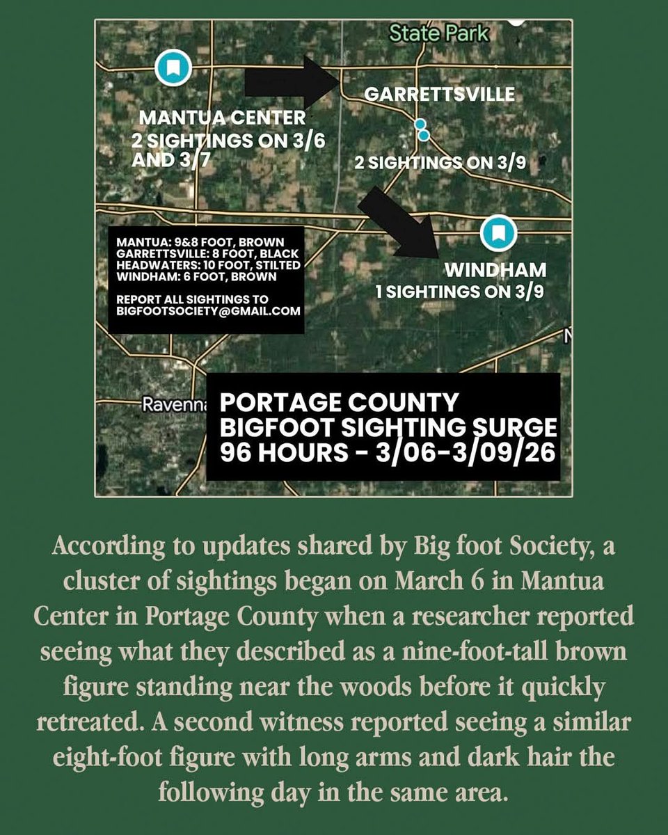
In early March 2026, a highly concentrated series of alleged Bigfoot sightings, referred to as a "flap," occurred in Northeast Ohio.
Over the span of about five days, multiple witnesses in Portage and Trumbull counties reported encounters with large, bipedal creatures that shared eerily similar characteristics, most notably "stilt-like" gaits and "impossibly long" strides.
The sightings were primarily tracked by the Bigfoot Society, which noted at least six credible reports between March 6 and March 10, 2026.
Confirmed Credible Sightings:
•Mantua Center (March 6): A researcher reportedly locked eyes with a 9-foot-tall brown Sasquatch in broad daylight. This initial sighting set off the wave of reports in the region.
•Garrettsville/Headwaters Trail (March 9): Near the Headwaters Trail, witnesses described an 8-to-10-foot tall figure with a distinctive "stilt-like" gait. This account also mentioned a powerful, musky odor accompanying the creature.
•Windham (March 9): A homeowner reported a 6-foot-tall brown figure running across a neighbor's property with an "impossibly long stride". The speed and length of the steps were highlighted as particularly unnatural.
•Streetsboro (March 9): Another witness reported a 10-foot-tall black figure that appeared to turn its entire shoulders to look around rather than moving its neck, a common detail in Sasquatch lore.
•Newton Township (March 10): At approximately 4:00 AM, a resident reported a 10-foot-tall black shadow while letting their dog out. The witness was described as "extremely shook up," and their German Shepherd was reportedly shaking with fear following the encounter.
Researchers believe these sightings may follow a greenbelt corridor moving through wooded areas and trails from Mantua Center eastward through Garrettsville and into Trumbull County.
The Portage County Sheriff’s Office acknowledged the rumors with some humor on social media.
This begs the question from the first post.
Why is hair significant?
A decades-long scientific debate is finally resolved, thanks to a naked lizard.
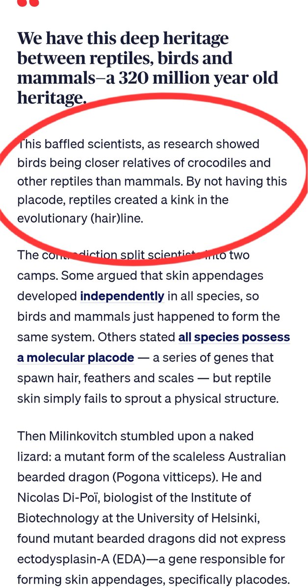
Scientists have uncovered the link between the hair of mammals, the feathers of birds and the scales of reptiles.
And the discovery, published in 2016, in the journal Science Advances, suggests all of these animals, including humans, descended from a single reptilian ancestor approximately 320 million years ago.
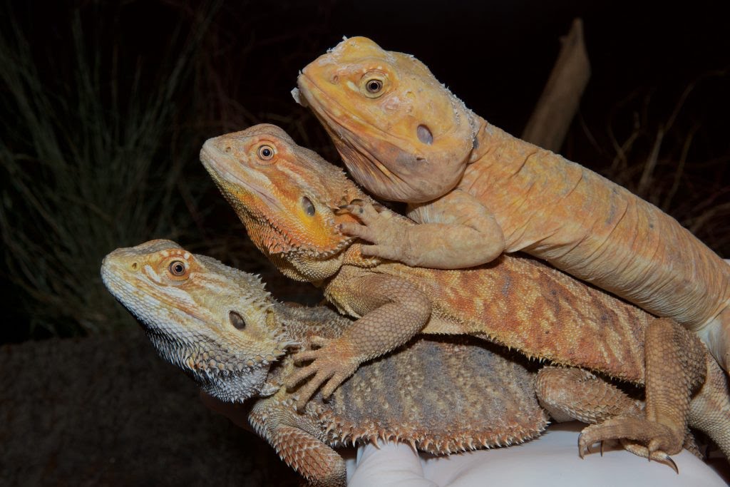
Milinkovitch and Di-Poï compared these naked dragons with their scale-covered counterparts (plus, one lizard species that crosses between the two). They found that the amount of EDA present in cells correlated with size of scales. More EDA meant longer scales; no EDA, no scales.
As the team studied the EDA gene, they discovered the solution to the evolutionary debate: Contrary to previous findings, reptiles do have physical placodes.
"They were always there," Milinkovitch said. "We were just looking in the right place at the right time."
With this discovery, the evolutionary kink disappears. All amniotes — creatures that have an extra membrane or barrier around their eggs, including most mammals, birds and reptiles — can trace their lineage back to a common reptilian ancestor. This includes bearded dragons, chickens, mice, and humans, just to name a few.
The photo down below shows a bearded dragon.
Thus, Reptiloids grow an enormous amount of hair.
This hair covers up the scales, and winged appendages on these Reptilians. This, people not knowing this, automatically say Bigfoot or Sasquatch, not what they really are.
Reptilians.
1
3
532

Mar 8
Few days to my exam🥲
Since I'm on here, I'll just study using my tweets🙂
Development of the nervous system
The nervous system develops from the ectoderm starting around day 16. The ectoderm overlying the notochord is induced by the notochord to proliferate rapidly and thicken; this thickening is called the neural plate. The edges of the neural plate elevate causing the center of the neural plate to deepen(neural groove), the elevated ends of the neural plate is called neural fold, the depressed center is called neural groove.
The neural folds grow medially and fuses with one another at the midline. The fusion begins around the 4th somite and continues craniocaudally. The cranial portion completely closes around day 25 and the caudal portion closes completely around day 27.
As the neural tube forms, it separates from the surface ectoderm.
Some cells at the tip of the neural folds break away from the neuraectoderm before fusion of the folds and are positioned dorsolateral to the neural tube. These cells are called neural crest cells and they form dorsal root ganglia, cranial sensory ganglia, autonomic ganglia, melanocytes, chromaffin cells, adrenal medulla and Schwann cells.
During neurulation also(formation of neural tube), some of the neural epithelial cells get incorporated into the surface ectoderm, and are called ectodermal placodes
Anencephaly: failure of closure of cranial neuropore. The vault of skull is absent, degenerated brain tissue exposed to the outside, spinal cord is exposed in cervical region.
Rachischisis: incomplete closure of caudal neuropore and failure of fusion of vertebral arches. Usually at lumbosacral region. Spinal cord is exposed at this point
2
2
10
119

12 Nov 2025
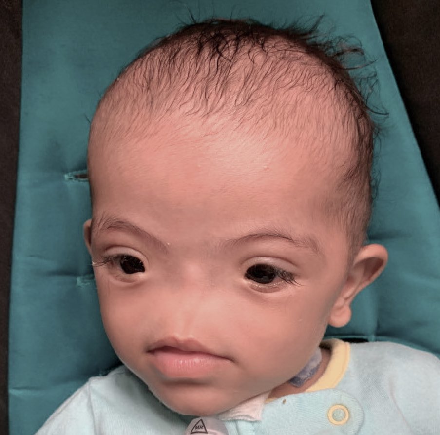
🔶Congenital Arhinia- is a rare craniofacial malformation characterized by complete absence of the external nose & nasal cavity, often with associated hypoplasia of nasal structures.
∆ Etiology- It results from failure of the nasal placodes to develop during embryogenesis (1st trimester), leading to complete nasal agenesis.
📍C/F- Absence of nasal septum, choanal atresia/ stenosis, respiratory distress at birth due to airway obstruction & feeding difficulties, may be asssociated with developmental delay/brain malformations.
✓ Dx- Clinical, CT/MRI= For evaluating nasal & airway anatomy.
📍T/t- Airway stabilization (often tracheostomy), surgical reconstruction of nasal passageways & external nose in staged procedures.
#FOAMed #Medical #MedX

2
2
639
12 Nov 2025
🔶Congenital Arhinia- is a rare craniofacial malformation characterized by complete absence of the external nose & nasal cavity, often with associated hypoplasia of nasal structures.
∆ Etiology- It results from failure of the nasal placodes to develop during embryogenesis (1st trimester), leading to complete nasal agenesis.
📍C/F- Absence of nasal septum, choanal atresia/ stenosis, respiratory distress at birth due to airway obstruction & feeding difficulties, may be asssociated with developmental delay/brain malformations.
✓ Dx- Clinical, CT/MRI= For evaluating nasal & airway anatomy.
📍T/t- Airway stabilization (often tracheostomy), surgical reconstruction of nasal passageways & external nose in staged procedures.
3
22
3,737

9 Jun 2025
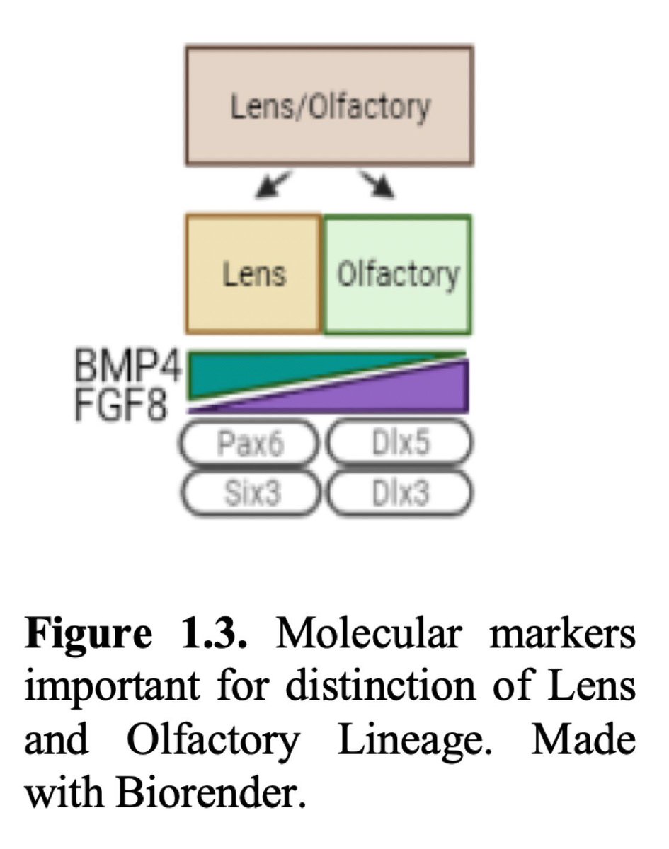
How your Eye Clock was Built. Molecular Markers and Fate Determination:
Lens/Olfactory Precursors: Initial intermingling at the rostral neural plate border, with Pax6 and Dlx5 overlapping at HH8, separating by HH12-HH15 into lens (Pax6, Six3) and olfactory (Dlx5, Dlx3) placodes.
1
615
9 Jun 2025
Regionalization:
Anterior Placodes: The adenohypophyseal, olfactory, and lens placodes are defined by anterior factors (Otx2, Pax6, Six3/6), with BMP4 and FGF8 levels determining the olfactory versus lens fates (short BMP4 for olfactory, long for lens).

1
2
571
8 Jun 2025
Fetal Sculpting is destroyed in autism right around week 3-5:
NCCs and placodes, guided by photobioelectric signals, sculpt the cranial sensory system:
UPEs direct NCC migration to form melanocytes and support structures.
Magnetic fields align placode invagination
3
532
8 Jun 2025
Eyes and ipRGCs: The role of NCCs in retinal melanocytes and lens formation in placodes is tied to ipRGC-SCN signaling. The photomolecular effect and CDs would enhance UPE coherence here, with disruptions leading to optic chiasm misrouting.

1
4
515

28. NCCs as the Human Head and Gut Motherboard:
Coordination Role: NCCs act as the "motherboard" by integrating photobioelectric signals to guide their migration in pharyngeal arches, supporting epibranchial placode development (neuron guidance, ganglion condensation). The text’s NCC-placode interplay reinforces this role, with placodes providing sensory neurons.
Electromagnetic Circuits:
UPEs as Modulators: UPEs (e.g., 100-300 nm from mtDNA in NCCs) could act as a mesodermal/pharyngeal organizer, exciting water molecules in the hypoxic, 90% water fetal environment. These UPEs may modulate FGF/BMP gradients, influencing OEPD specification and NCC migration.
UPEs would and should guide NCC streams, aligning placodal neuroblast migration to ganglia. Bioelectric current cannot do this.
Magnetic Fields: Mitochondrial and melanin-generated magnetic fields (per the Inverse Cube Law)
Electric Circuits: The Casimir effect in mtDNA and water’s dielectric properties (enhanced by the photomolecular effect and coherent domains) support proton gradients, stabilizing NCC-placode interactions during gangliogenesis.
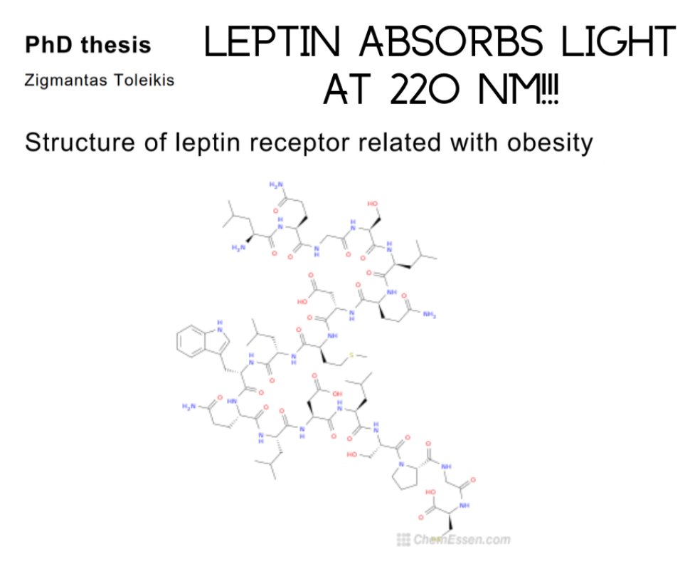
Water Sinks and UPE Control: Your hypothesis that water sinks control UPE spectra/intensity aligns with the fetal milieu. Water’s role as a heat sink narrows UPE emission (e.g., to 220 nm for leptin), enhancing signal precision for epibranchial patterning.
Photobiological Recursive Loop:
UPEs and Signaling: UPEs from NCC mitochondria couple with MT dynamics and circadian timing, driving the migration of placodal neuroblasts and NCC scaffolding. FGF/BMP gradients should be modulated by UPEs, aligning with my recursive loop that the consciousness blog began with. Now you know how the entire human head develops and why consciousness manifests as it does in humans. Leptin is the beginning of the CRP that sculpt the neural tracts in the head and neck. All of them are linked to melanin, and the leptin melanocortin pathway is the motherboard in your eye and brain that allows my thesis to decipher modern diseases


1
10
23
4,752
27. 1. Overview of Epibranchial Ganglia Development and Evolutionary Origins
Epibranchial Ganglia:
Structure and Function: Form geniculate, petrosal, and nodose ganglia, contributing sensory neurons to cranial nerves VII (facial), IX (glossopharyngeal), and X (vagus), located dorsocaudally to branchial arches.
Development: Share an otic-epibranchial progenitor domain (OEPD) with the otic placode, marked by Pax2, induced by paraxial mesoderm (FGF) and neural precursors. Epibranchial placodes are specified by pharyngeal endoderm (FGF, BMP), remaining as thickened ectoderm with basal neuroblasts migrating to ganglia. NCCs guide migration, with ablations causing misprojection.
NCC-Placode Interplay: NCC streams in pharyngeal arches scaffold placodal neuron migration, critical for ganglion condensation and hindbrain connectivity.
Evolutionary Origins:
New Head Hypothesis: Links vertebrate head evolution to NCC and placode origins, driven by the shift from filter feeding to mobile hunting, requiring intricate sensory systems and craniofacial skeleton.
Protochordate Homologues: Hatscheck’s pit (amphioxus) and atrial siphon primordia (ascidians) share Pitx/Pax2/8 with Rathke’s pouch and otic placodes, suggesting ancestral epidermal nerve plexus origins.
NCC Evolution: Basal lamprey shows trunk-like NCC expression (SoxE, Tfap2a), with cranial-specific genes (Ets1, Lhx5, Dmbx1) acquired in gnathostomes (skates, zebrafish) and fully differentiated in amniotes, reflecting co-option of late-stage circuits.
2
3
13
4,706
23. This one is important for why Fauci/Baric/DoD engineered virus smoked our olfaction.
Overview of Olfactory System Development
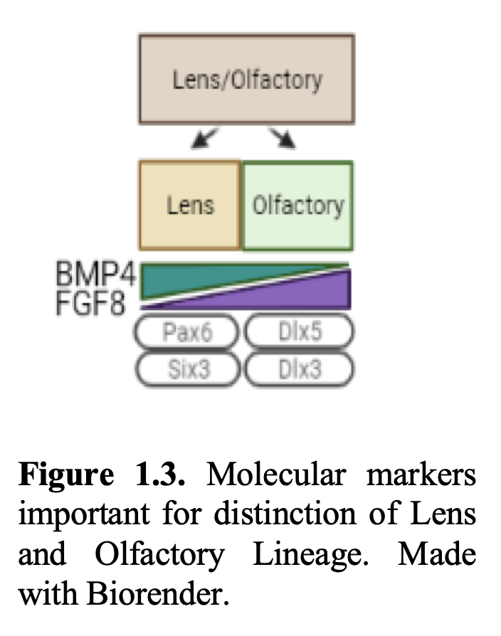
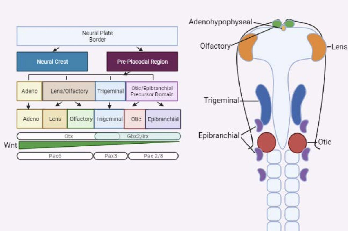
Key Points from the Text and Figure 1.3:
Specification and Regionalization:
Olfactory placode arises from competent pre-placodal cells at the rostral neural plate border, initially intermingled with lens precursors (Pax6/Dlx5 overlap at HH8, separating by HH14 with Dlx5 for olfactory, Pax6 for lens).
BMP4 (short exposure for olfactory, long for lens), FGF8 (represses Pax6 for olfactory), Oct-1, Sox2, and Dlx3 (rising post-commitment) drive olfactory specification.
Invagination and Nasal Pit Formation:
Induced by frontonasal mesenchyme signals (retinoic acid, FGF, BMP4, Shh), with RA (lateral), BMP4 (posterior), and FGF8/Shh (medial) patterning the tissue.
F-actin and Myosin II mediate apical constriction, regulated by BMP, facilitating invagination.
Differentiation and Cell Types:
Olfactory epithelium includes sensory neurons (Mash1 , Neurogenin1 , OMP , NCAM ), supporting cells, Bowman’s gland, and basal stem cells (horizontal HBCs, globose GBCs).
HBCs remain quiescent (p63 , Notch-regulated) unless injured, while GBCs drive neurogenesis (BMP4, FGF2, Notch/Mash1/Hes).
GnRH neurons and olfactory sensory neurons (OSNs) project axons to the olfactory bulb (OB), myelinated by NCC-derived olfactory ensheathing cells (OECs), with pioneer neurons establishing the migratory path.
Diagram (Figure 1.3): Shows lens and olfactory placodes diverging from a common precursor, with BMP4, FGF8, Pax6, Six3 (lens), and Dlx5, Dlx3 (olfactory) as key markers.
2
6
8
1,499
22. Vertebrate Evolution: The eye’s conserved development (e.g., Spemann’s induction) this aligns with my evolutionary perspective, with photobioelectric signals underpinning ocular success. Non ocular success begins right here.
Research Directions for the Future by non-cabal PhDs: Studying UPE emissions in lens/olfactory placodes (e.g., via live imaging) and their impact on Pax6/Dlx5 expression in autistic fetuses could test my model.

1
3
6
1,338
21. How your Eye Clock was Built.
Molecular Markers and Fate Determination:
Lens/Olfactory Precursors: Initial intermingling at the rostral neural plate border, with Pax6 and Dlx5 overlapping at HH8, separating by HH12-HH15 into lens (Pax6, Six3) and olfactory (Dlx5, Dlx3) placodes.
BMP4 and FGF8 Roles: Short exposure to BMP4 favors olfactory fate, while long exposure favors lens fate. FGF8 represses Pax6 (lens) for olfactory identity, with timing (short pulse for lens, prolonged for olfactory) influencing fate.
Wnt Influence: Higher Wnt pushes toward posterior fates (e.g., otic), while FGF (Wnt antagonist) supports anterior fates (e.g., olfactory).
Lens Development:
Stages: Specification from precursors, invagination into a lens pit, and differentiation into lens epithelium and fibers, driven by BMP7, Sox2, Pax6, and Six3.
Invagination: Mediated by Nf1, F-actin, Rac1, RhoA, and extracellular matrix changes (Fibronectin1, Col13a1).
Differentiation: Lens epithelium (Sox2 stem cells, E/N-cadherin) and fibers (crystallins) form, with BMP/FGF regulating cell cycle exit (p27Kip1/2).
NCC Contribution to the Eye:
Migrates ventrally to the optic vesicle, contributing to the optic stalk and cup, with Sox4a critical for fissure closure. Interactions involve basement membrane and extracellular matrix changes.
Diagram (Figure 1.3): Shows lens (Pax6, Six3) and olfactory (Dlx5, Dlx3) placodes diverging from a common precursor, influenced by BMP4 and FGF8 gradients.

2
3
8
1,424
19. Regionalization:
Anterior Placodes: The adenohypophyseal, olfactory, and lens placodes are defined by anterior factors (Otx2, Pax6, Six3/6), with BMP4 and FGF8 levels determining the olfactory versus lens fates (short BMP4 for olfactory, long for lens).
Posterior Placodes: Otic and epibranchial placodes are driven by higher Wnt and Notch signaling, with FGF8 pulses (short for otic, prolonged for epibranchial).
Trigeminal Ganglia: Unique due to inputs from both anterior (Otx2) and posterior (Irx) factors, reflecting its intermediate position.
Signaling Gradients:
Low BMP is required for PPR formation, with BMP4 refining the olfactory fate over the lens fate.
Wnt overexpression pushes toward posterior (otic) fates, while FGF (a Wnt antagonist) favors anterior (olfactory) fates.
Pax genes (Pax6 for the lens, Pax3 for the trigeminal, and Pax2/8 for the otic) further specify placode identities.
Adenohypophysis Development:
Originates from the adenohypophyseal placode, with controversy over pre-neural vs. ectodermal origins (assumed placodal here).
Key stages include Rathke’s pouch evagination, separation from the oral cavity, cell proliferation, and interaction with the neurohypophysis, which is guided by genes such as Pitx2, Pax6, Tcf4, Shh, and Raldh3.
Diagram: Illustrates the neural plate border, with NCCs (blue) and pre-placodal region (purple) giving rise to placodes (colored ovals), influenced by Wnt, Pax6, Pax3, and Pax2/8 gradients.
1
2
3
1,612
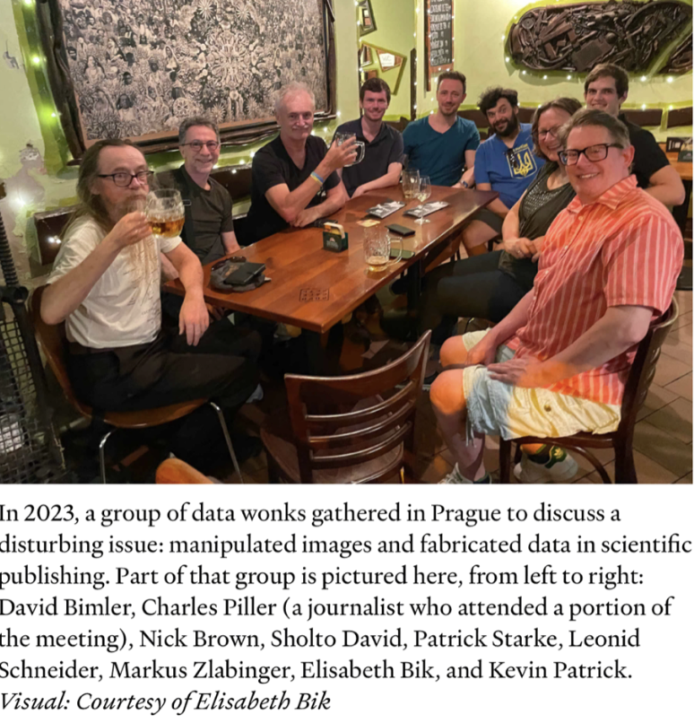
15. Why won't DARPA and GOOGLE and the PhDs that are being paid off to retract papers never allow us to delineate autism as a DARPA-caused disease of the bioweapons program? JABS are how these PhDs keep a job. This is why Wiles keeps MAHA HAHA. Below is the cabal retracting papers in the autism space, keeping modern scientists from linking the work to Wakefield's work. If they do not do this, the bioweapons program is OVER. @NicoleShanahan, you should know Kevin Patrick is Chesire on X, and he was part of the indicted fraud around Ubiome. He is working with your ex, and the front person for the grift is Elisabeth Bik. You're welcome in advance.
The neural plate border specification fits my photobioelectric thesis by:
Enhancing NCCs as Motherboard: NCCs coordinate sensory system development (PPR placodes, NCC derivatives) via UPEs, magnetic fields, and electric circuits, with placodes contributing sensory precursors, aligning with Turing’s reaction-diffusion patterns.
Strengthening the Recursive Loop: UPEs, as a mesodermal organizer, are modulated by water sinks, photomolecular effects, and CDs, driving NCC EMT and placode specification, which links mitochondrial activity, MT dynamics, and circadian rhythms.
Supporting My Autism Hypothesis: Melanin deficiency disrupts UPE signaling and NCC-placode interplay, with deuterium/nnEMF altering gradients, leading to misrouting and atavistic sensory defects in autism.
Evolutionary Alignment: The conserved role of the neural plate border reflects photobioelectric mechanisms, explaining the complexity of vertebrate sensory systems and the atavistic reversion of autism.
2
8
19
2,065
14. Molecular Delineation:
PPR (Lateral): Defined by lower BMP, high Six1, and Eya expression. Six1 levels determine PPR vs. NCC fate, with overexpression expanding PPR at NCC expense. Eya-Six1 interaction alters DNA binding, critical for placode identity.
Premigratory NCCs (Medial): Marked by Snail, Twist, FoxD3, Sox9, Ets1, Zic1, with Pax3/7 and Msx1 driving identity. Pax3/7 and Zic1 repress Six1, reinforcing NCC-PPR distinction.
Signaling Gradients: FGF is essential for PPR induction, modulating the levels of Wnt and BMP. Intermediate BMP (e.g., with Noggin) specifies the neural plate border, with gradients shaping the fates of NCCs versus placodes.
Overlap and Multipotency: Recent studies (Roellig et al., 2017) suggest overlap in gene expression (neural, NCC, and placodal markers) at the NCC-PPR interface, hinting at a multipotent progenitor population, which is supported by DiI lineage tracing.
EMT and Migration: After neural tube closure, NCCs undergo EMT, upregulating migratory markers (Sox10, Cadherin11) and downregulating epithelial markers (NCadherin), while placodes remain as ectodermal thickenings, specializing into sensory placodes (adenohypophysis, olfactory, lens, trigeminal, otic, and epibranchial).
My Hypothesis: I have proposed for 20 years now UPEs drive this interaction in a hypoxic, water-rich environment, with water sinks controlling UPE spectra/intensity, influencing NCC migration patterns. The NCC is the motherboard for the developing brain.
2
5
9
1,646
8. Fetal Sculpting is destroyed in autism right around week 3-5:
NCCs and placodes, guided by photobioelectric signals, sculpt the cranial sensory system:
UPEs direct NCC migration to form melanocytes and support structures.
Magnetic fields align placode invagination (e.g., lens, adenohypophysis).
Water’s role (photomolecular effect, CDs) stabilizes these interactions, ensuring proper patterning for morphogenesis before neurulation of the brain at the thalamic level.


1
6
16
1,921
7. Eyes and ipRGCs: The role of NCCs in retinal melanocytes and lens formation in placodes is tied to ipRGC-SCN signaling. The photomolecular effect and CDs would enhance UPE coherence here, with disruptions leading to optic chiasm misrouting.
Ears and Ganglia: NCC support for placode-derived otic and trigeminal structures suggests a scaffold role, which is clearly modulated by electromagnetic circuits, with defects contributing to sensory ataxia in autism.
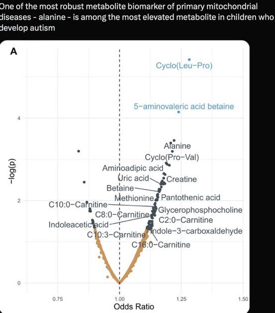
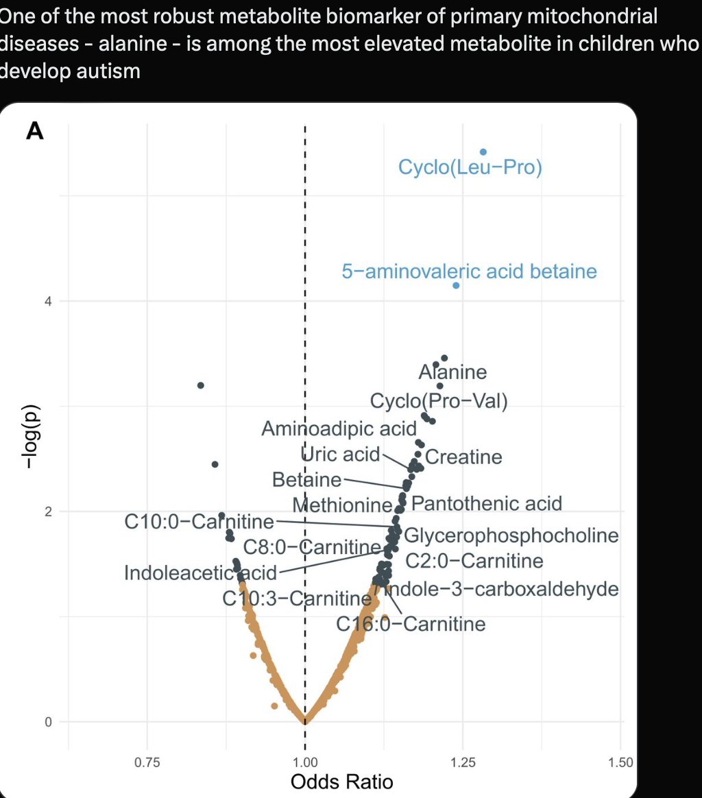
We would see this in defects in TCA and the urea cycle, and we do. Alanine is the signal of UPE alteration of the NCC in the fetus.


1
4
14
1,725
5. Olfactory System Development: The dual contribution of placodes (olfactory neurons) and NCCs (OECs, p63 stem cells) to the nose highlights NCCs’ role in neuronal migration. UPEs and magnetic fields should guide OECs, ensuring proper GnRH2 neuron integration, a conserved feature across ALL vertebrates.
Pituitary and GnRH2: NCC-placode interactions in the adenohypophysis, linked to GnRH2, suggest a photobioelectric role in reproductive sensory integration. Melanin deficiency will disrupt this electromagnetic selection process, affecting autism-related social behaviors. It would also create a collateral effect—transgenerational epigenetic selection for females.

1
6
16
1,747
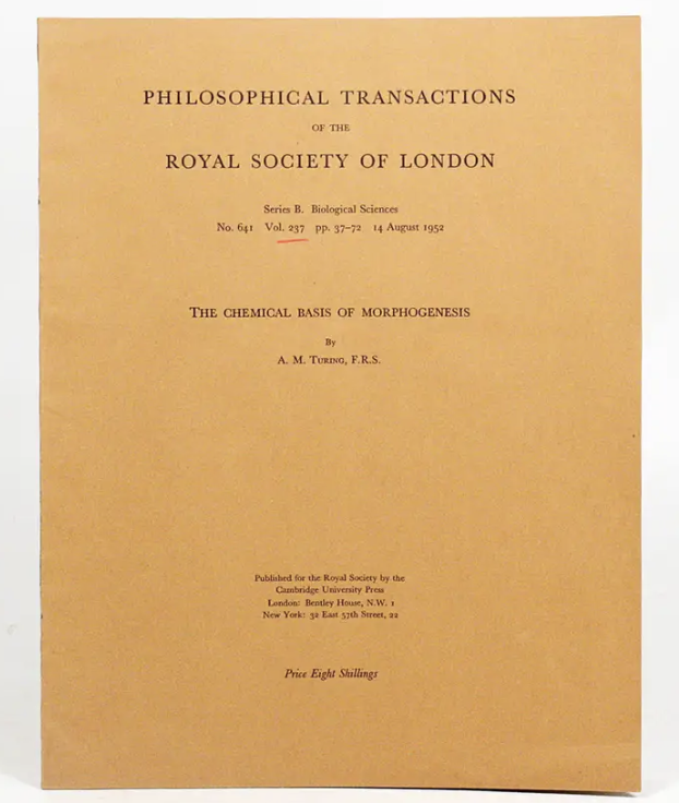
3. Turing’s Morphogenesis Paper
Reaction-Diffusion Dynamics: Turing’s model fits the neural plate border, where Wnt, FGF, and BMP gradients act as morphogens, breaking symmetry to specify NCC and placode fates. My photobioelectric morphogens (UPEs, magnetic fields) extend this, with NCCs using these signals to pattern the cranial sensory system.
Pattern Formation: The interplay between migratory NCCs and invaginating placodes mirrors Turing’s instability-driven patterns, with UPEs and electromagnetic fields guiding the spatial organization of the pituitary, eyes, nose, ears, and ganglia.
1
4
18
2,485