
ANATOMY NEET PG OFFLINE CLASSES - Coimbatore, June 2026
#anatomy #neetpg #inicet #fmge #medicaleducation #offlineclasses #education #anatomylearning


153

Apr 9
“教学用医学三维动画 仅限学习用途 非真实影像·非实景镜头
#medicaleducation #3danimation,
#anatomylearning #sciencevisuals
#healthknowledge”
2
2,429

Enhance anatomy teaching with real-specimen, 3D rotation videos based on Dr. Acland’s proven approach. Clear, accurate, and easy to use across programs. Explore now: ow.ly/mMCa50YxPAa
#MedEd #HealthSciences #AnatomyLearning
3
141

Feb 27
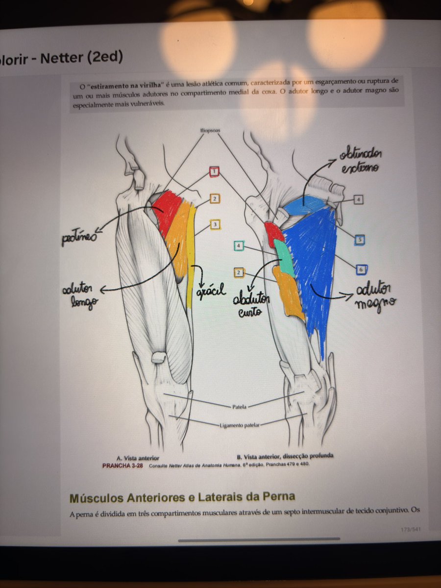
netter para colorir anatomylearning = mediciner mto feliz estudando anato
3
3
82
3,703

24 Nov 2025
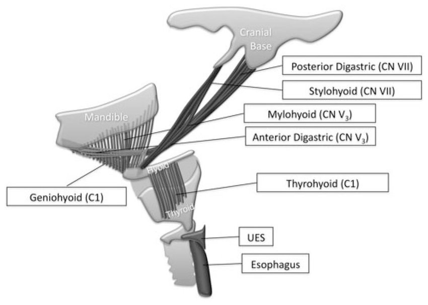
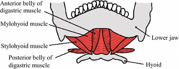
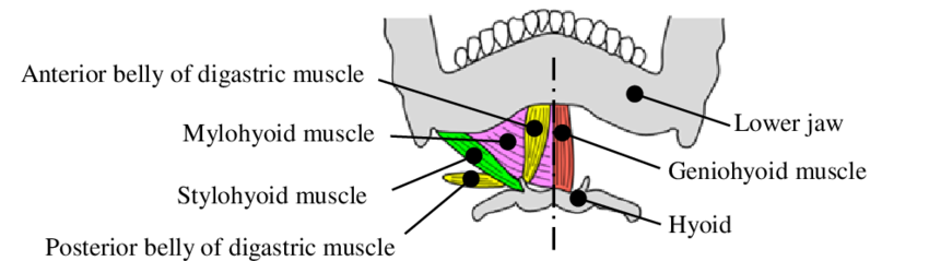
The suprahyoid muscles are a group of four small muscles located above the hyoid bone that connect the mandible and tongue to the upper neck. Their primary function is to elevate the hyoid bone and floor of the mouth, playing a key role in swallowing, speech, and stabilizing the airway. Clinically, dysfunction or weakness in these muscles can contribute to dysphagia, speech difficulties, and complications in airway management.
Read: wikism.org/Suprahyoid_Muscle…
Watch: youtube.com/shorts/sawbMdzo7…
#Anatomy #AnatomyLearning #AnatomyEducation #AnatomyOfTheDay #SportsMedicine #Orthopedics #PhysicalTherapy #AthleticTraining #Rehab #InjuryPrevention #Physio #SportsInjury #SportsRehab #PhysioTherapy #Meded #FOAMed
1
6
22
845
23 Nov 2025
The suprahyoid muscles are a group of four small muscles located above the hyoid bone that connect the mandible and tongue to the upper neck. Their primary function is to elevate the hyoid bone and floor of the mouth, playing a key role in swallowing, speech, and stabilizing the airway. Clinically, dysfunction or weakness in these muscles can contribute to dysphagia, speech difficulties, and complications in airway management.
Read: wikism.org/Suprahyoid_Muscle…
Watch: youtube.com/shorts/sawbMdzo7…
#Anatomy #AnatomyLearning #AnatomyEducation #AnatomyOfTheDay #SportsMedicine #Orthopedics #PhysicalTherapy #AthleticTraining #Rehab #InjuryPrevention #Physio #SportsInjury #SportsRehab #PhysioTherapy #Meded #FOAMed
1
17
41
2,158
21 Nov 2025
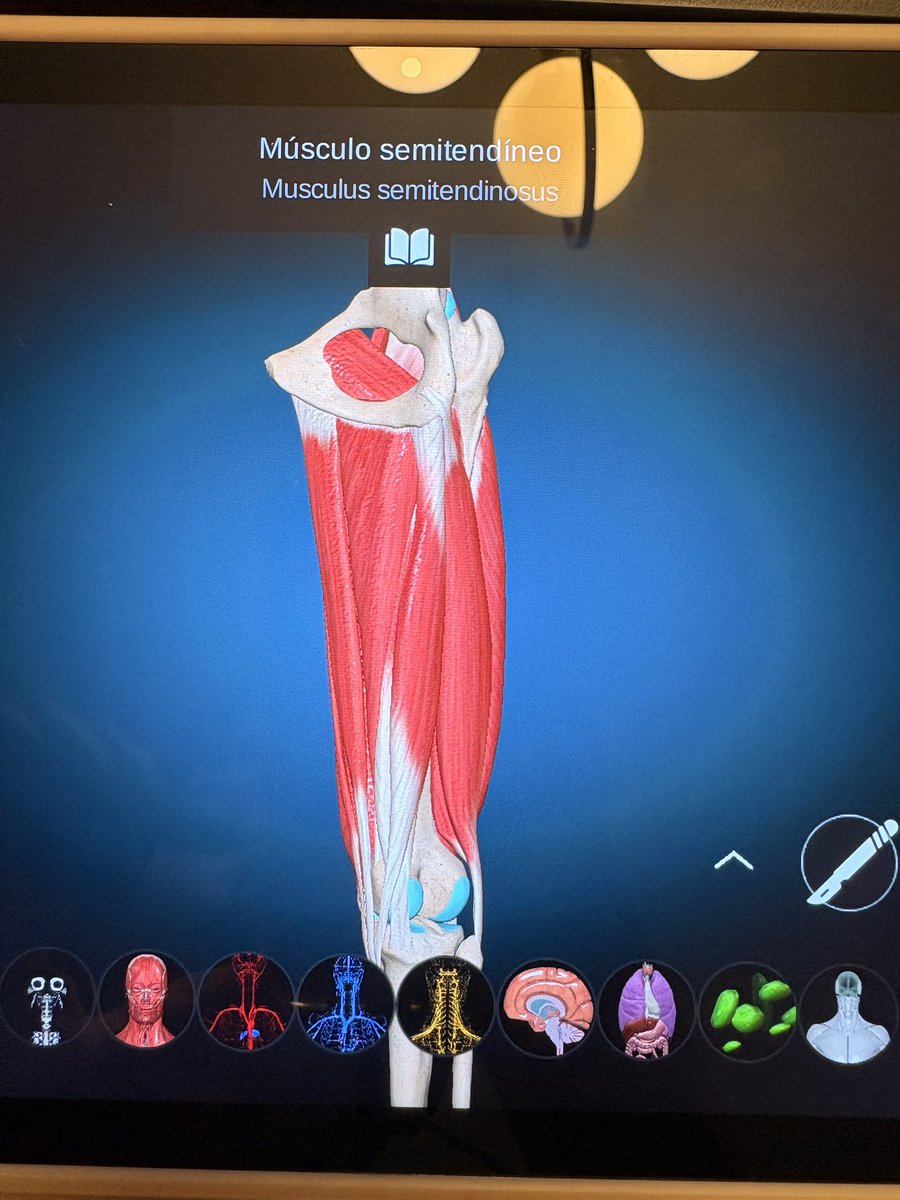
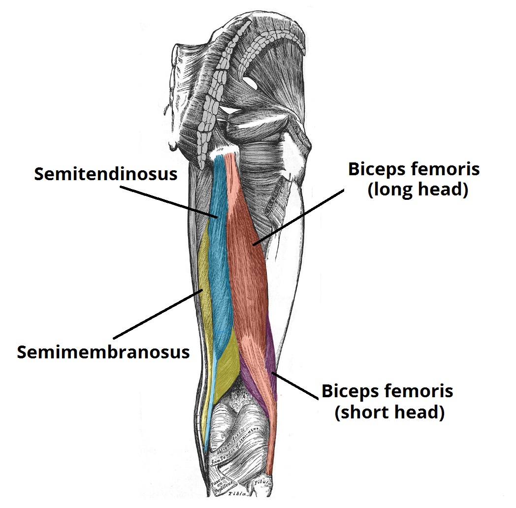
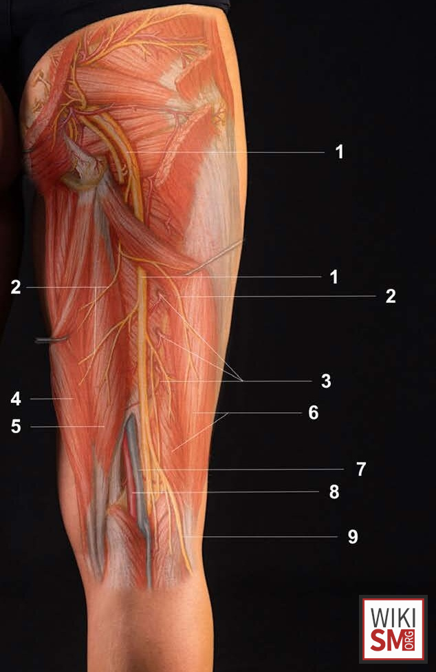
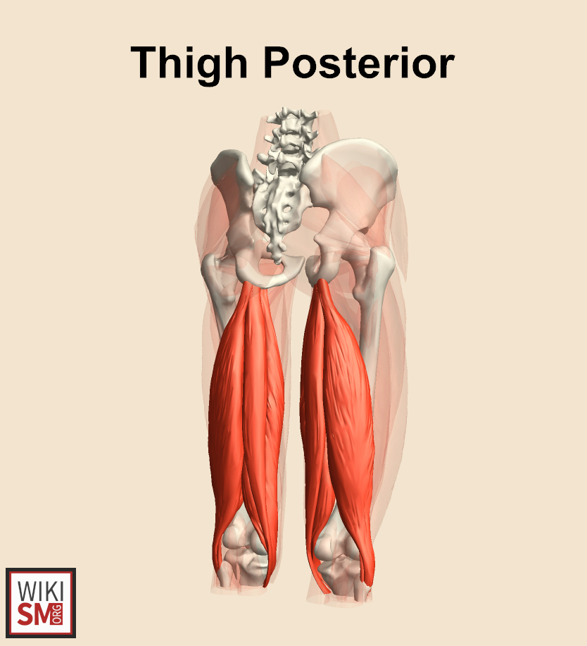
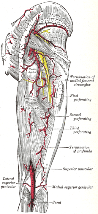
The posterior compartment of the thigh contains the hamstring muscles, which originate from the ischial tuberosity and are primarily innervated by the tibial division of the sciatic nerve. These muscles function to extend the hip and flex the knee, contributing to gait and dynamic lower-limb stability. Common pathologies include hamstring strains, tendinopathy, and proximal avulsion injuries, often caused by sudden acceleration or overuse.
Read: wikism.org/Posterior_Compart…
Watch: youtube.com/shorts/RoZxbGX0b…
#Anatomy #AnatomyLearning #AnatomyEducation #AnatomyOfTheDay #SportsMedicine #Orthopedics #PhysicalTherapy #AthleticTraining #Rehab #InjuryPrevention #Physio #SportsInjury #SportsRehab #PhysioTherapy #Meded #FOAMed
1
22
72
2,774
20 Nov 2025
The posterior compartment of the thigh contains the hamstring muscles, which originate from the ischial tuberosity and are primarily innervated by the tibial division of the sciatic nerve. These muscles function to extend the hip and flex the knee, contributing to gait and dynamic lower-limb stability. Common pathologies include hamstring strains, tendinopathy, and proximal avulsion injuries, often caused by sudden acceleration or overuse.
Read: wikism.org/Posterior_Compart…
Watch: youtube.com/shorts/RoZxbGX0b…
#Anatomy #AnatomyLearning #AnatomyEducation #AnatomyOfTheDay #SportsMedicine #Orthopedics #PhysicalTherapy #AthleticTraining #Rehab #InjuryPrevention #Physio #SportsInjury #SportsRehab #PhysioTherapy #Meded #FOAMed

1
20
76
2,876
17 Nov 2025
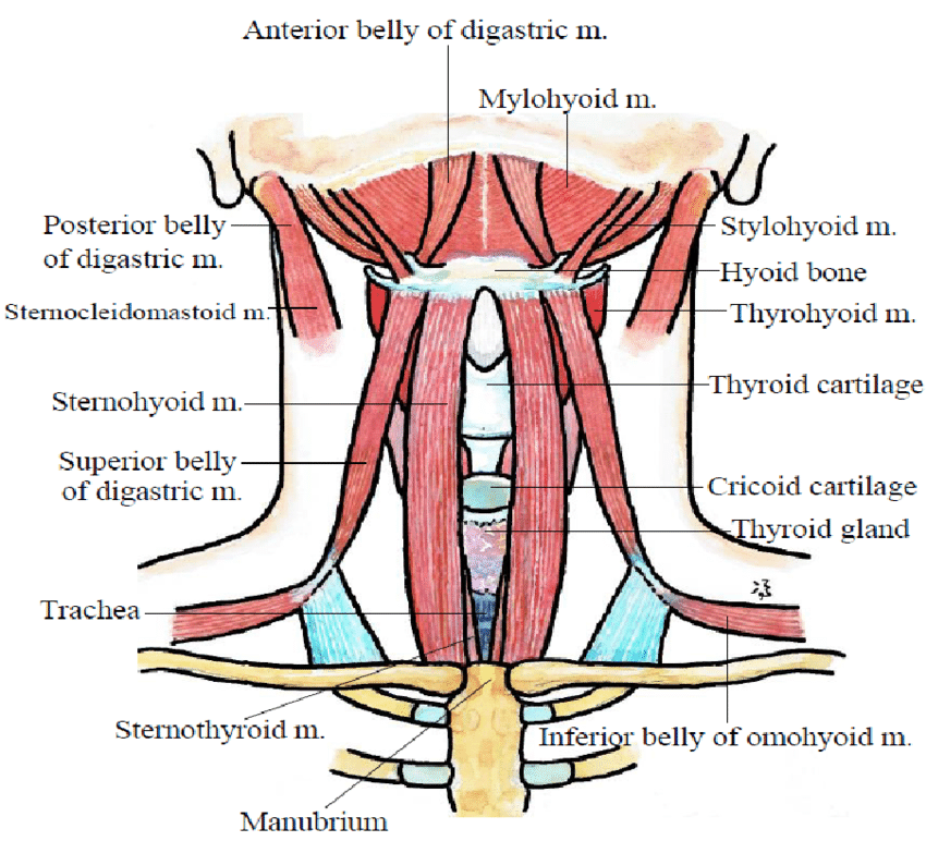
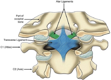
The cervical spine consists of seven vertebrae, labeled C1 through C7, which support the skull and enable a wide range of head and neck movements. Between each vertebra are intervertebral discs that act as shock absorbers and allow flexibility, while facet joints guide motion and provide stability. The cervical spine also houses the spinal cord and nerve roots, which transmit signals between the brain and the rest of the body.
Read: wikism.org/Cervical_Spine_An…
Watch: youtube.com/shorts/UqSHjomLC…
#Anatomy #AnatomyLearning #AnatomyEducation #AnatomyOfTheDay #SportsMedicine #Orthopedics #PhysicalTherapy #AthleticTraining #Rehab #InjuryPrevention #Physio #SportsInjury #SportsRehab #PhysioTherapy #Meded #FOAMed
1
8
48
1,510
16 Nov 2025
The cervical spine consists of seven vertebrae, labeled C1 through C7, which support the skull and enable a wide range of head and neck movements. Between each vertebra are intervertebral discs that act as shock absorbers and allow flexibility, while facet joints guide motion and provide stability. The cervical spine also houses the spinal cord and nerve roots, which transmit signals between the brain and the rest of the body.
Read: wikism.org/Cervical_Spine_An…
Watch: youtube.com/shorts/UqSHjomLC…
#Anatomy #AnatomyLearning #AnatomyEducation #AnatomyOfTheDay #SportsMedicine #Orthopedics #PhysicalTherapy #AthleticTraining #Rehab #InjuryPrevention #Physio #SportsInjury #SportsRehab #PhysioTherapy #Meded #FOAMed


14
53
2,594
13 Nov 2025
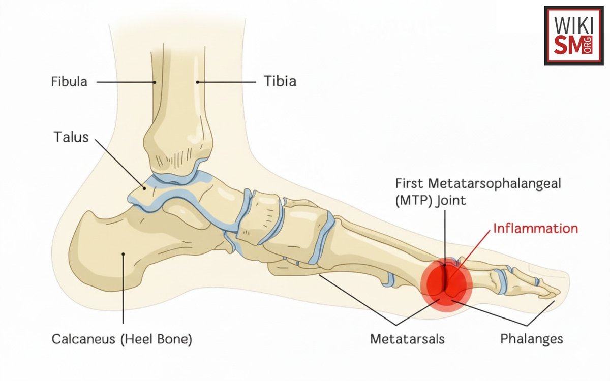
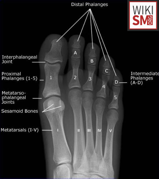
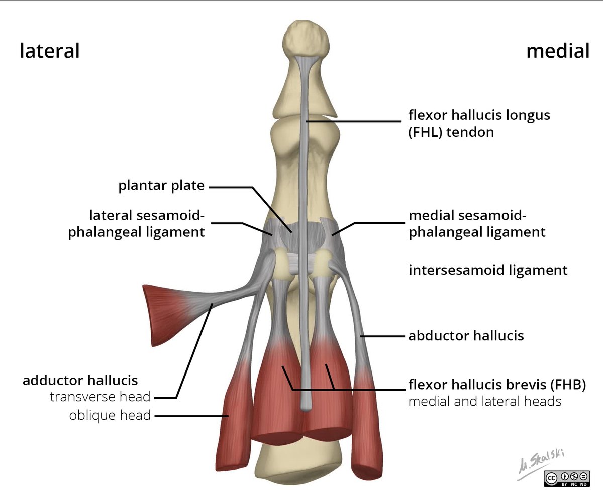
The metatarsophalangeal joints are the articulations between the metatarsal heads and the proximal phalanges of the toes, with the first MTP joint connecting the first metatarsal to the proximal phalanx of the great toe. These joints allow for flexion, extension, abduction, adduction, and play a critical role in weight-bearing and propulsion during gait. Pathologies of the first MTP joint commonly include hallux valgus, hallux rigidus, and osteoarthritis, leading to pain, reduced mobility, and gait abnormalities.
Read: wikism.org/Metatarsophalange…
Watch: youtube.com/shorts/iWrP4k03V…
#Anatomy #AnatomyLearning #AnatomyEducation #AnatomyOfTheDay #SportsMedicine #Orthopedics #PhysicalTherapy #AthleticTraining #Rehab #InjuryPrevention #Physio #SportsInjury #SportsRehab #PhysioTherapy #Meded #FOAMed
1
9
42
1,059
13 Nov 2025
The metatarsophalangeal joints are the articulations between the metatarsal heads and the proximal phalanges of the toes, with the first MTP joint connecting the first metatarsal to the proximal phalanx of the great toe. These joints allow for flexion, extension, abduction, adduction, and play a critical role in weight-bearing and propulsion during gait. Pathologies of the first MTP joint commonly include hallux valgus, hallux rigidus, and osteoarthritis, leading to pain, reduced mobility, and gait abnormalities.
Read: wikism.org/Metatarsophalange…
Watch: youtube.com/shorts/iWrP4k03V…
#Anatomy #AnatomyLearning #AnatomyEducation #AnatomyOfTheDay #SportsMedicine #Orthopedics #PhysicalTherapy #AthleticTraining #Rehab #InjuryPrevention #Physio #SportsInjury #SportsRehab #PhysioTherapy #Meded #FOAMed

2
23
62
2,302
12 Nov 2025
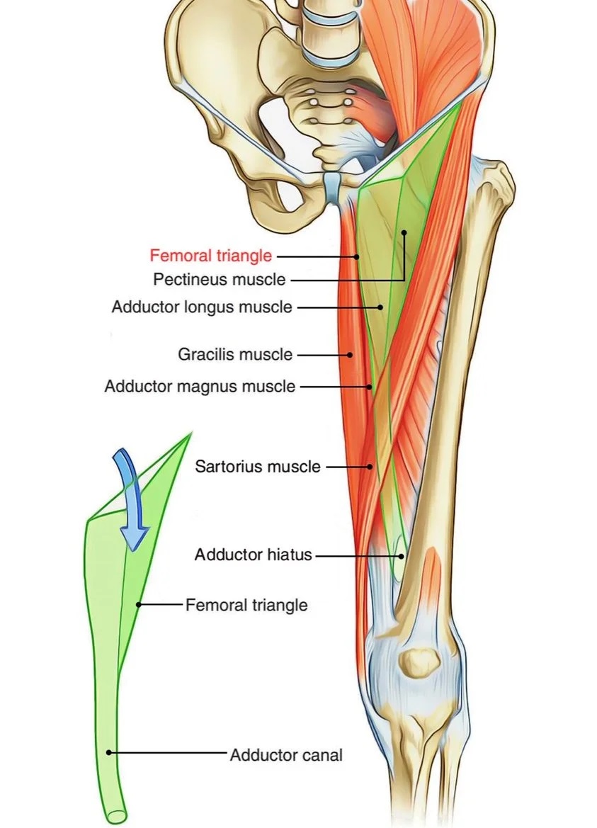
The adductor hiatus is an opening in the distal portion of the adductor magnus muscle, located just above the knee. It serves as a passage for the femoral artery and vein as they transition into the popliteal vessels within the popliteal fossa. Clinically, it is significant because vascular injuries or occlusions near this site can lead to popliteal artery entrapment or thrombosis.
Read: wikism.org/Adductor_Hiatus
Watch: youtube.com/shorts/SdYYkw-to…
#Anatomy #AnatomyLearning #AnatomyEducation #AnatomyOfTheDay #SportsMedicine #Orthopedics #PhysicalTherapy #AthleticTraining #Rehab #InjuryPrevention #Physio #SportsInjury #SportsRehab #PhysioTherapy #Meded #FOAMed
22
71
2,184
12 Nov 2025
The adductor hiatus is an opening in the distal portion of the adductor magnus muscle, located just above the knee. It serves as a passage for the femoral artery and vein as they transition into the popliteal vessels within the popliteal fossa. Clinically, it is significant because vascular injuries or occlusions near this site can lead to popliteal artery entrapment or thrombosis.
Read: wikism.org/Adductor_Hiatus
Watch: youtube.com/shorts/SdYYkw-to…
#Anatomy #AnatomyLearning #AnatomyEducation #AnatomyOfTheDay #SportsMedicine #Orthopedics #PhysicalTherapy #AthleticTraining #Rehab #InjuryPrevention #Physio #SportsInjury #SportsRehab #PhysioTherapy #Meded #FOAMed


14
54
2,245
11 Nov 2025
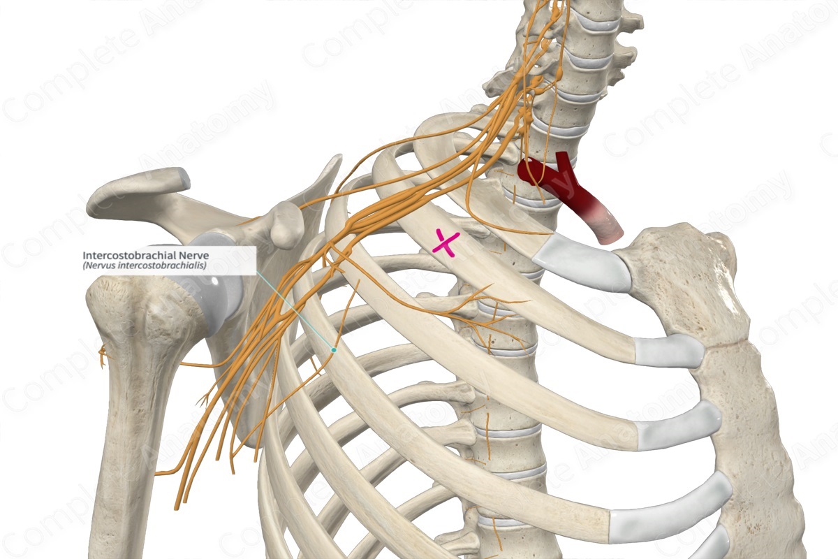
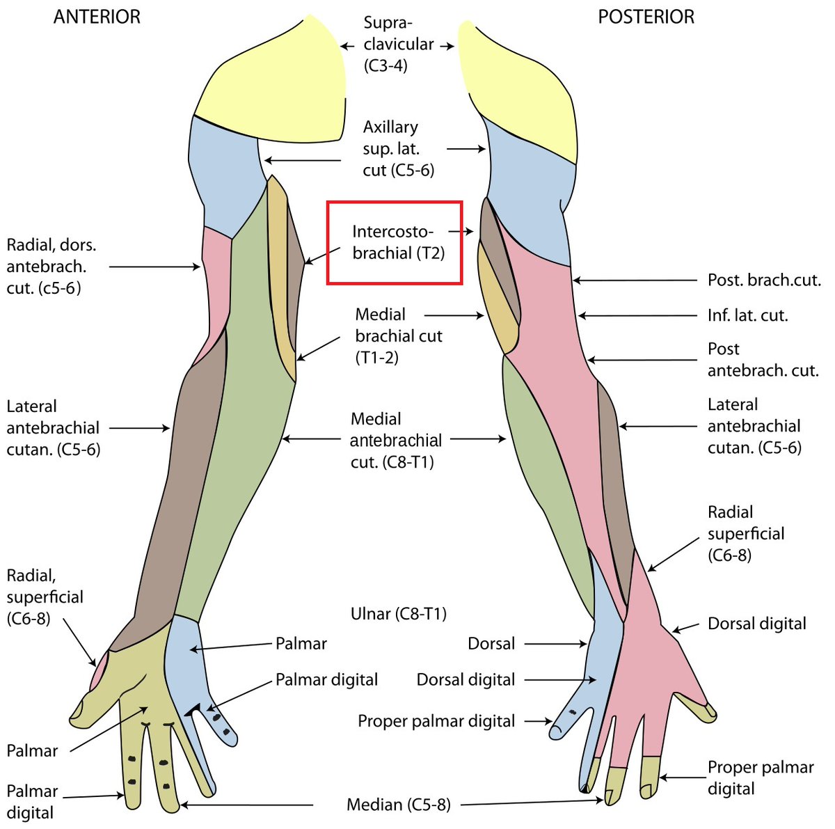
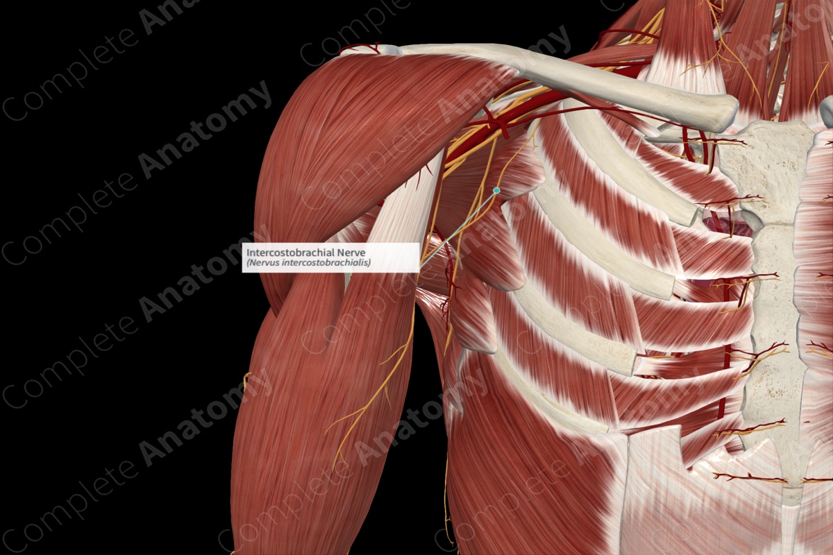
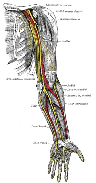
The intercostobrachial nerve is the lateral cutaneous branch of the second intercostal nerve (T2) that exits the intercostal space and crosses the axilla to supply sensation to the upper medial arm and axillary skin. Its primary function is providing sensory innervation to the skin of the upper medial arm, axilla, and sometimes the upper lateral chest wall. Pathology or injury to this nerve, commonly during axillary lymph node dissection or breast surgery, can result in numbness, paresthesia, or neuropathic pain in its sensory distribution.
Read: wikism.org/Intercostal_Brach…
Watch: youtube.com/shorts/hb3vO7Nth…
#Anatomy #AnatomyLearning #AnatomyEducation #AnatomyOfTheDay #SportsMedicine #Orthopedics #PhysicalTherapy #AthleticTraining #Rehab #InjuryPrevention #Physio #SportsInjury #SportsRehab #PhysioTherapy #Meded #FOAMed
7
28
861
10 Nov 2025
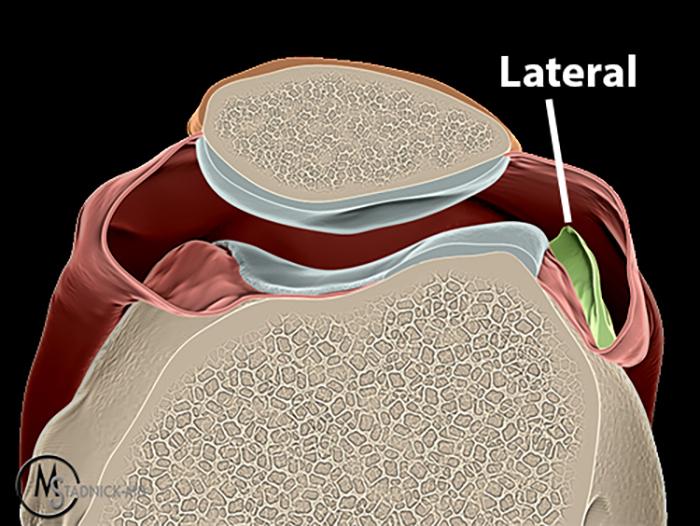
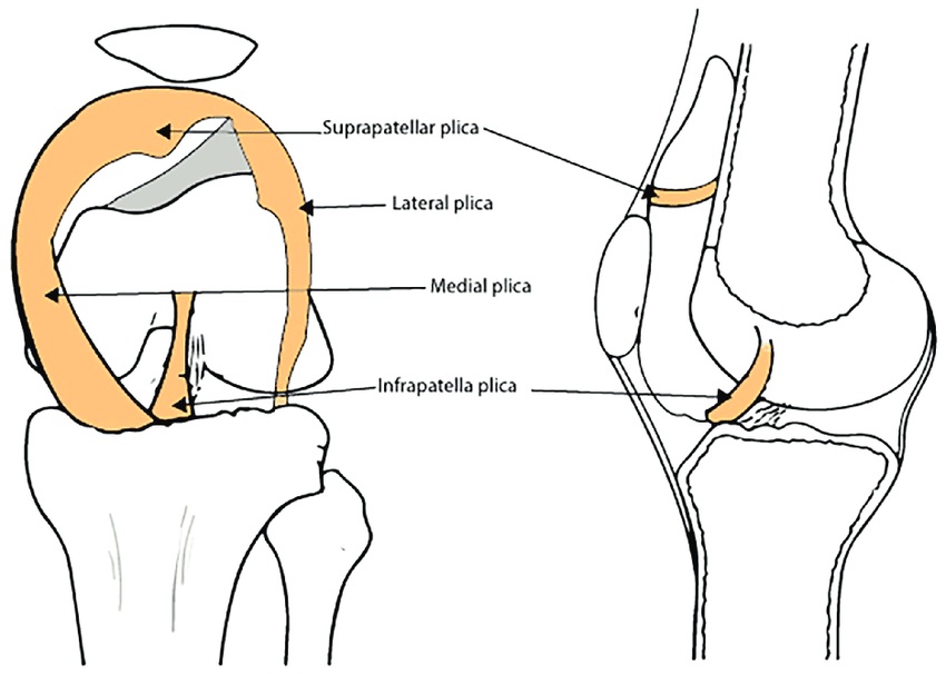
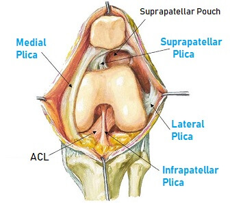
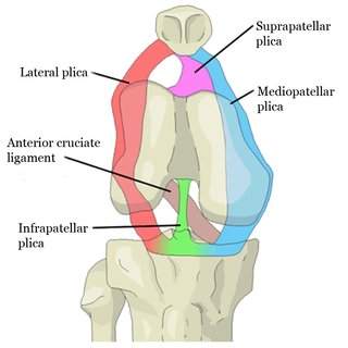
The lateral patellar plica is a fold of synovial tissue located on the lateral aspect of the knee joint, running along the inner surface of the lateral patellar facet. Its function is generally considered minor, serving as a synovial fold that allows smooth gliding of the patella during knee movement. Clinically, it becomes significant when thickened or inflamed, potentially causing lateral knee pain, catching, or symptoms of plica syndrome, which may require conservative or surgical management.
Read: wikism.org/Lateral_Patellar_…
#Anatomy #AnatomyLearning #AnatomyEducation #AnatomyOfTheDay #SportsMedicine #Orthopedics #PhysicalTherapy #AthleticTraining #Rehab #InjuryPrevention #Physio #SportsInjury #SportsRehab #PhysioTherapy #Meded #FOAMed
3
35
131
3,809
10 Nov 2025
The lateral patellar plica is a fold of synovial tissue located on the lateral aspect of the knee joint, running along the inner surface of the lateral patellar facet. Its function is generally considered minor, serving as a synovial fold that allows smooth gliding of the patella during knee movement. Clinically, it becomes significant when thickened or inflamed, potentially causing lateral knee pain, catching, or symptoms of plica syndrome, which may require conservative or surgical management.
Read: wikism.org/Lateral_Patellar_…
#Anatomy #AnatomyLearning #AnatomyEducation #AnatomyOfTheDay #SportsMedicine #Orthopedics #PhysicalTherapy #AthleticTraining #Rehab #InjuryPrevention #Physio #SportsInjury #SportsRehab #PhysioTherapy #Meded #FOAMed
1
34
162
5,593
6 Nov 2025
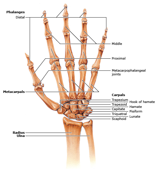
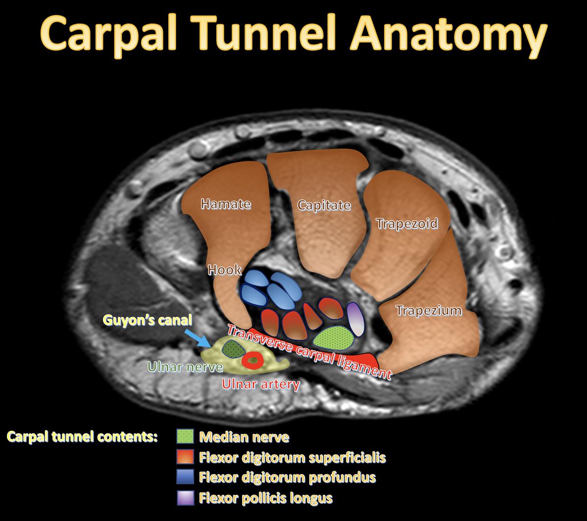
The hand and wrist are composed of 27 bones that provide both strength and dexterity for fine motor movements. The wrist contains eight carpal bones arranged in two rows, forming the connection between the forearm and the hand. The metacarpal bones extend from the wrist to the fingers, supporting the palm, while the phalanges form the fingers and thumb. Numerous muscles, tendons, ligaments, and nerves work together to allow precise movements such as gripping, flexion, extension, and rotation. Common pathologies of the hand and wrist include carpal tunnel syndrome, tendonitis, fractures, and osteoarthritis, which can impair strength, sensation, and range of motion.
Read: wikism.org/Hand_And_Wrist_An…
Watch: youtube.com/shorts/REIY6X4Dx…
#Anatomy #AnatomyLearning #AnatomyEducation #AnatomyOfTheDay #SportsMedicine #Orthopedics #PhysicalTherapy #AthleticTraining #Rehab #InjuryPrevention #Physio #SportsInjury #SportsRehab #PhysioTherapy #Meded #FOAMed
1
16
41
914
6 Nov 2025
The hand and wrist are composed of 27 bones that provide both strength and dexterity for fine motor movements. The wrist contains eight carpal bones arranged in two rows, forming the connection between the forearm and the hand. The metacarpal bones extend from the wrist to the fingers, supporting the palm, while the phalanges form the fingers and thumb. Numerous muscles, tendons, ligaments, and nerves work together to allow precise movements such as gripping, flexion, extension, and rotation. Common pathologies of the hand and wrist include carpal tunnel syndrome, tendonitis, fractures, and osteoarthritis, which can impair strength, sensation, and range of motion.
Read: wikism.org/Hand_And_Wrist_An…
Watch: youtube.com/shorts/REIY6X4Dx…
#Anatomy #AnatomyLearning #AnatomyEducation #AnatomyOfTheDay #SportsMedicine #Orthopedics #PhysicalTherapy #AthleticTraining #Rehab #InjuryPrevention #Physio #SportsInjury #SportsRehab #PhysioTherapy #Meded #FOAMed


24
98
3,574
5 Nov 2025
The brachialis muscle is a strong flexor of the elbow located deep to the biceps brachii in the anterior compartment of the arm, originating from the distal half of the anterior humerus and inserting on the coronoid process and tuberosity of the ulna. It is primarily innervated by the musculocutaneous nerve, with occasional contribution from the radial nerve, and functions to flex the forearm regardless of wrist position. Pathologies associated with the brachialis include muscle strains, tendinopathy, and compression of the musculocutaneous nerve, leading to pain, tenderness, or weakness in elbow flexion.
Read: wikism.org/Brachialis
Watch: youtube.com/shorts/Px1n2a_Vj…
#Anatomy #AnatomyLearning #AnatomyEducation #AnatomyOfTheDay #SportsMedicine #Orthopedics #PhysicalTherapy #AthleticTraining #Rehab #InjuryPrevention #Physio #SportsInjury #SportsRehab #PhysioTherapy #Meded #FOAMed
1
5
36
1,095