
Jun 7
What a Guilty electrogram. Do you complete anterior isthmus block?
1
2
231

This live case explores one of the enduring questions in Brugada syndrome: are the observed abnormalities driven by depolarisation, repolarisation, or a combination of both?
During epicardial mapping, Roderick Tung identifies characteristic high-frequency electrograms and local J-wave activity within the RVOT substrate. As mapping progresses, the team uncovers a broader area of abnormal electrograms than initially anticipated, highlighting the value of detailed epicardial assessment.
The discussion also touches on the challenges of mapping in regions with significant epicardial fat and the importance of correlating electrogram findings with anatomical imaging and clinical understanding of Brugada substrate.
#EPeeps #Brugada #Electrophysiology
5
12
1,282

May 26
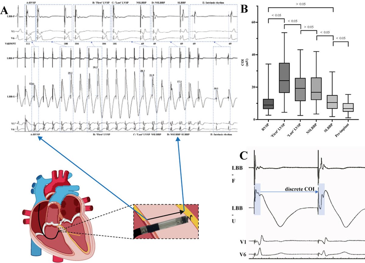
Electrophysiological characteristics of lead-position-dependent electrogram uninterrupted transition during left bundle branch pacing #OpenAccess
@JiaboS89929
heartrhythmjournal.com/artic…
13
37
3,837
Posterior wall mapping is only the beginning ⚡
Jérôme Lacotte and Vladimir Manenti discuss a key question in complex AF ablation:
Empirical anatomical lesions… or targeted electrogram-guided substrate modification?
All while avoiding unintended appendage disconnection.
#EPeeps #AFib #Ablation
3
13
1,514

May 14
🫀 #EHRATopicWeek Focal or reentrant? Modern insights into atrial tachycardia
What we once called “focal” AT is often far more complex.
Traditionally, focal AT (FAT) was defined as activation spreading centrifugally from a discrete site. Today, high-resolution mapping has revealed that many apparently focal tachycardias are actually tiny localized reentrant circuits — micro-reentrant ATs (MiRATs).
📌 True focal AT is mainly driven by:
• enhanced automaticity
• triggered activity
Meanwhile, localized reentrant ATs (LRATs) commonly occur in scarred atria or after previous ablation procedures and are characterized by slow conduction and long fractionated electrograms.
⚡ Modern EP increasingly relies on:
• high-density mapping
• entrainment mapping
• electrogram interpretation
• detailed anatomical understanding
Common AT locations include the crista terminalis, pulmonary veins, left atrial ridge, septal regions, and prior ablation scars.
💡 The more we map atrial tachycardias, the more we understand that “focal” does not always mean focal.
📖 Read more in the #EHRA_ESC Consensus document on Atrial Tachycardias 👉bit.ly/4wk5sBn
@escardio @EuropaceEiC


1
17
55
3,670

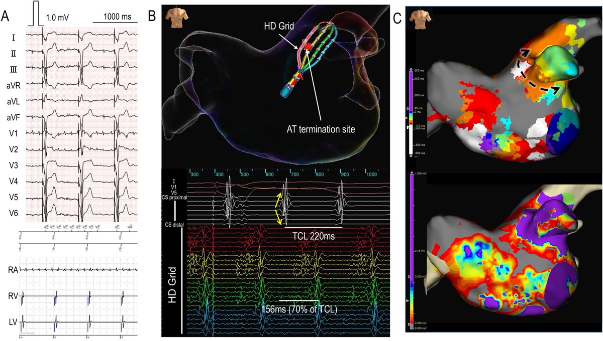
“Pure” Self-Reference Omnipolar Electrogram–Based Mapping for Localization of the Reentrant Circuit in the Dissociated Right Atrium
heartrhythmcasereports.com/a…
1
9
52
9,248

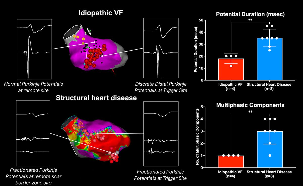
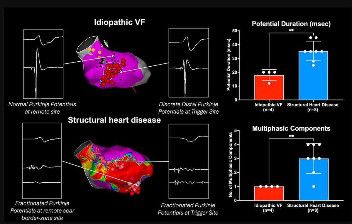
#Purkinje #electrogram morphology at VF-triggering sites differs between #idiopathic and #structural #heart #disease 👇 heartrhythmjournal.com/artic…
1
4
425
May 3
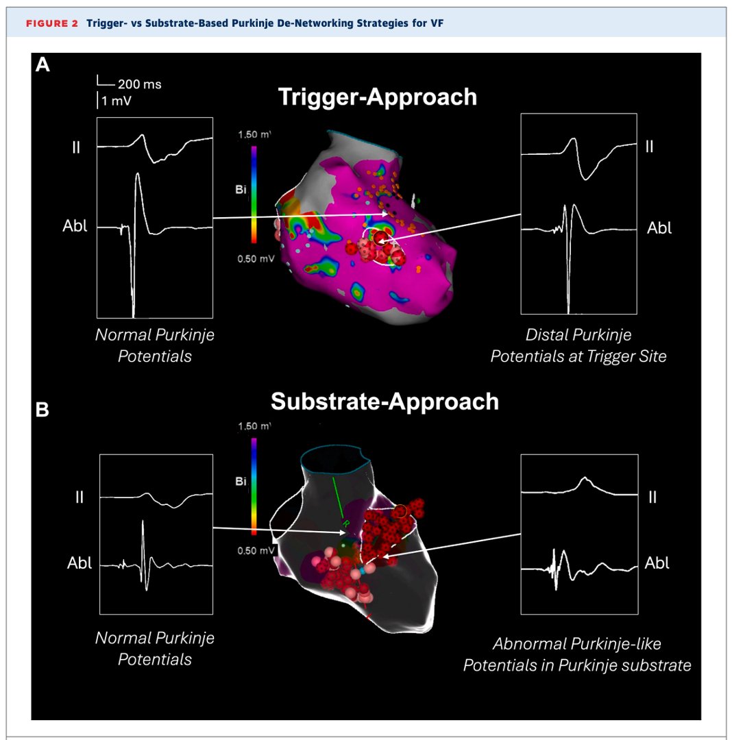
Distinct Purkinje Electrogram Phenotypes at Ventricular Fibrillation Trigger Sites in Idiopathic and Structural Heart Disease
@RobNKerley
heartrhythmjournal.com/artic…

1
18
54
3,582

Apr 28
More data from our multicenter cohort of VF ablation patients. In this paper, @RobNKerley describes Purkinje electrogram characteristics seen in idiopathic VF vs. VF associated with structural heart disease.
heartrhythmjournal.com/artic…
1
8
38
2,684
Apr 28
This HRS award winning work builds upon previously published VT Symposium award winning work by @RobNKerley and team.
Latest VF ablation paper: authors.elsevier.com/a/1m~…
Prequel ICD electrogram guided VF ablation free read is here: rdcu.be/ePCbs
Apr 27

Multicenter data on Purkinje de-networking for VF
⚡️Substrate-based ablation → greater freedom from VT/VF
⚡️Trigger-based ablation → lower risk of conduction injury
Balancing efficacy and safety remains key.
Free read through this author share link: secure-web.cisco.com/1sVkbGL…
12
31
5,314

Most of the EP lab systems can show the last pacing stimulus of the consecutive pacing trains at the same position of the window. Therefore, the NPP evaluation requires only observing the timing of the electrogram of the return cycle. 2/n
1
1
2
603

Apr 23
New in Heart Rhythm 🫀
Amiodarone reshapes VT circuits & expands abnormal electrogram regions during substrate mapping.
Key lesson for the EP lab when AMD can't be stopped before ablation 👇
doi.org/10.1016/j.hrthm.2026…
#VTablation #DigitalTwin #EPeeps @HopkinsEngineer @JHUBME
1
15
45
3,010

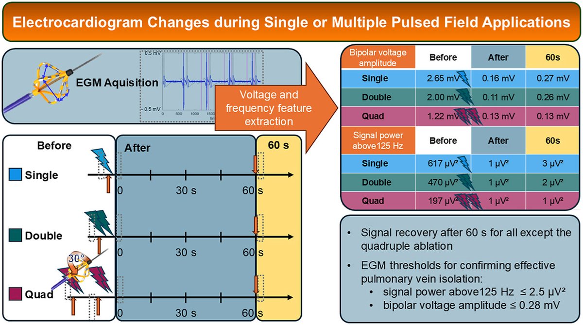
Single or multiple pulsed field applications: Intracardiac electrogram changes and implications for procedural end points
@UniSpitalBasel @inselgruppe #EPeeps
heartrhythmopen.com/article/…
4
18
2,422

Apr 1
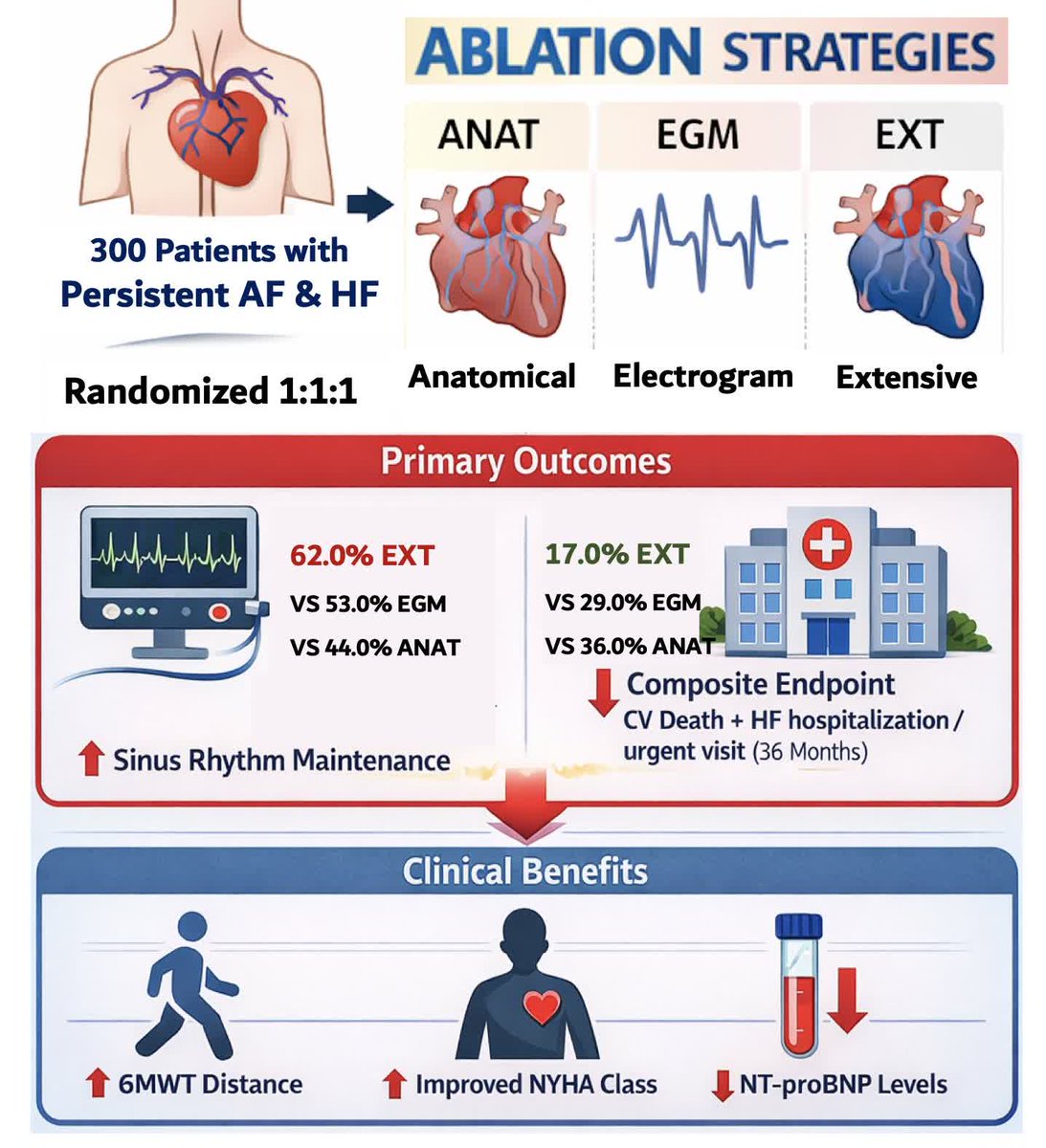
In patients with persistent AF and HF, extensive ablation improved sinus rhythm and reduced CV events vs anatomical or electrogram-guided ablation @KaigeLi #Epeeps ahajrnls.org/4mf1Xr5
3
23
2,732
Mar 25
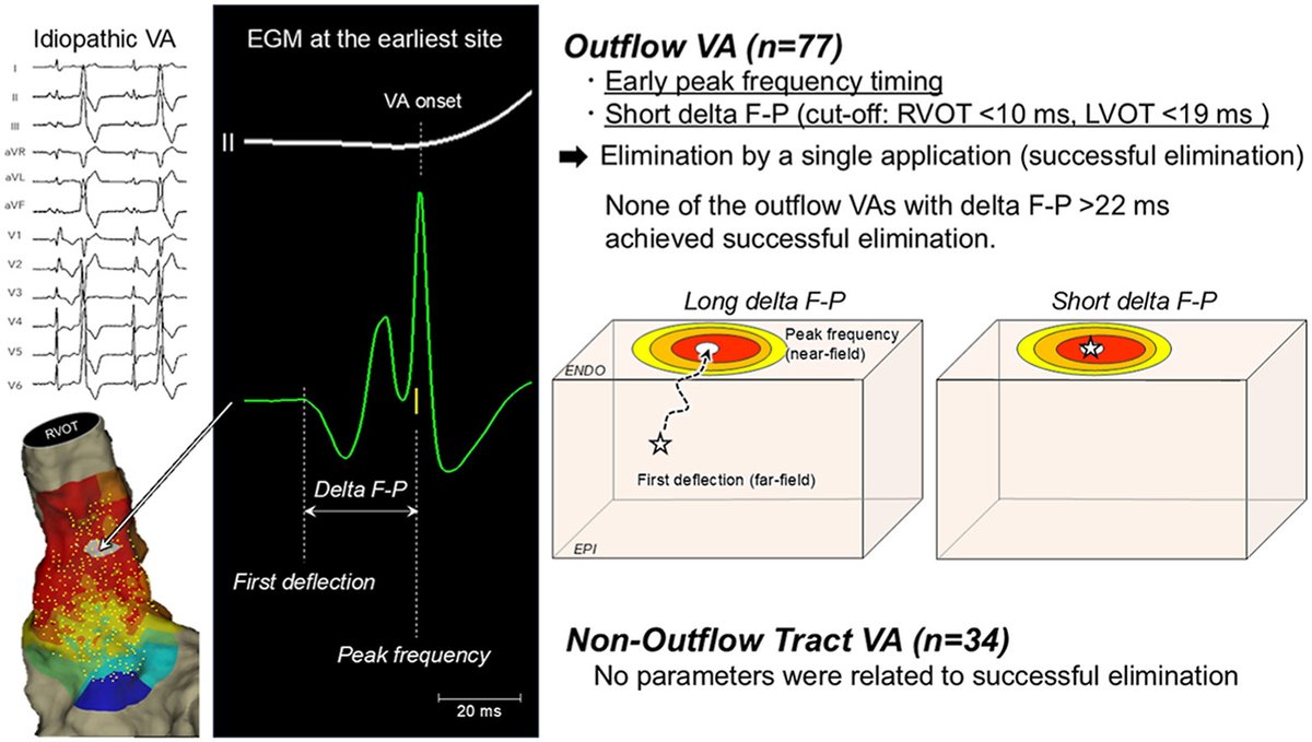
Comparison of Far-Field and Peak Frequency Electrogram Characteristics at the Earliest Activation Sites during Idiopathic Ventricular Arrhythmias: A novel index to predict ablation success #OpenAccess
@TakuroNishimu @ISCT_cvm
heartrhythmjournal.com/artic…
2
32
101
6,230
Mar 15
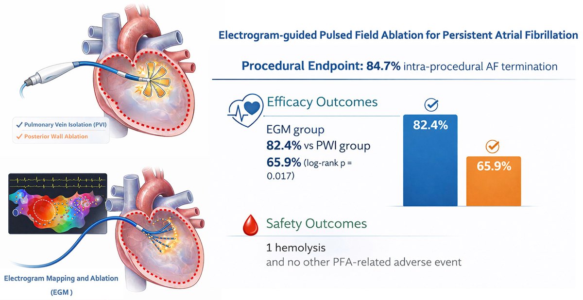
Electrogram-guided Pulsed Field Ablation for Persistent Atrial Fibrillation: A Prospective Cohort Study
heartrhythmjournal.com/artic…
2
14
55
8,735

Mar 15
#DePlaatVandaag
‘MUSIC OF MY LIFE’
Ik heb van de nummers die mij in mijn leven zijn opgevallen een TOP 20.000 gemaakt. Ik plaats elke dag nummers uit de TOP!
17.967 Friet Met Maynaise - Mike Vincent & DVO Electrogram
youtube.com/watch?v=3UOguFxr…

2
4
114

Mar 1
Distinct intracardiac electrogram waveforms with perforation during left bundle branch area pacing implantation academic.oup.com/europace/ar…
4
17
1,786
This live case highlights precision mapping and line assessment in a challenging ablation case, led by Jérôme Lacotte and Vladimir Manenti.
The mitral line is reassessed after incomplete block is identified, with possible wavefront collision suggested by colour convergence on the activation map. The updated Affera activation histogram displays 100% of the cycle length on the left side, supporting interpretation of the circuit.
Real-time electrograms reveal fragmented signals along the mitral line, indicating residual conduction and the need for further refinement.
This segment demonstrates how advanced activation analytics and electrogram interpretation enhance control and precision during complex line validation.
#EPeeps
19
2,271

💡Continue your electrophysiology education with Essentials of EP, Part 2!
This intensive course is designed to strengthen complex procedural skills, clinical decision-making, and electrogram analysis, building on the foundations established in Part 1.
📅 April 10 – 12, 2026
📍 Westin Arlington, Virginia
🔗 Secure your spot today: ow.ly/C2xV50Ye1Hx
#JnJInstitute #ECG #Electrophysiology


1
8
15
11,354