
Pathologist who loves a good diagnostic puzzle 🔬 | Educator at heart | Here to share cases, tips & the occasional plot twist under the microscope
- Tweets 236
- Following 874
- Followers 939
- Likes 980
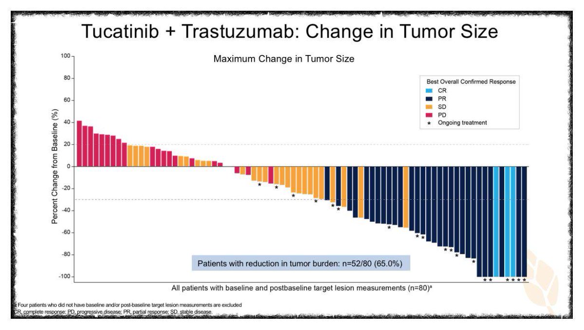
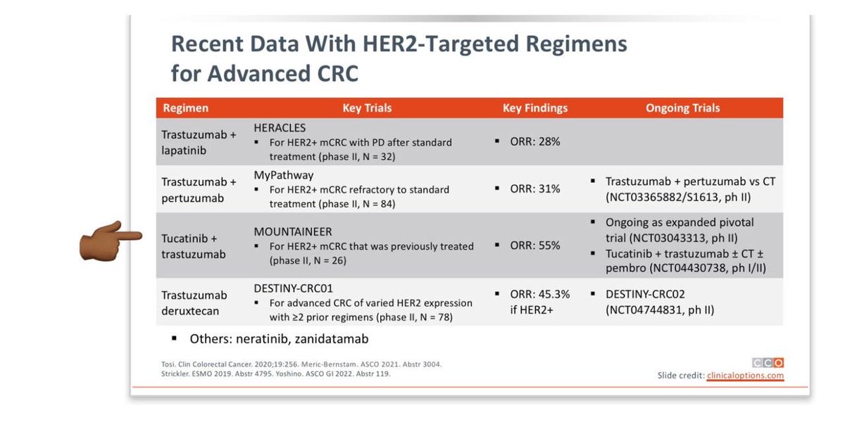
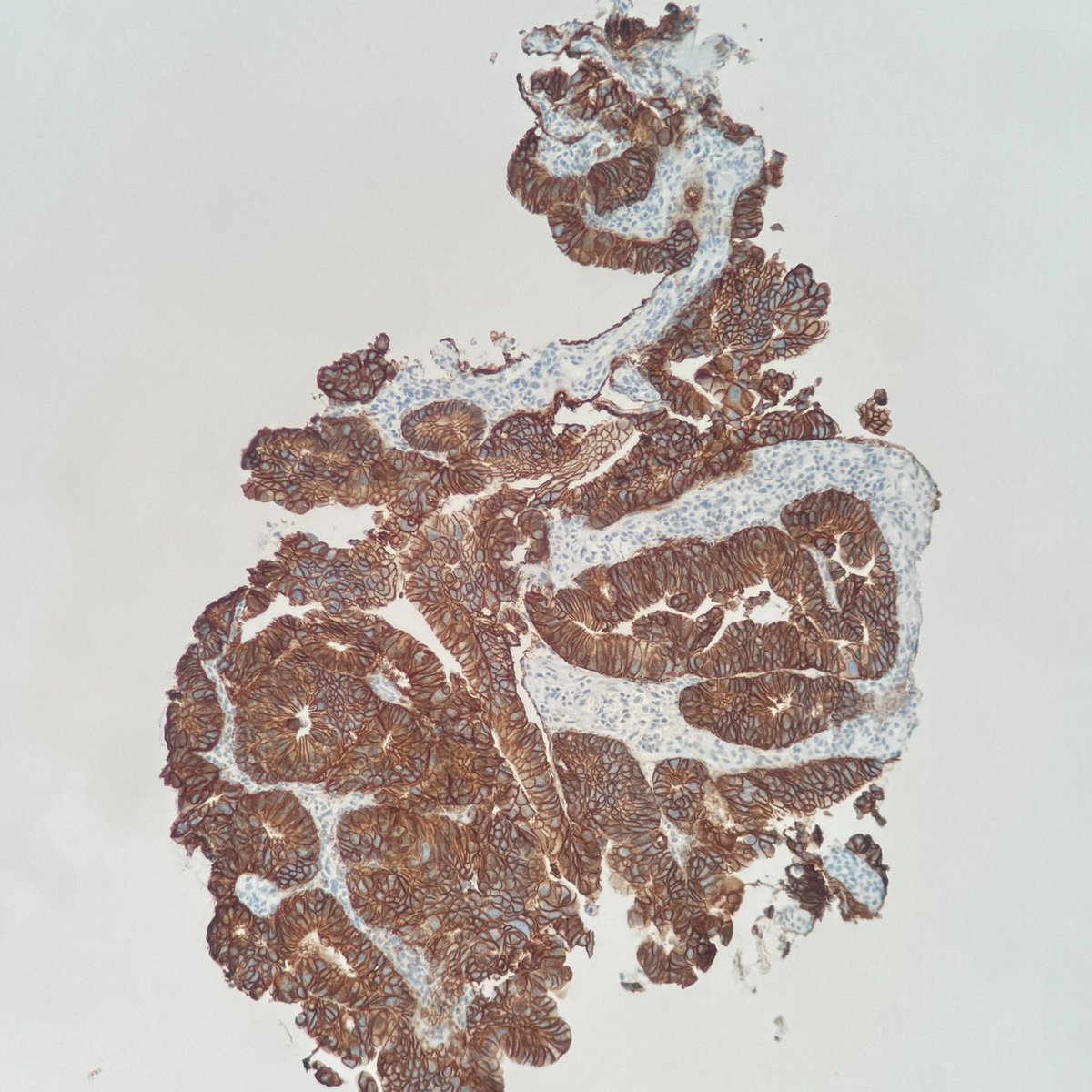


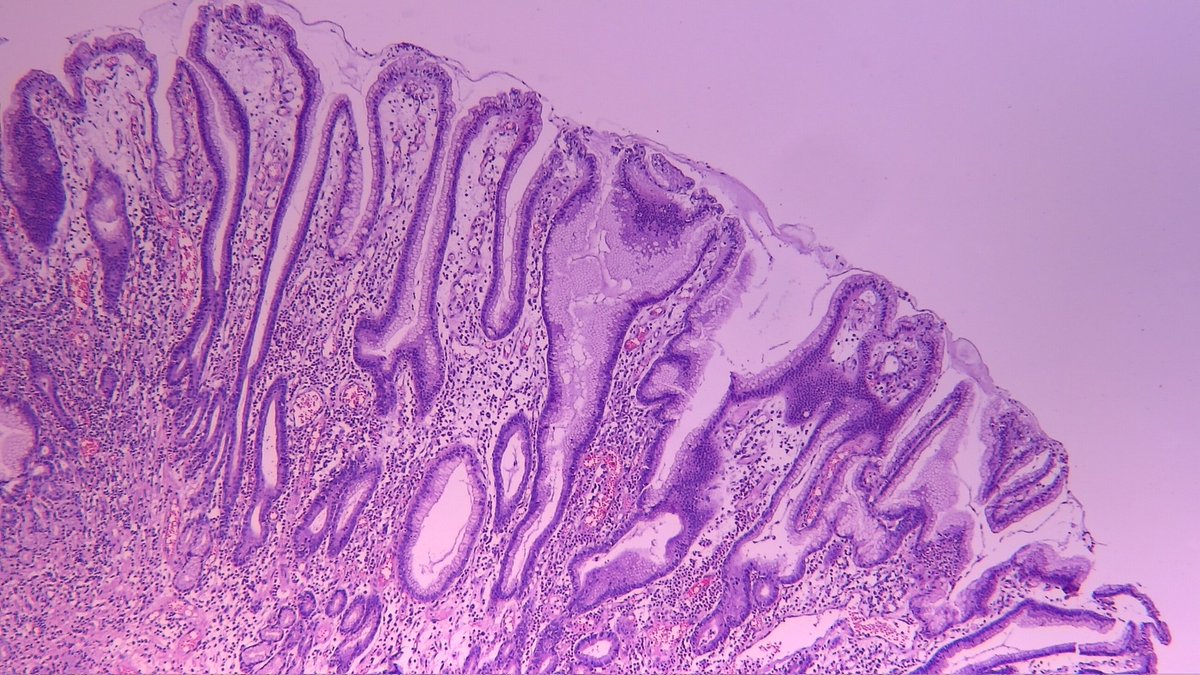
ALT colorectal adenocarcinoma with HER2 expression

ALT colorectal adenocarcinoma with HER2 expression
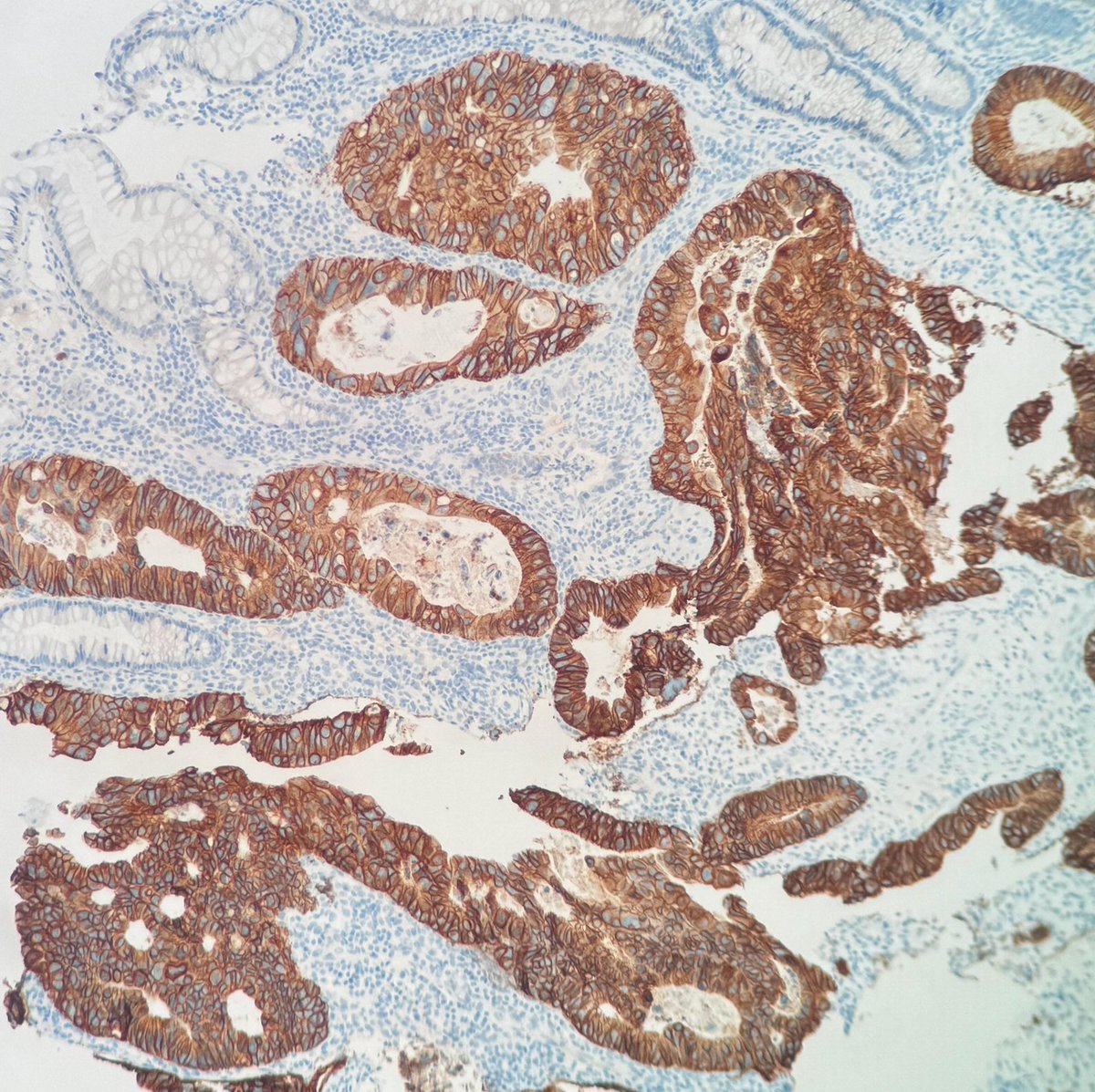
ALT colorectal adenocarcinoma with HER2 expression

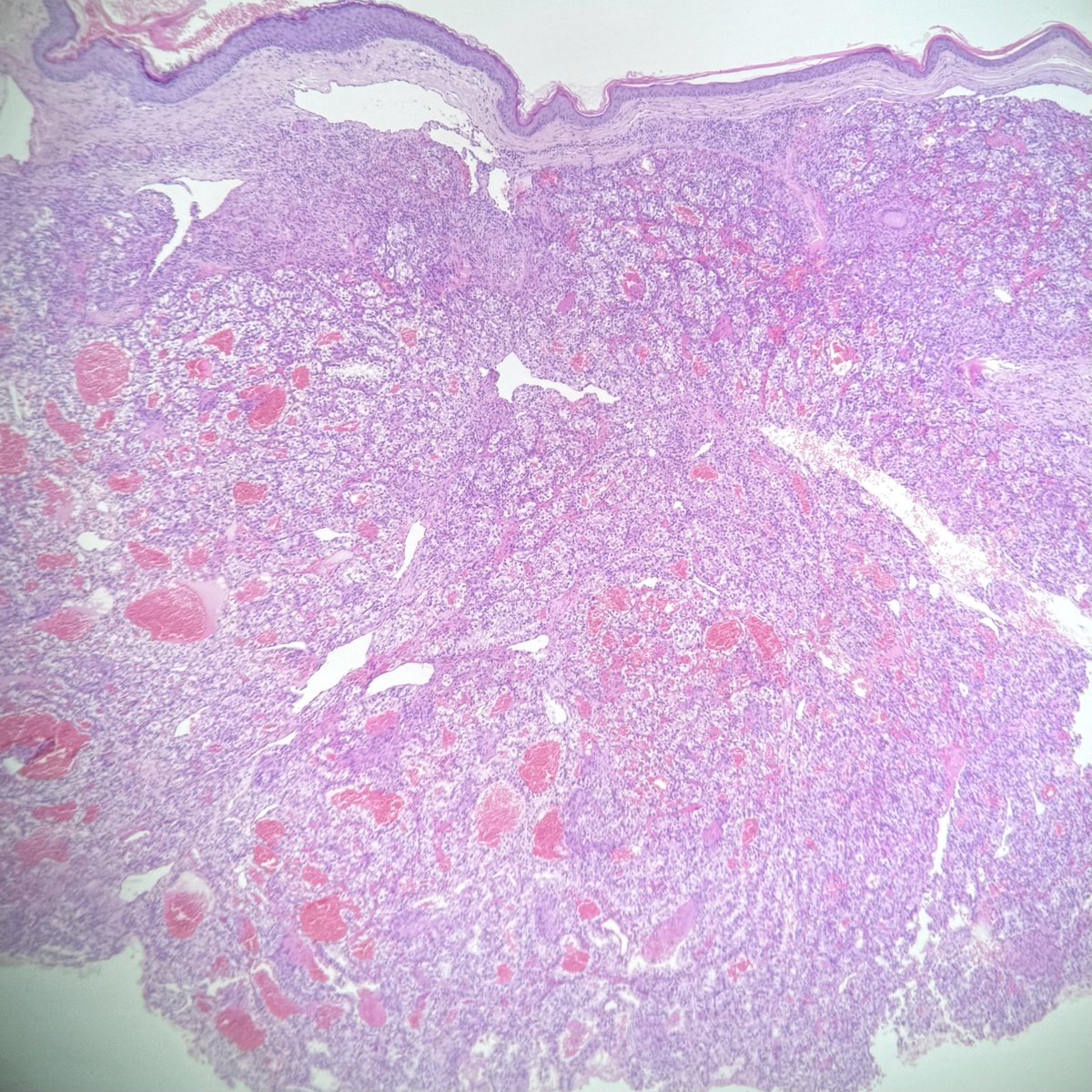
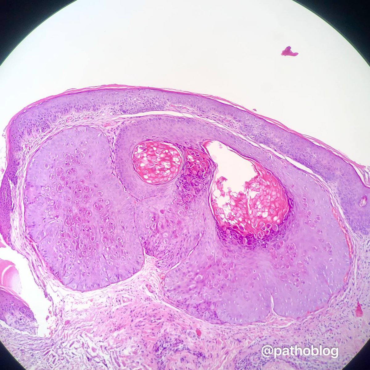
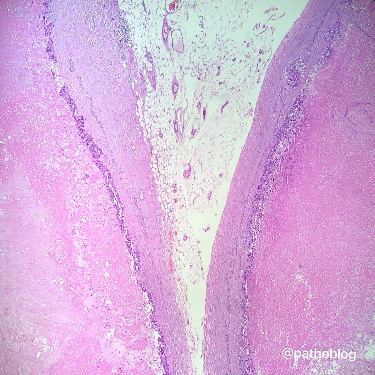
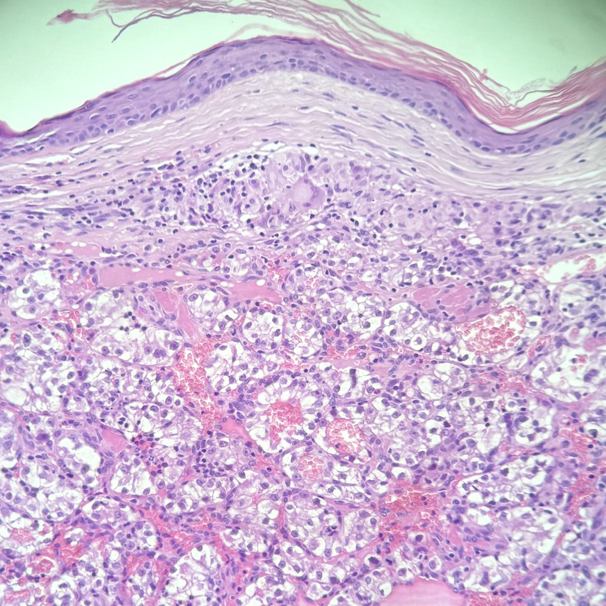
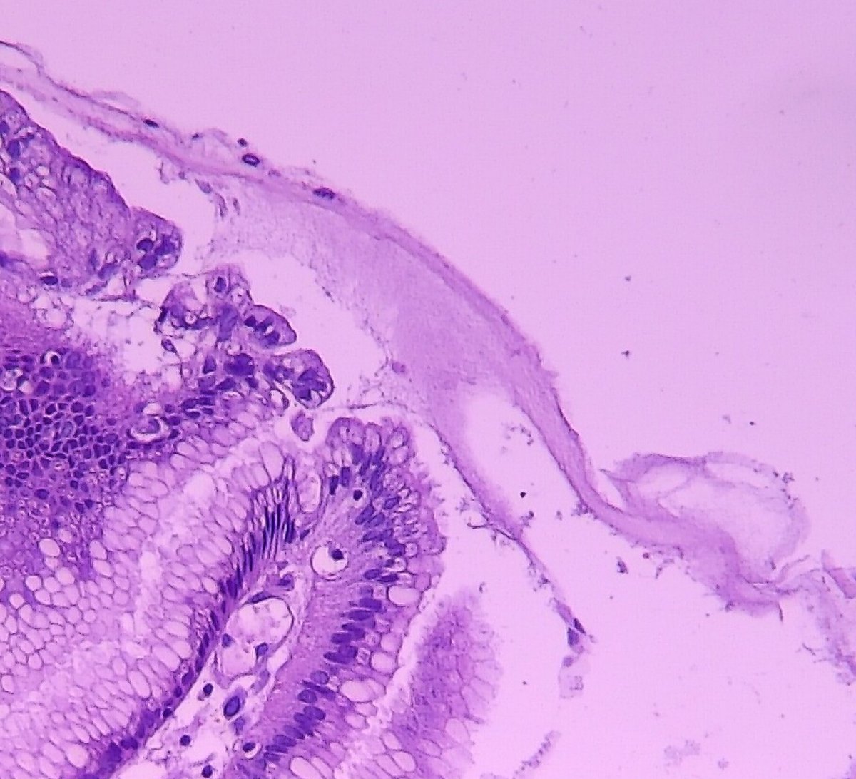
ALT H&E staining. Skin biopsy: intact epidermis and a dermal tumor composed of clear cells, separated by a prominent network of thin-walled vessels.
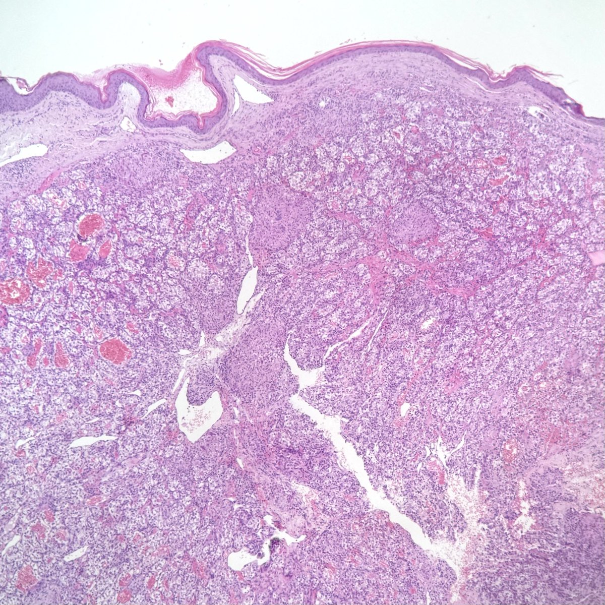
ALT H&E staining. Skin biopsy: intact epidermis and a dermal tumor composed of clear cells, separated by a prominent network of thin-walled vessels.
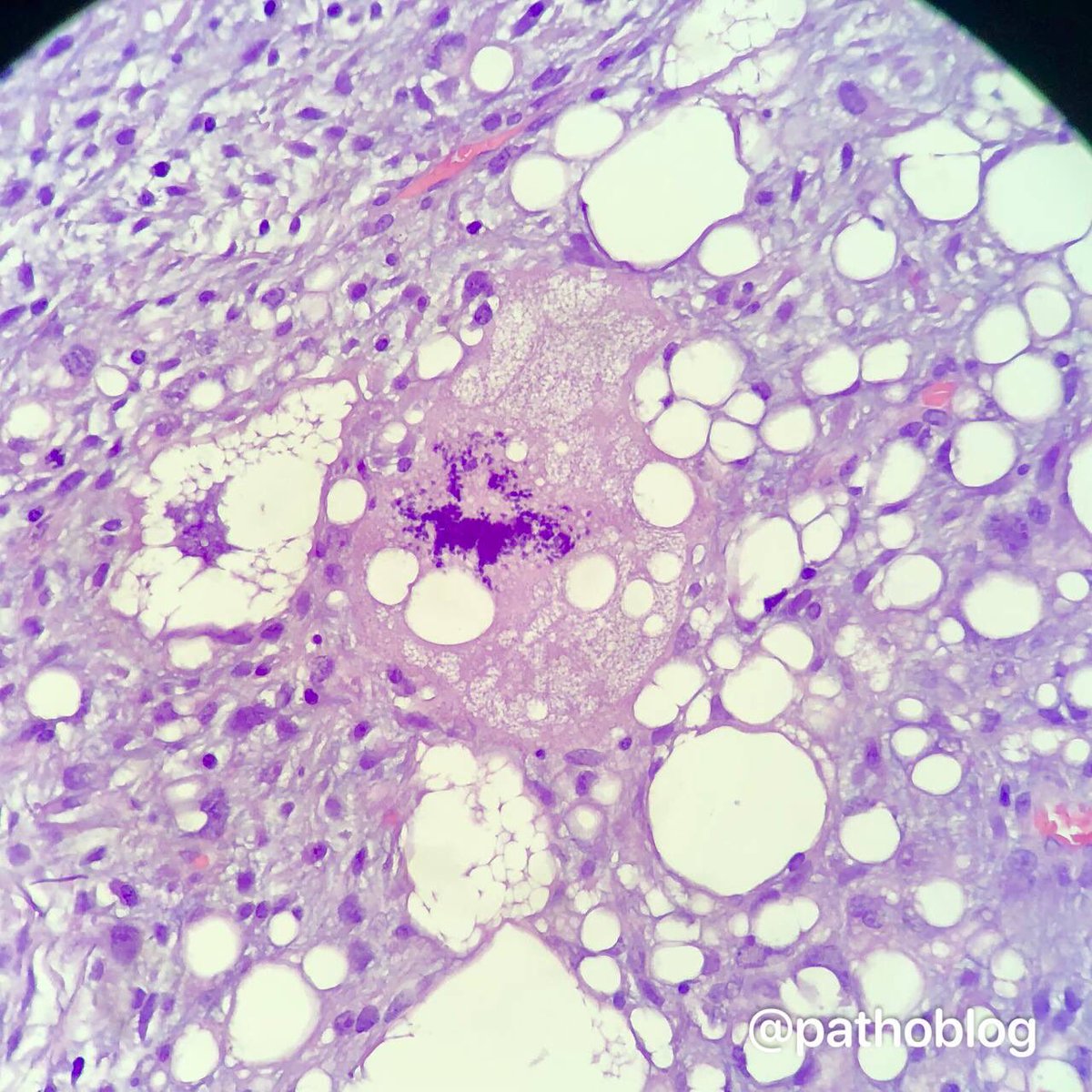
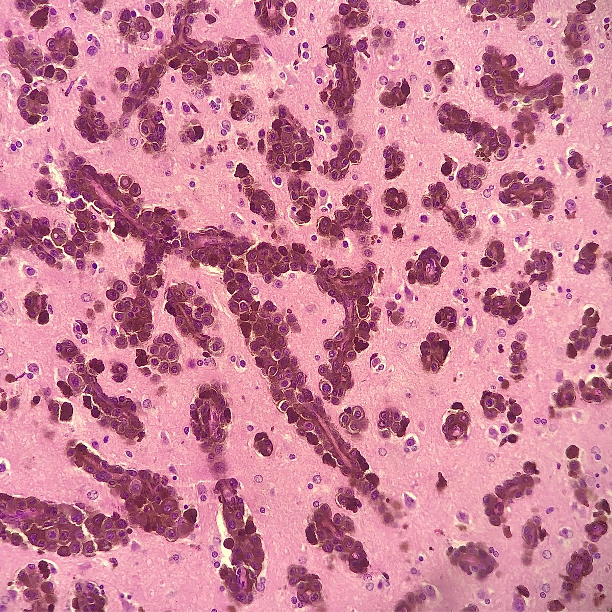
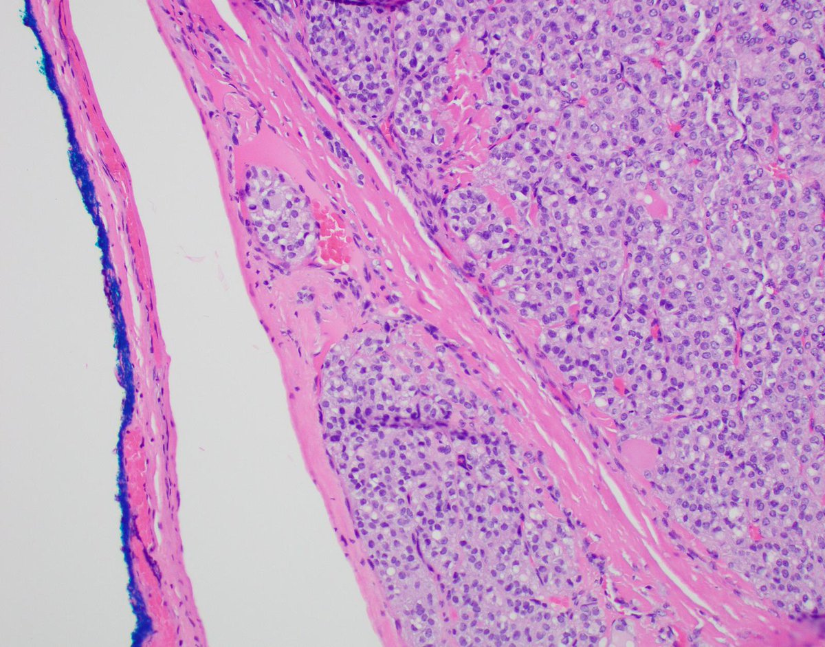
ALT H&E stain. High-power view showing tumor cells with abundant clear cytoplasm and small, round, uniform nuclei. Thin-walled vessels are visible between tumor cell nests.
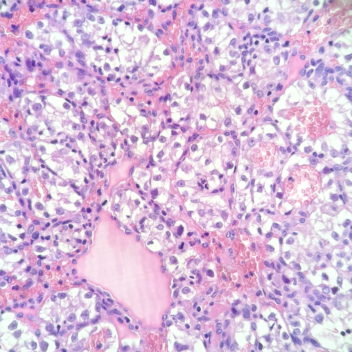
ALT H&E stain. High-power view showing tumor cells with abundant clear cytoplasm and small, round, uniform nuclei. Thin-walled vessels are visible between tumor cell nests.


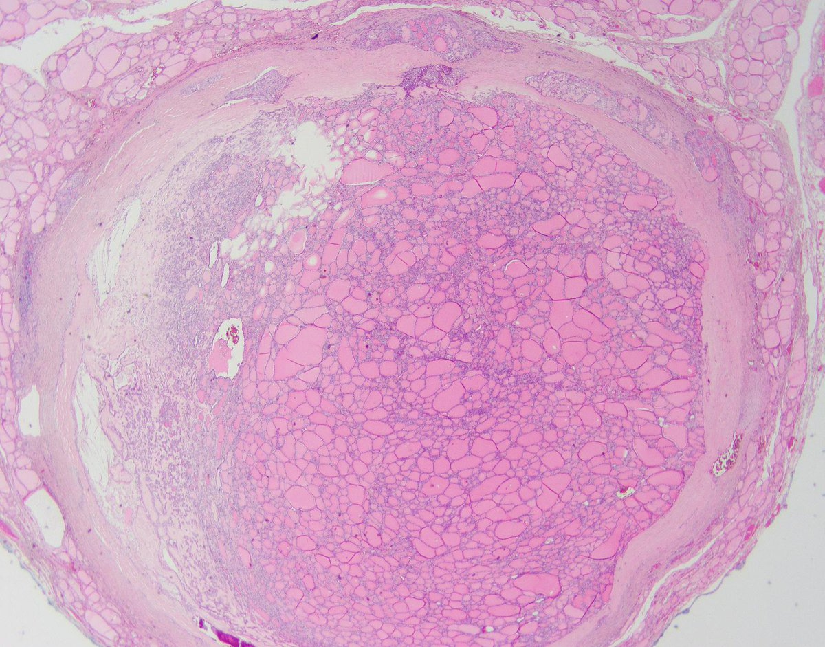
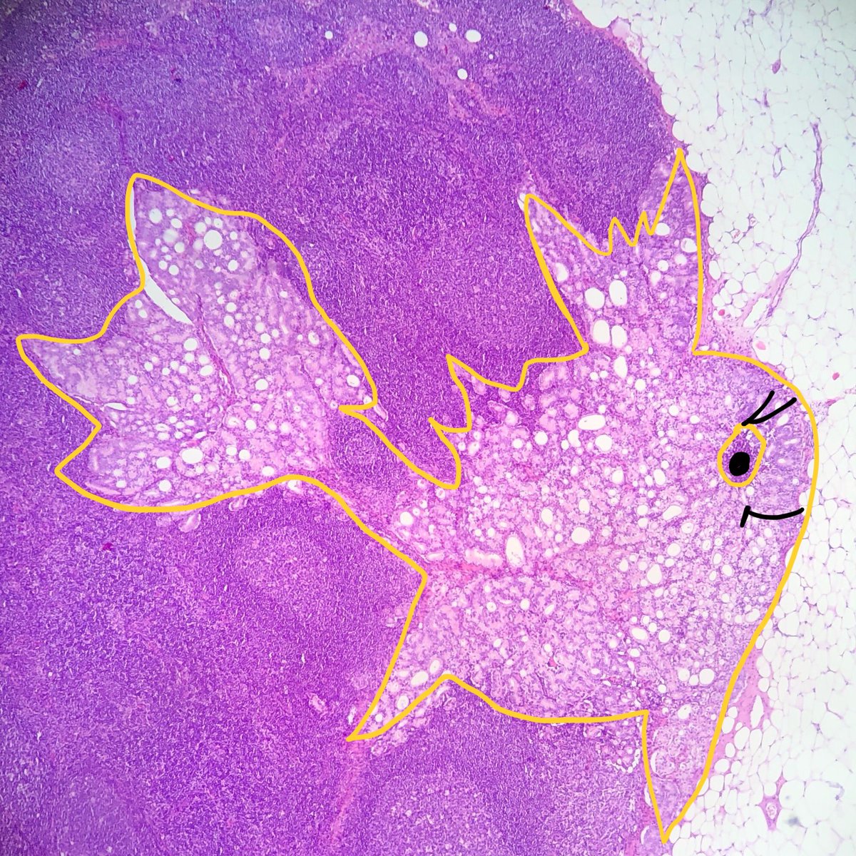
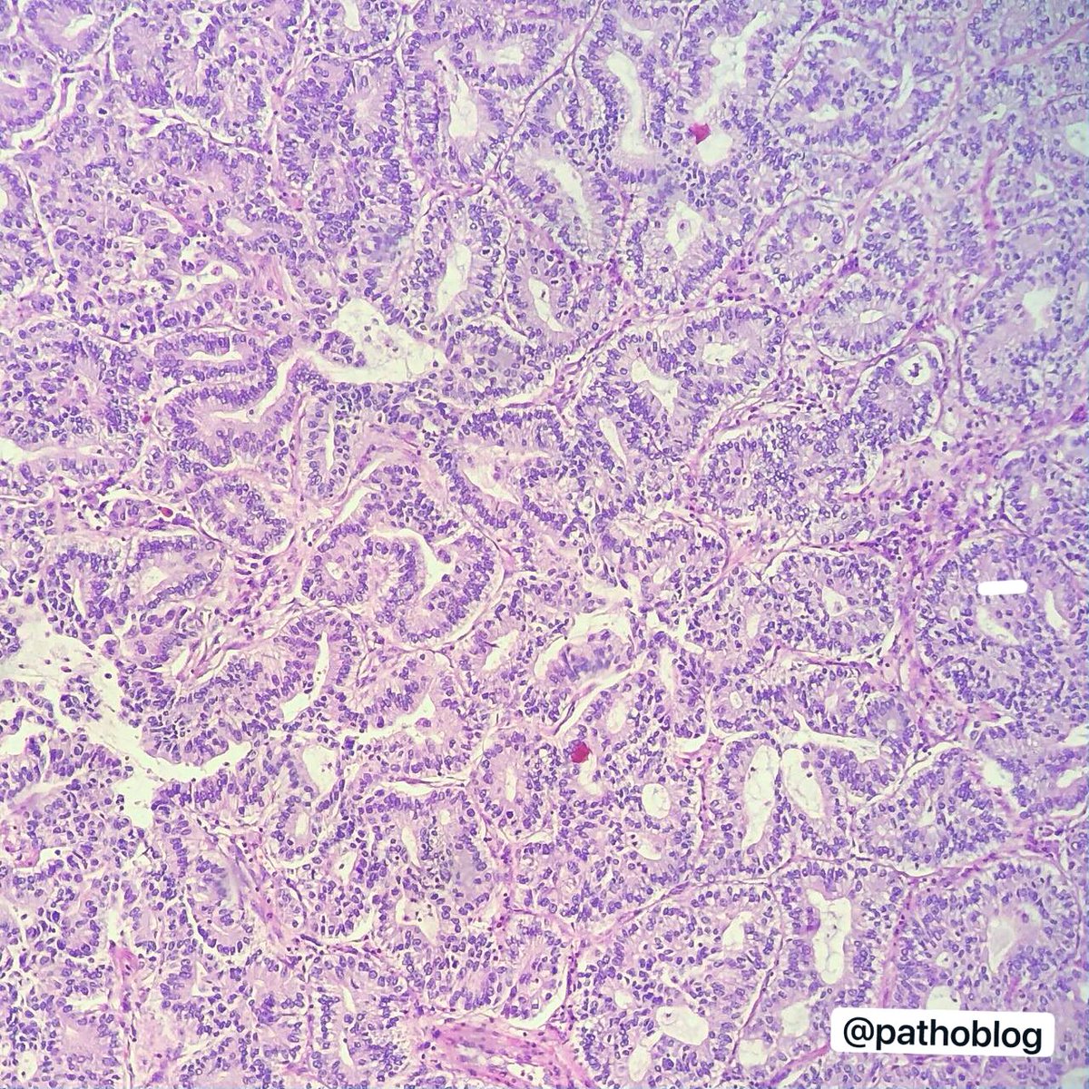
ALT A 48-year-old woman presents with a solitary, well-demarcated thyroid mass. Capsular invasion. @IARCWHO
ALT Note the unequivocal penetration through the thick fibrous capsule and into adjacent vascular spaces.

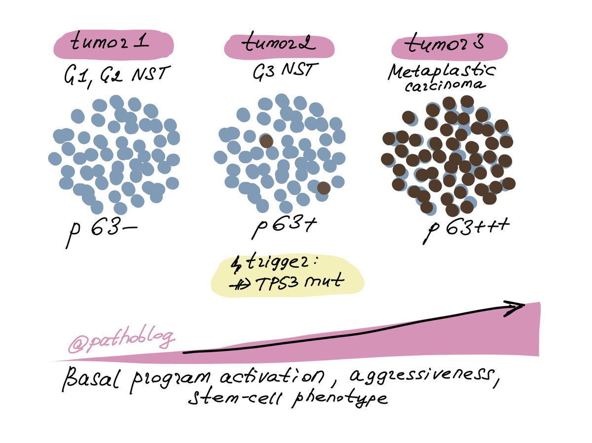
ALT Illustrated diagram showing p63 expression across three breast tumor types. Tumor 1 (G1/G2 NST): uniformly blue cells, p63-negative. Tumor 2 (G3 NST): predominantly blue cells with scattered brown cells, p63-positive, triggered by TP53 mutation. Tumor 3 (Metaplastic carcinoma): predominantly brown cells, p63-strongly positive. An arrow along the bottom indicates increasing basal program activation, aggressiveness, and stem-cell phenotype from left to right.