
Int'l open-access journal by Biophysical Society of China. Publishes novel theories, methods & protocols in bio/biomed research. Est. 2015.
- Tweets 353
- Following 99
- Followers 35
- Likes 245


ALT Figure 1. Illustration of stem cell-driven oto-regenerative prospect in hearing loss therapy. This figure was generated using FigDraw online platform (www.figdraw.com)


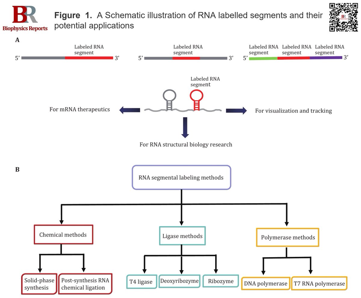
ALT Figure 1. A Schematic illustration of RNA labelled segments and their potential applications. B Classification of RNA segmental labeling methods


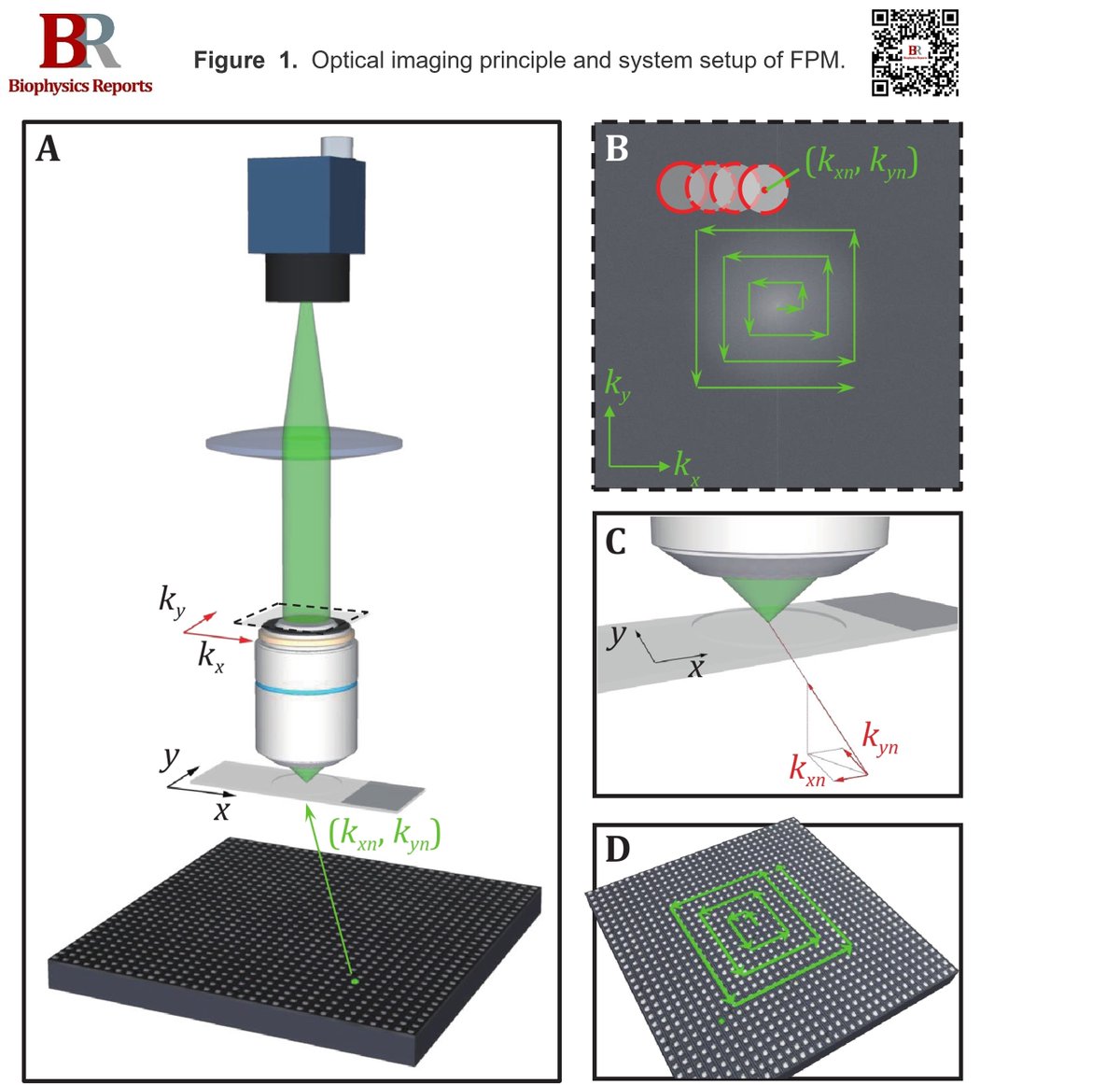
ALT Figure 1. Optical imaging principle and system setup of FPM. A System setup of FPM. A programmable LED matrix is placed beneath the sample and sequentially illuminated, one LED at a time. The nth LED illuminates the sample with a wave-vector (kxn, kyn). B The object’s finite spatial frequency support, determined by the microscope's numerical aperture (NA) in the Fourier domain (red circle), is shifted to reflect each unique LED illumination angle. The Fourier transforms of multiple shifted low-resolution measurements (each represented by a circle) are combined to extend the resolution of the complex sample spectrum beyond the objective lens’s cutoff. C Light emitted from a single LED illuminates a small sample area with a wave-vector (kxn, kyn). D LEDs are sequentially activated during FPM image acquisition