
Flagship journal of @Intlsocdiff. Published by #Elsevier for 40 years. All things relating to differentiation, development, stem cells and regeneration
- Tweets 234
- Following 334
- Followers 346
- Likes 147
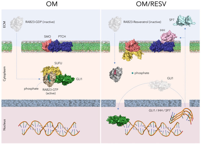


ALT Fig. 6. Proposed model of the IHH signaling pathway modulation by RAB23-GDP, -GTP and -Resveratrol molecular interactions. The rendered images represent the functional state of RAB23 protein (active or inactive) according of molecular docking with GDP, GTP and Resveratrol, and its impact in the IHH gene transcription (promotion or inhibition) induced by GLI1 transcription factor, in l-PDLCs cultivated in osteogenic medium with and without resveratrol pretreatment (OM and OM/RESV groups, respectively).


ALT Call for papers! Submit to the Splicing in Development and Disease special issue by June 30, 2026.
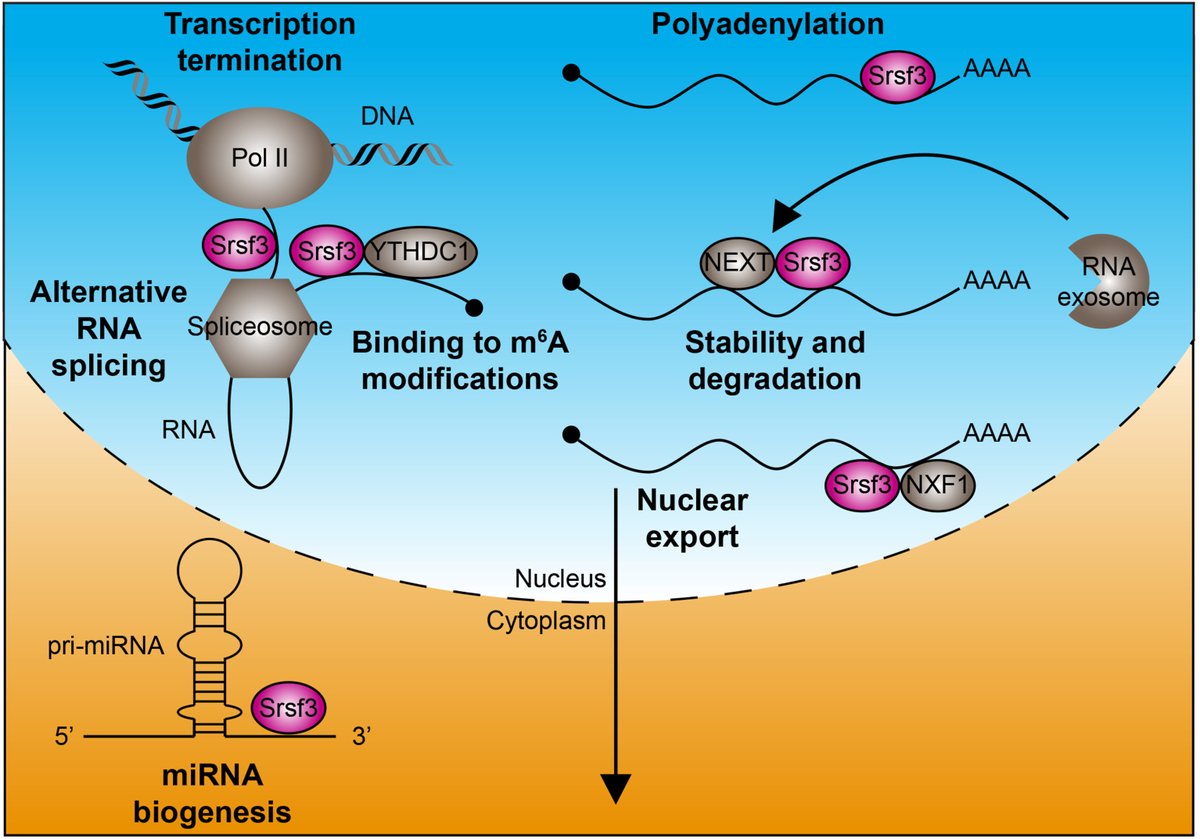
ALT Fig. 2. Roles of SRSF3 in RNA metabolism. SRSF3 (magenta) contributes to alternative RNA splicing, binding to m6A modifications, transcription termination, polyadenylation, transcript stability, nuclear export and miRNA biogenesis. Pol II, RNA polymerase II; YTHDC1, YTH domain-containing protein 1; NEXT, nuclear exosome targeting complex; NXF1, nuclear RNA export factor 1.
ALT Fig. 1. The core mechanisms of NNMT-mediated fibrosis: NAD depletion, methylation imbalance, inflammation, and oxidative stress NAD , Nicotinamide adenine dinucleotide; MAPK, Mitogen-activated protein kinase; SAH, S-adenosyl-L-homocysteine; SAA, Serum amyloid A; NAM, Nicotinamide; ROS, Reactive oxygen species; SAM, S-Adenosylmethionine; NNMT, Nicotinamide N-methyltransferase; MNA, 1-Methylnicotinamide; Hcy, Homocysteine; PARPs, Poly (ADP-ribose) polymerases.
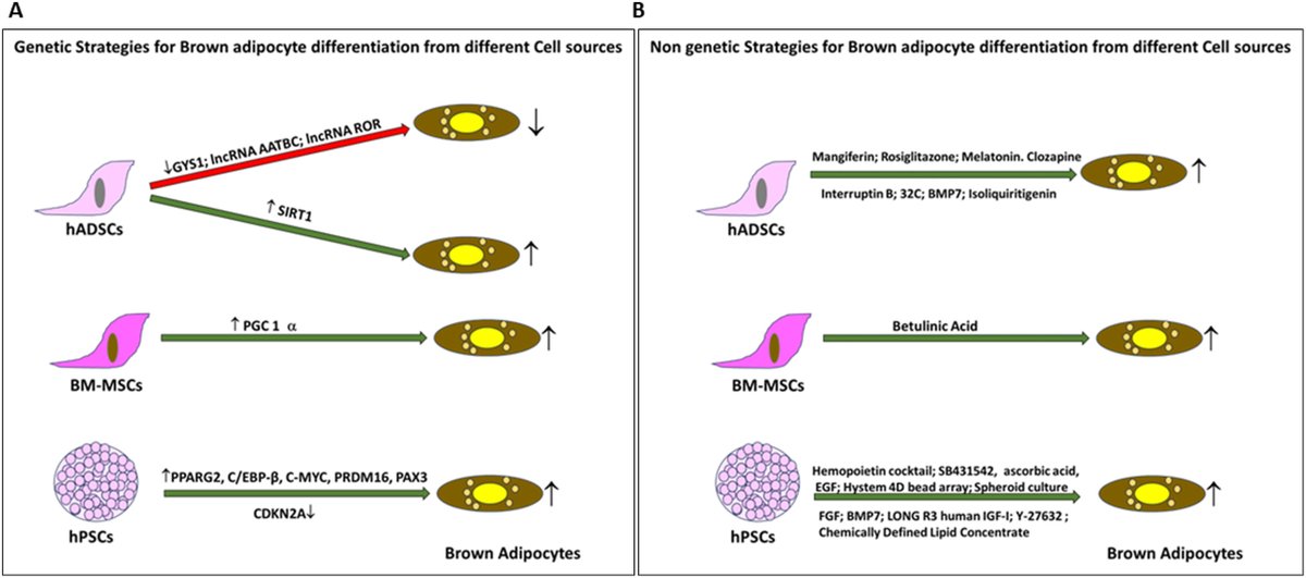
ALT Fig. 3. Differentiation strategies employed for brown adipocyte induction from human stem cells. (A) Schematic representation of genetic factors and their mode of modulation (overexpression or knockdown) across different cell sources, indicating whether each manipulation enhanced or reduced brown adipocyte differentiation. (B) Diagrammatic summary of non-genetic factors applied to various human cell sources, depicting the range of stimuli such as growth factors, small molecules, hormones, phytochemicals, and physiological or developmental cues reported to promote brown adipocyte differentiation.
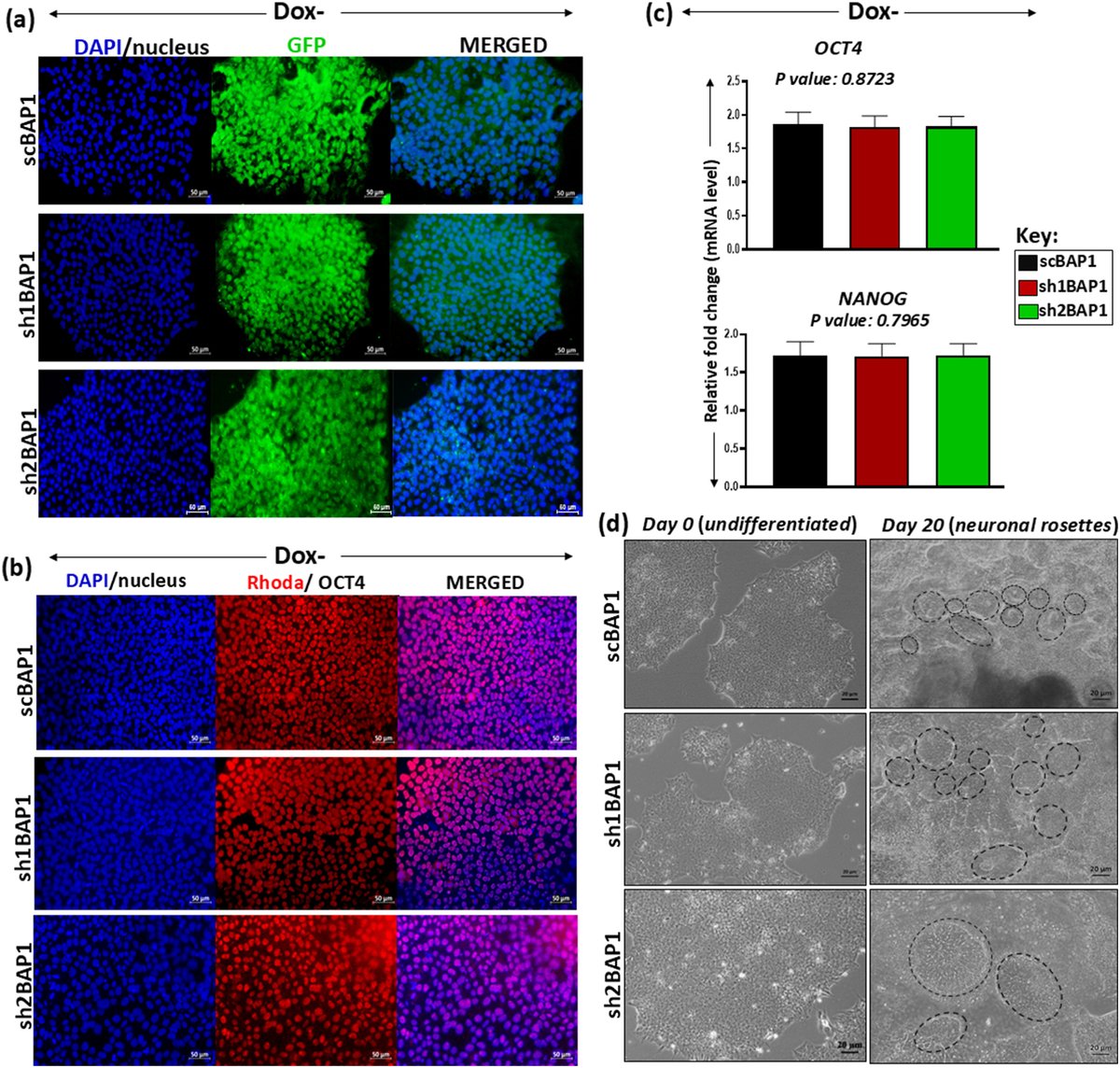
ALT Fig. 1. | Generation of scBAP1, sh1BAP1, sh2BAP1 stable cell lines and differentiation into neuronal progenitor cells (a) Post lentiviral transduction of human embryonic stem cells (hESCs) with scBAP1, sh1BAP1 and sh2BAP1 lentiviruses, was verified by enhanced green fluorescence protein (EGFP) at day 4, in absence of doxycycline (Dox-). DAPI (blue) was used to stain the nucleus. (Magnification: 20X)
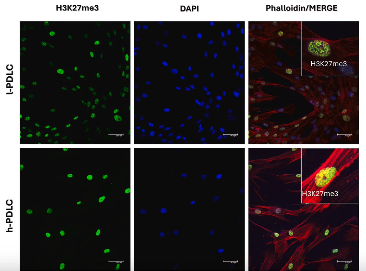
ALT Fig. 1. Basal characterization of the epigenetic mark H3K27me3, the epigenetic enzymes associated with H3K27me3 levels and key osteogenic genes.
ALT Fig. 2. Characterization of bone fragments A. Representative H&E stained section of bone fragment showing abundant adipocytes in the bone marrow next to mineralized bone with osteocytes. Bar = 100 μm. B. Representative histological slides of BGLAP (Bone Gamma-Carboxyglutamate Protein) immunoreactivity in human bone samples. The black arrows point out the presence of lining osteoblasts expressing BGLAP. Bar = 50 μm.
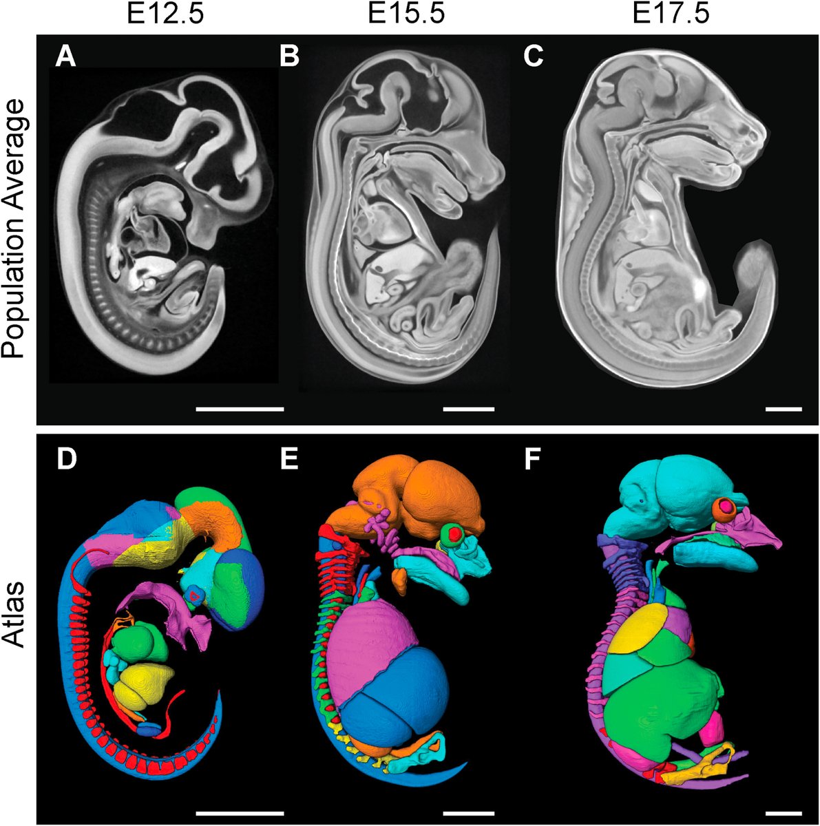
ALT Fig. 1. Creation of three new embryonic mouse PTA-based population averages and matching atlases. (A) Sagittal view of E12.5 population average created from 11 wild type embryos at a voxel size of 7 μm3, (B) E15.5 population average created from 32 wild type embryos at voxel size of 14 μm3, (C) E17.5 population average created from 22 wild type embryos at voxel size of 14 μm3 which was resized to 28 μm3. (D–F) Corresponding atlases at (D) E12.5 with 43 labels, at (E) E15.5 with 47 labels and at (F) E17.5 with 46 labels. Only a subset of labels are shown for illustrative purposes. Scale bar = 2 mm.
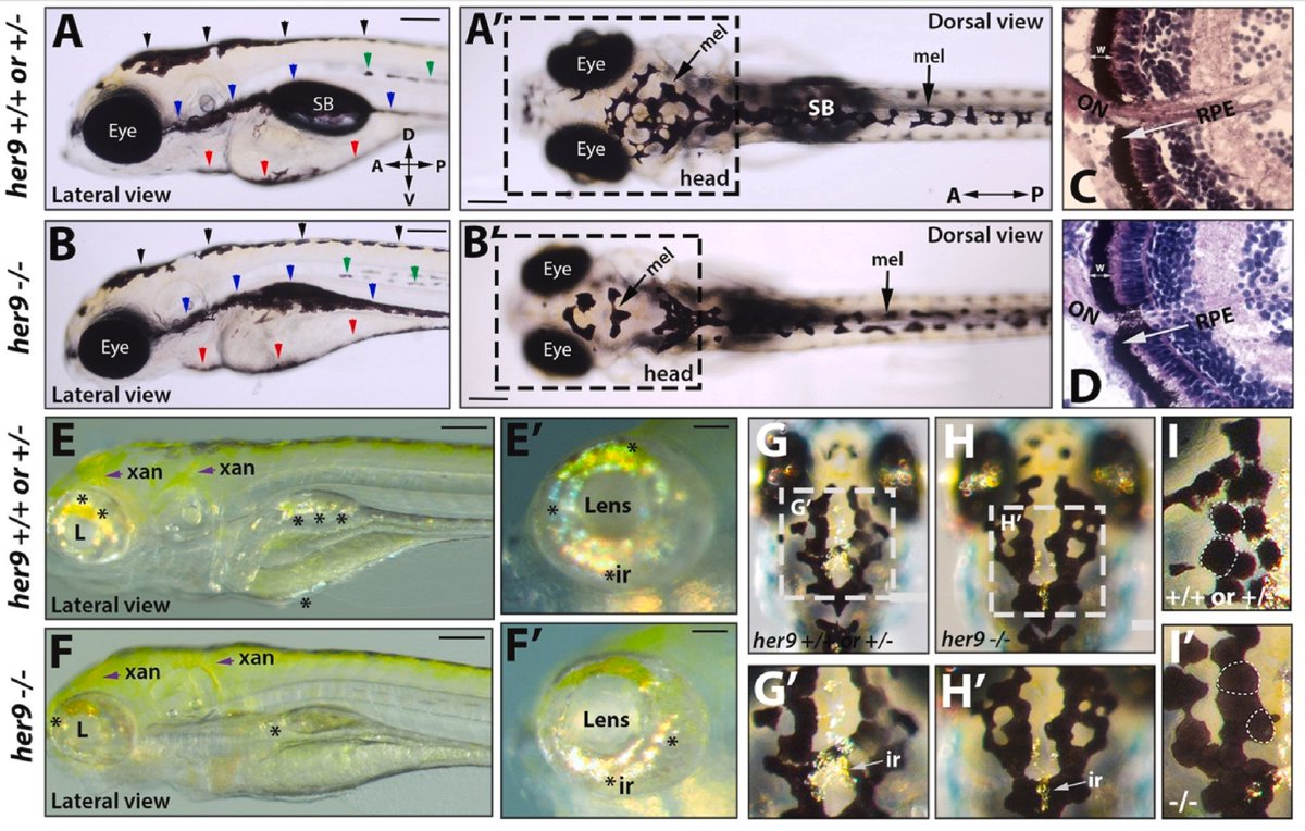
ALT Fig. 3. Her9 mutants display pigmentation defects. A-B′) Lateral (A-B) and dorsal (A′-B′) views of melanophore patterning in WT/heterozygous and her9 mutant larvae at 6 dpf. Black arrowheads, dorsal stripe; green arrowheads, lateral stripe; blue arrowheads, ventral stripe; red arrowheads, yolk stripe; SB, swim bladder. C-D) H&E staining of WT/heterozygous and her9 mutant retina at 7 dpf; the retinal pigmented epithelium (RPE) is indicated with a white arrow. E-F′) Xanothophore (xan) and iridophore (ir) patterning in WT/heterozygous and her9 mutant larvae. Her9 mutants have reduced abundance of iridiphores (asterisks) and greater spread of xanthophores (yellow) than WT or heterozygous siblings. G-I′) Dorsal view of iridiphores and melanophores on the heads of WT/heterozygous and her9 mutant larvae.
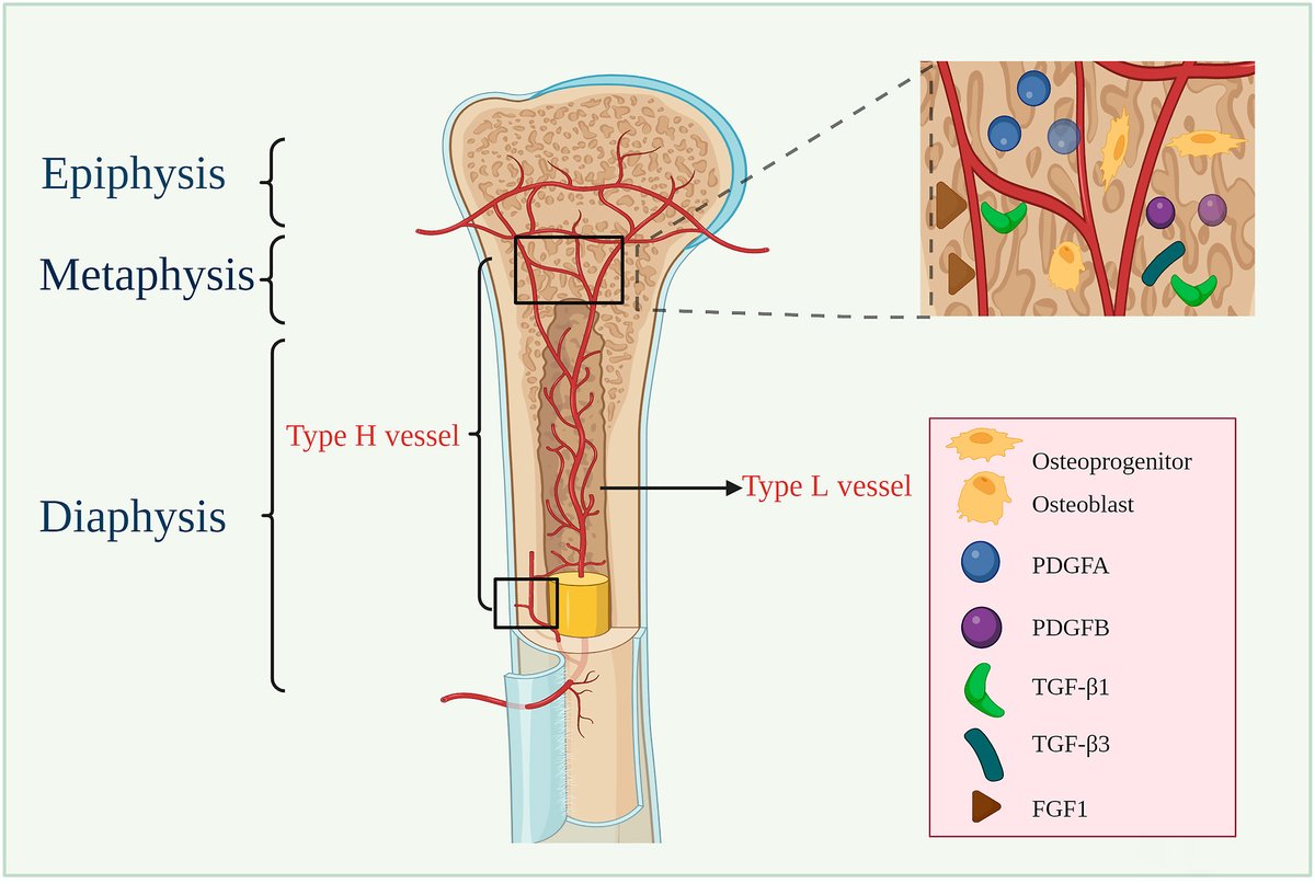
ALT "Type H vessels in osteogenesis, homeostasis, and related disorders," by Xiaoru Qin et al. Fig. 1. Vascular distributions in long bones. Type H vessels are located in the metaphysis and along the periosteal and endosteal surface, whereas type L vessels are located in the diaphysis's bone marrow cavity. Secretion of PDGFA, PDGFB, TGF-β1, TGF-β3, and FGF1 from type H ECs is significantly higher than that from type L ECs. More RUNX2 and OSX osteoprogenitors and osteoblasts surround type H vessels.
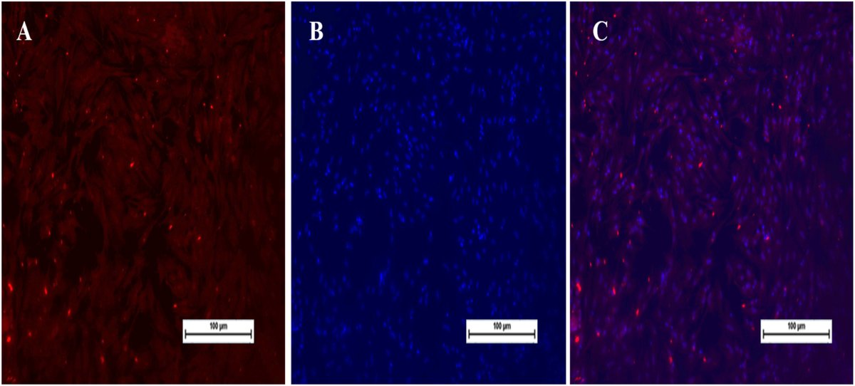
ALT Fig. 9. Immunofluorescence staining for MyoD at passage 15th, Scale bar: 100 μm. (A) Cells stained with MyoD (Red fluorescence); (B) Cells stained with DAPI; (C) Merged (MyoD & DAPI).
ALT Call for Papers! Submit to the Splicing in Development and Disease special issue
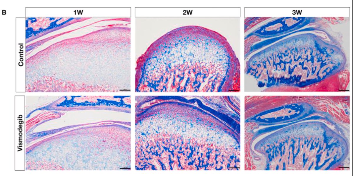
ALT Fig. 3. Histological analysis of TMJ in fetal mice from the experimental and control groups at postnatal weeks 1–3 using H&E and Masson's trichrome staining. B: Masson's trichrome staining; red dashed lines indicate the boundary between the chondrocyte layer and prechondrocyte layer, while yellow dashed lines indicate the boundary between the chondrocyte layer and hypertrophic chondrocyte layer. (N = 6. Scale bar = 100 μm)
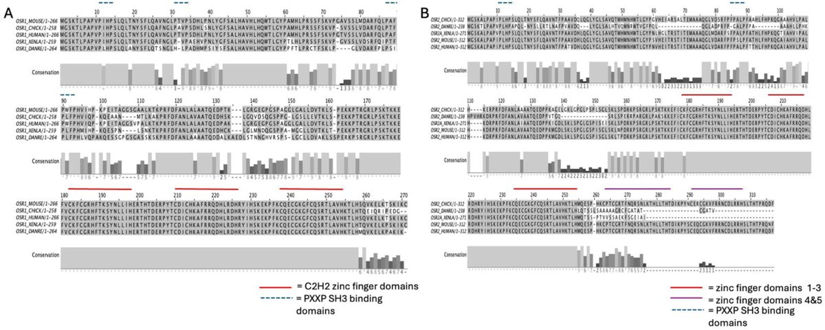
ALT Fig. 1. Conservation of zinc finger domains of Osr1 and Osr2 in vertebrates. (A) Amino acid sequence alignment of Osr1 using MafftWS sequence alignment reveals high conservation of the zinc finger binding domains. B) Amino acid sequence alignment of Osr2 using MafftWS sequence alignment reveals high conservation of zinc fingers 1–3 in all species. Zinc fingers 4 and 5 are only conserved in chick, mouse, and humans. Zinc finger domains 1–3 and 4–5 are indicated by red and purple bars, respectively, shown above the amino acid sequence of interest. OSR2B, the alternatively spliced protein product has only been identified in mammals and was therefore omitted from the protein conservation analysis. Conservation is depicted below the corresponding amino acid sequence.

ALT Fig. 1. Illustration of the triad system in tissue engineering, representing the interaction between cell types, scaffolds, and external factors.
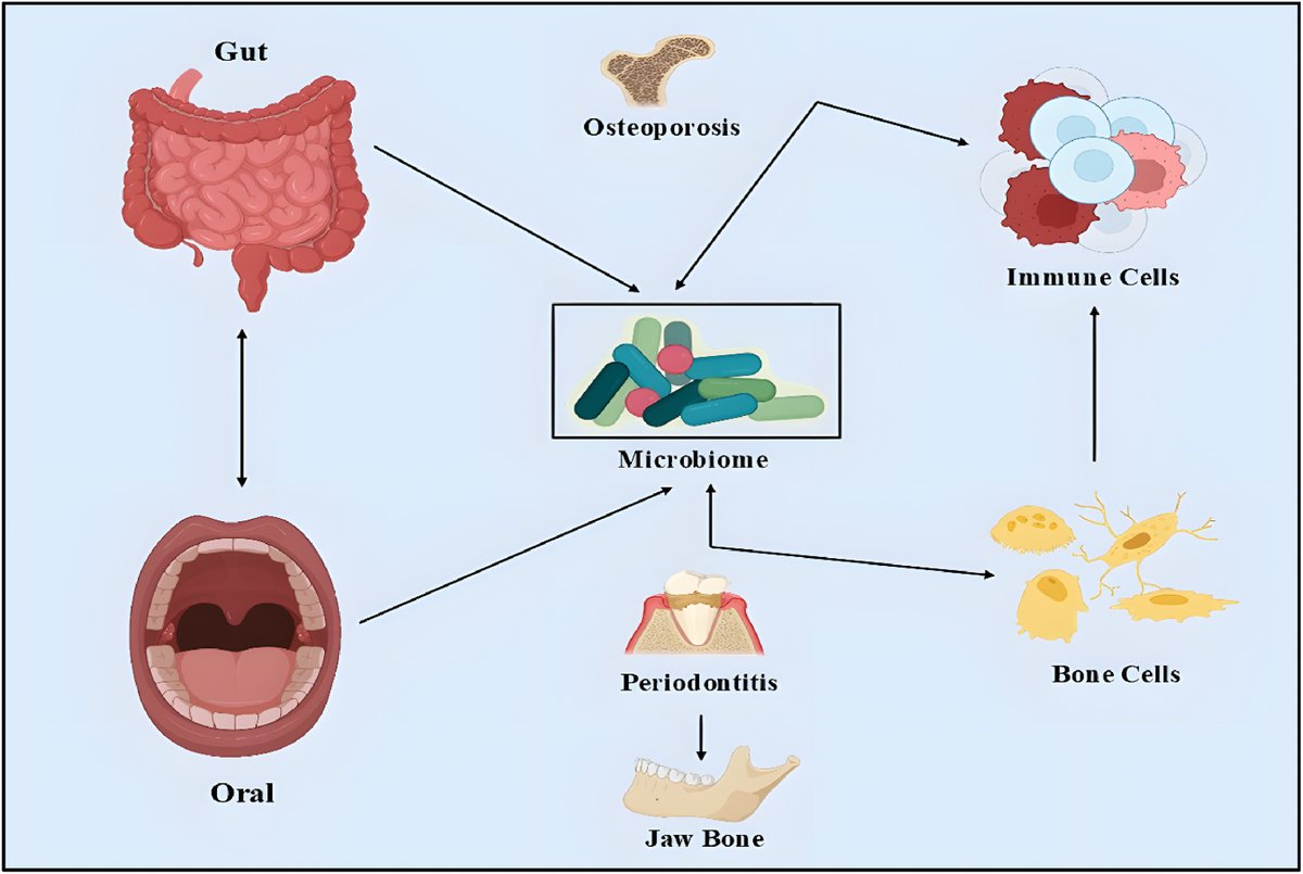
ALT Fig. 2. Interconnection between gut and oral microbiota in regulating bone and immune function. Dysbiosis in either site contributes to systemic and local inflammatory responses, influencing bone remodelling and promoting diseases such as periodontitis and osteoporosis through immune signalling pathways.