
@AIOpticalBioLab. Group Leader @itqbnova & H. Prof @LMCB_UCL. #Nanoscopy & #AI for #LifeSciences. #SRRF sensei. #ERCCoG, #EMBO member. Dad. Opinions my own
Joined August 2009
- Tweets 21,635
- Following 5,197
- Followers 15,328
- Likes 69,968
2,913 Photos and videos


Pinned Tweet

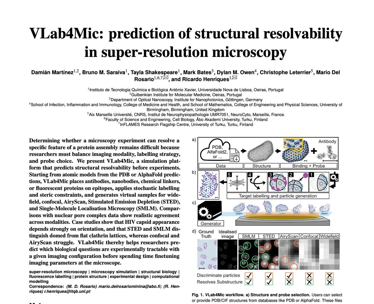
🔬🚀 It's out! Run 100s of imaging experiments before touching a microscope. #VLab4Mic reproduces them in silico from PDB/AlphaFold to help you image right first time. From rockstars @JDamianMtzR & @Bruno_MSaraiva, inaugural preprint of @_mariod's new lab
biorxiv.org/content/10.64898…

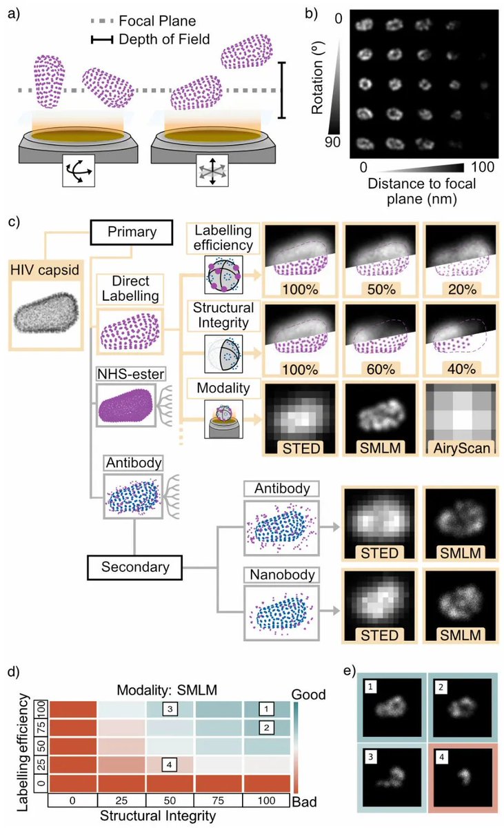
How do you know if a microscopy experiment can actually resolve a biological structure?
Introducing VLab4Mic, a platform that predicts structural resolvability before you go to the microscope
Led by @JDamianMtzR and @Bruno_MSaraiva at @HenriquesLab
biorxiv.org/content/10.64898…

1
6
13
2,151
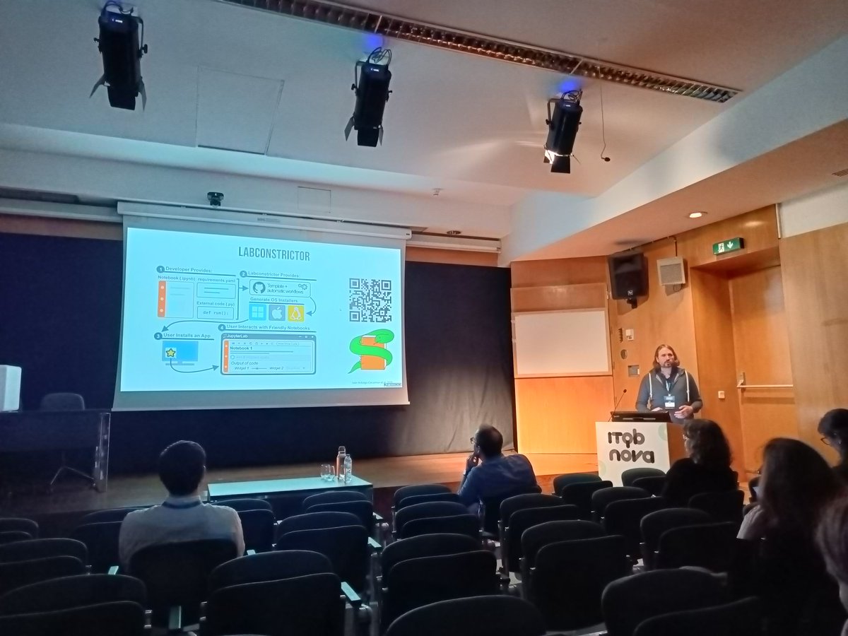
🔬🚀 Preprint update for #VLab4Mic - our super-resolution experiments virtual simulation system. Now installs as a one-click desktop app: no Python, no setup, thanks to the magic of the Jacquemet's lab #LabConstrictor packaging engine
biorxiv.org/content/10.64898…
How do you know if a microscopy experiment can actually resolve a biological structure?
Introducing VLab4Mic, a platform that predicts structural resolvability before you go to the microscope
Led by @JDamianMtzR and @Bruno_MSaraiva at @HenriquesLab
biorxiv.org/content/10.64898…
4
15
2,111
Ricardo Henriques retweeted

Congrats to @JDamianMtzR on the preprint release of his project #VLab4Mic, a tool able to virtually predict whether a biological structure will be resolved by a chosen microscopy or labeling.🎉
Congrats @Bruno_MSaraiva @_mariod @HenriquesLab
Read at: shorturl.at/vnQy1

3
7
499
Ricardo Henriques retweeted

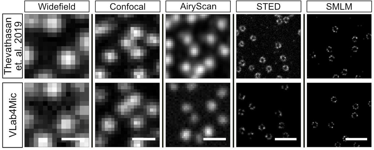
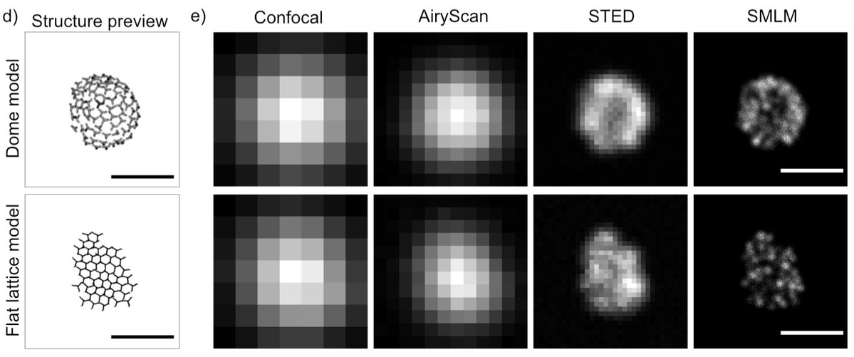
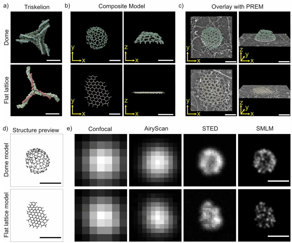
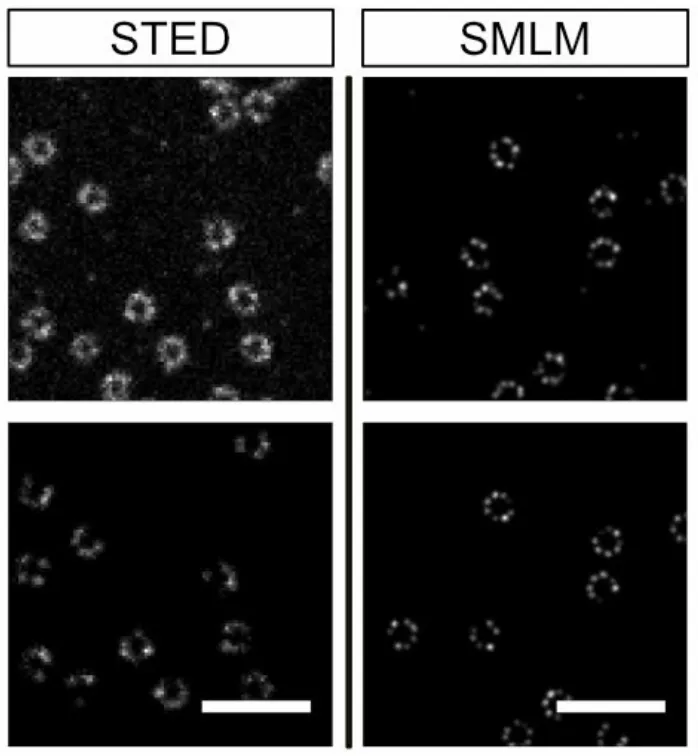
Can microscopy distinguish a flat from a domed clathrin lattice?
VLab4Mic predicts:
❌ Confocal
❌ AiryScan
✅ STED
✅ SMLM
A simple example of how simulation can guide modality selection before spending microscope time
1
2
3
272
Ricardo Henriques retweeted
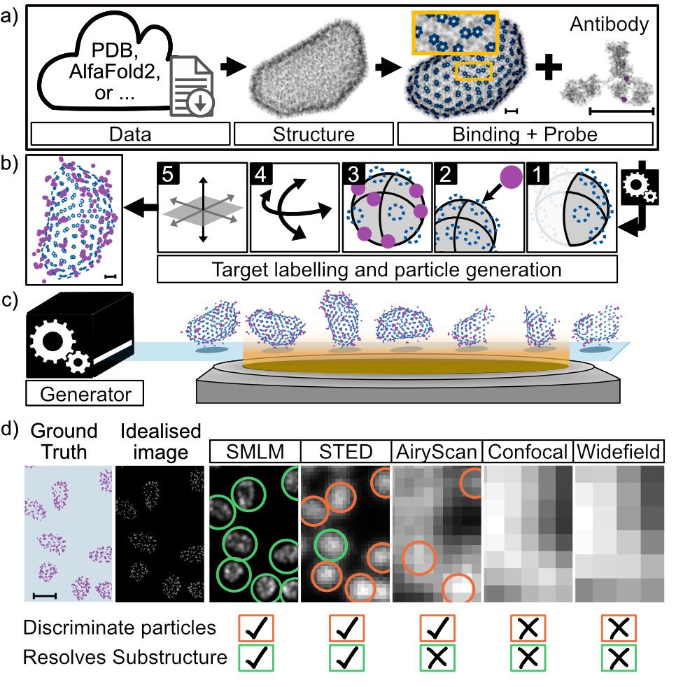
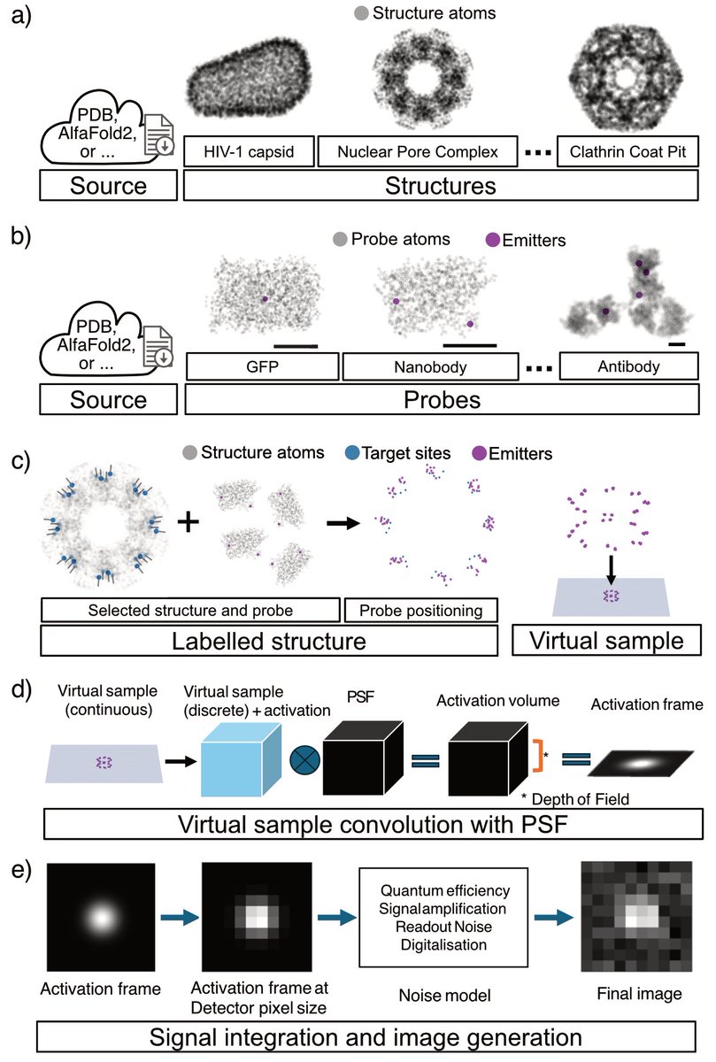
Workflow:
🧬 Import structure
🏷️ Choose labelling strategy
🎲 Simulate realistic stochastic labelling
🔬 Generate virtual images for Widefield, Confocal, AiryScan, STED and SMLM
Helping researchers test experiments before collecting data vlab4mic.henriqueslab.org
1
2
3
273
Ricardo Henriques retweeted
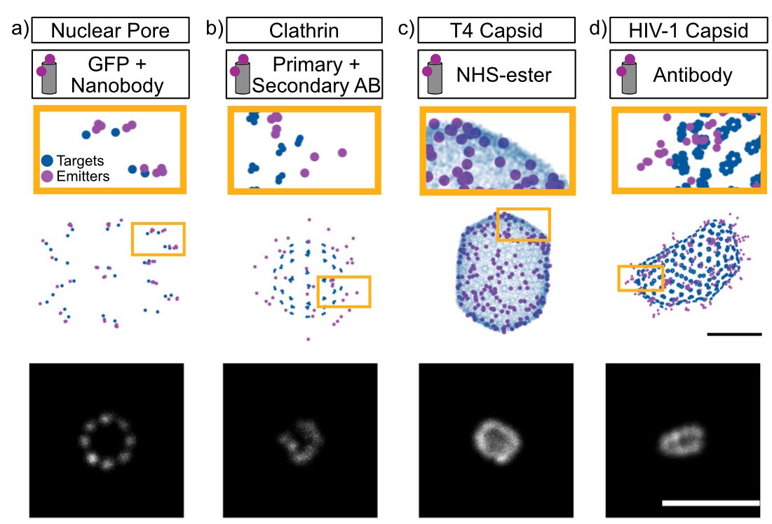
Resolution isn't just optics. Structural resolvability also depends on probe size, linkage error, labelling efficiency, steric hindrance and, molecular organisation. VLab4Mic models all of these starting from PDB or AlphaFold structures
1
2
2
742
Ricardo Henriques retweeted

Our preprint is out!! We present VLab4Mic, a platform that predicts structural resolvability for diverse biological structures before any experiments!! biorxiv.org/content/10.64898…
How do you know if a microscopy experiment can actually resolve a biological structure?
Introducing VLab4Mic, a platform that predicts structural resolvability before you go to the microscope
Led by @JDamianMtzR and @Bruno_MSaraiva at @HenriquesLab
biorxiv.org/content/10.64898…
2
6
28
4,610
🔬🚀 It's out! Run 100s of imaging experiments before touching a microscope. #VLab4Mic reproduces them in silico from PDB/AlphaFold to help you image right first time. From rockstars @JDamianMtzR & @Bruno_MSaraiva, inaugural preprint of @_mariod's new lab
biorxiv.org/content/10.64898…
How do you know if a microscopy experiment can actually resolve a biological structure?
Introducing VLab4Mic, a platform that predicts structural resolvability before you go to the microscope
Led by @JDamianMtzR and @Bruno_MSaraiva at @HenriquesLab
biorxiv.org/content/10.64898…
1
6
13
2,151
Huge thank you to the labs of @christlet, Dylan Owen and Mark Bates, and to @T_Shakespeare, who helped us make it happen. #VLab4Mic also heavily uses data from RCSB PDB, Jonas Ries & Justin Taraska's labs
1
5
8
550
This closes a loop we opened with #NanoJSQUIRREL (@SuperResoluSian et al., 2018), which mapped where super-resolution images go wrong relative to a reference. #VLab4Mic comes at it from the other end: predicting what's resolvable from known atomic structure before you even image
3
9
344
Ricardo Henriques retweeted
How do you know if a microscopy experiment can actually resolve a biological structure?
Introducing VLab4Mic, a platform that predicts structural resolvability before you go to the microscope
Led by @JDamianMtzR and @Bruno_MSaraiva at @HenriquesLab
biorxiv.org/content/10.64898…
3
12
28
11,032
Ricardo Henriques retweeted

1/ 🚀 Excited to release: napari-mcp - agentic control of napari from any MCP-capable AI assistant!
Use it for interactive image processing, analysis, and visualisation! Really cool project from @ilan_theodoro in my team!
#napari #bioimageanalysis #MCP @biohub
2
18
68
6,741
Ricardo Henriques retweeted

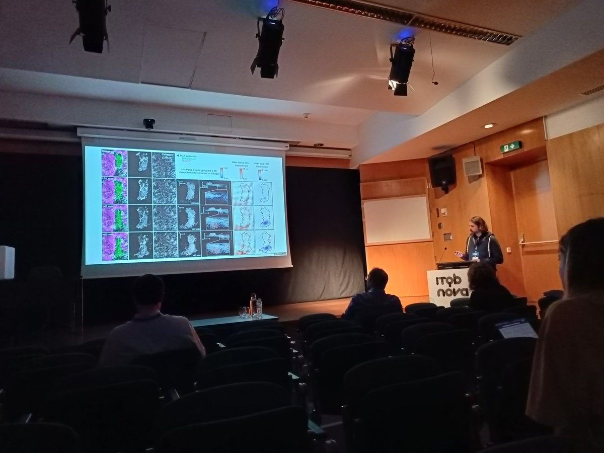
#IMS2026 D1 finishes with great @HenriquesLab and Guillaume Jacquemet pushing the limits of live-cell super-resolution, democratizing AI and quantitative microscopy, and using all these to study cancer cell transmigration and attachment 🌟
1
2
571
Ricardo Henriques retweeted
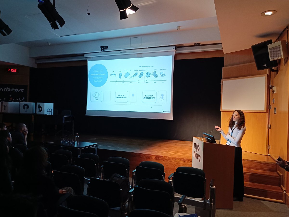
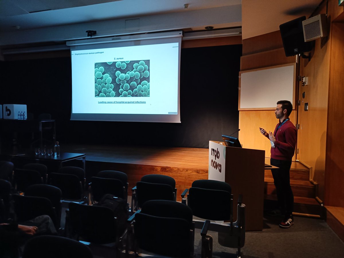
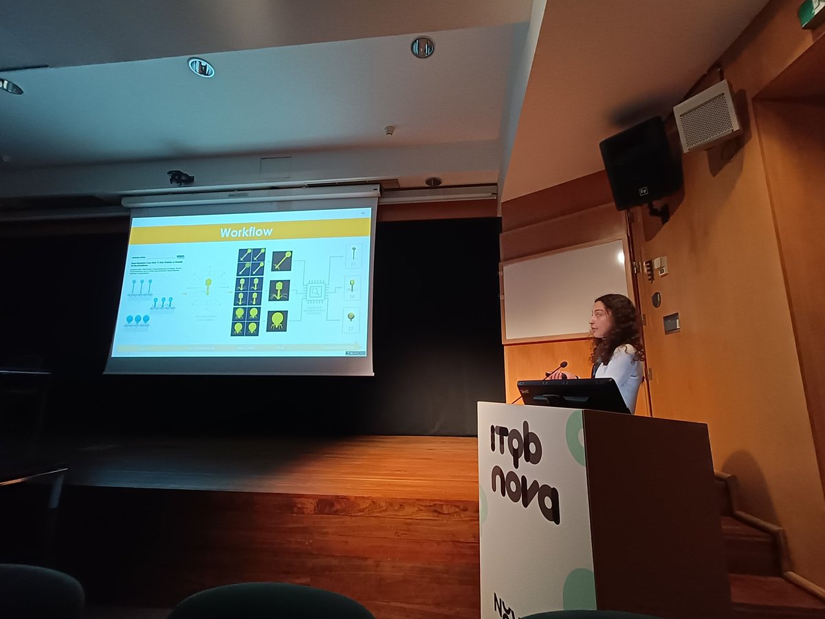
Students and postdocs from @HenriquesLab and Pedro Pereira's lab shining at #IMS2026 with intracellular infection, bacteriophages, expansion microscopy and lung on a chip🔥

1
4
539

Wuhuuuu!!

Big news🥂I'll start my research group at @NIMSB_NOVA!🔥🎉Hard to describe how
HAPPY EXCITED I am about bringing new #AI #imagedriven biomedical research growing together🌱I'm grateful to my supervisors,colleagues,collabs mentors who inspired encouraged me throughout 🤍

1
1
16
1,332
Ricardo Henriques retweeted
Absolutely delighted to share that we’ve been awarded by @wellcometrust with amazing collaborators John Barr, Juan Fontana & Martin Stacey! 🚀 An exciting adventure studying #bunyavirus viral factories with cutting-edge techniques is about to startI! 🔬🧪🦠🧬💻🌟

1
16
1,068
Ricardo Henriques retweeted
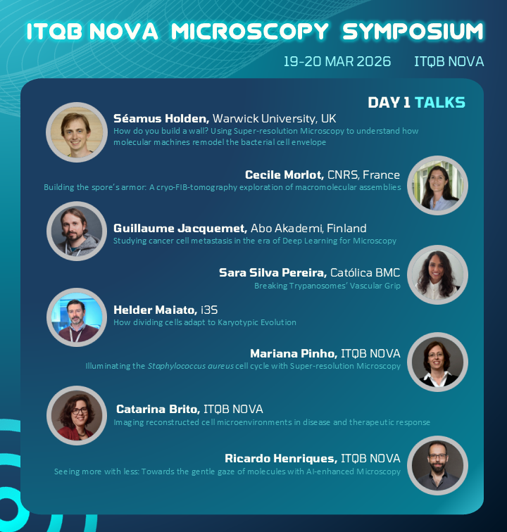
Last days to register for 𝐈𝐌𝐒𝟐𝟎𝟐𝟔 organized by @itqbnova.
DON´T miss these amazing researchers on 19/03: Seamus Holden, Cecile Morlot, Guillaume Jacquemet, @sara_spereira, Helder Maiato @i3S_UPorto, Mariana Pinho, Catarina Pinho, @HenriquesLab
🔗:ims2026.itqbnovacommunity.or…
4
7
821
Ricardo Henriques retweeted

Congrats to Sam in their paper

Scientists at Cambridge have developed a groundbreaking 3D molecular microscope that lets researchers watch life at work inside living cells. ch.cam.ac.uk/news/scientists…

ALT 3D ‘Molecular Microscope’ to Watch Life at Work Inside Cells
3
10
1,474
📯👨🚀 Calling all #bioimage analysis aficionados. #EZInput is out! We wanted to bring #ImageJ’s capacity to record settings to the #Python ecosystem 🐍. It is a declarative library for generating UIs that just work. Done with Guillaume Jacquemet's lab
arxiv.org/abs/2601.08859
1
1
5
335
We also needed something flexible for AI workflows 🤖. With EZInput, you get cross-environment support: same code, same interface. It works as a widget in Jupyter/Colab OR as a TUI in the terminal. 🖥️ Perfect for prototyping and then accelerating experimentation on the cluster 🚀
1
241
Huge kudos to the team: the brainchild of Bruno Saraiva, with contributions from Iván Hidalgo-Cenalmor, António Brito, Damián Martínez, and Tayla Shakespeare! 🙌 We built this to simplify how we interact with our own tools. Hope you find it useful too! 👇
arxiv.org/abs/2601.08859
1
256