
Pioneering end-to-end solutions for 3D spatial biology.
Joined June 2018
- Tweets 511
- Following 385
- Followers 509
- Likes 359
283 Photos and videos


We think #lightsheet microscopy can be more flexible, more scalable, and more intuitive. Meet up with us at #ELMI2026 next week to learn about our light sheet systems and our unique sample-to-data approach. 🔬🇵🇹 @PPBioImaging @EuroBioImaging @EuLiMI1
1
55
📽️ SYFP2 expression data from Dr. Tanya Daigle & Emily Kussick, imaged with #SmartSPIM!

Spinal motor neurons in dazzling detail. ✨
Each green dot is a spinal motor neuron - crucial but rare cells making up just 1% of neurons in the spinal cord. In diseases like ALS, they are selectively damaged, making them of special interest to neuroscientists. #SciShots
1
2
235
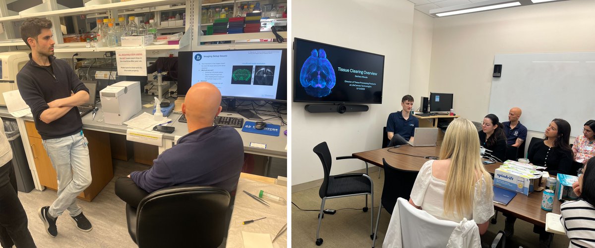
We got to introduce the LSFM community to #DALISPIM light sheet imaging at @MDIBL last week! The excitement was palpable as we had a packed demo schedule, with students getting to see firsthand how easy DALISPIM is to use.
#MicroscopyMonday
2
167
How can we get gene therapies to the heart more safely & effectively? 🫀 A research team led by @DJSiegwart & Eric Olson at @UTSWMedCenter has developed optimized lipid nanoparticles (LNPs) that demonstrate robust, targeted mRNA delivery & therapeutic potential 🧵 (1/3) @GDE_UTSW
1
3
206
Yehui Sun & @YuChungPien et al. screened LNPs using human iPSC-derived cardiomyocytes, then used Clear #tissueclearing with #SmartSPIM light sheet microscopy to characterize biodistribution of their lead candidate throughout the whole mouse heart. (2/3)
1
1
101
📖 Read the study to learn more: pnas.org/doi/10.1073/pnas.25… (3/3)
86
LifeCanvas Technologies retweeted

Jun 2
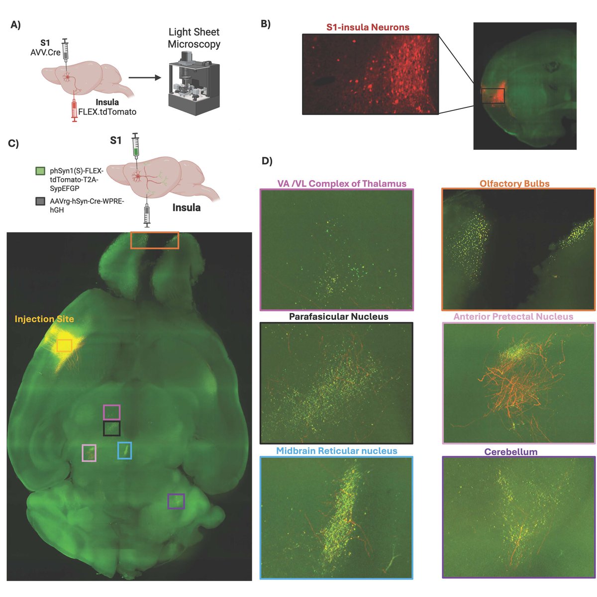
#JNeurosci: Adkins et al. explored how binge alcohol exposure and protracted abstinence impact the S1-insular circuit in mice, revealing how alcohol can reshape sensory processing to drive alcohol use disorder-related behavior.
doi.org/10.1523/JNEUROSCI.15…
6
19
3,118
Always exciting to see a SmartSPIM cover image 🤩
Jun 3

This Week in The Journal #JNeurosci | Exploring Backpropagating Signals in Foveal Cones; Why Do Cochlear Implants Lead to Residual Hearing Loss? jneurosci.org/content/46/22/…
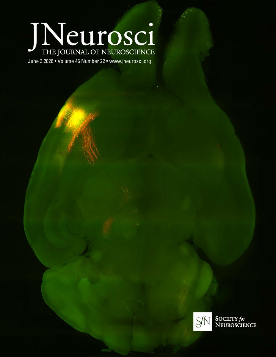
ALT Cover image of The Journal of Neuroscience featuring a fluorescent image of a brain. The cover displays vibrant green and orange colors under dark lighting, highlighting neural structures.
1
4
211
Calling all SmartBatch users! Have you validated a new antibody or tissue type?
Send us proof and receive 15% off your next reagent order for 1 new validation, or 25% off for 2 validations.
📆 Submit your data by June 17th: bit.ly/4ekYzsj
49
The quality of your #lightsheet 🔬 data is directly dependent on how your sample was prepared. This video shows a general comparison of some popular #tissueclearing methods, followed by Clear results from several mouse tissue types.
4
6
1,201
If you're going to @LSFM_NA at @MDIBL next week, you can check out DALISPIM in person and get a hands-on demo!

What tissue type do you study? DALISPIM makes #lightsheet imaging simple scalable for...
🧫 Organoids and other #NAMs
👥 Human clinical tissue samples
🐁 Rodents and other animal models
These were all imaged directly in multiwell plates! #FluorescenceFriday #microscopy
1
1
156
What tissue type do you study? DALISPIM makes #lightsheet imaging simple scalable for...
🧫 Organoids and other #NAMs
👥 Human clinical tissue samples
🐁 Rodents and other animal models
These were all imaged directly in multiwell plates! #FluorescenceFriday #microscopy
3
9
1,210
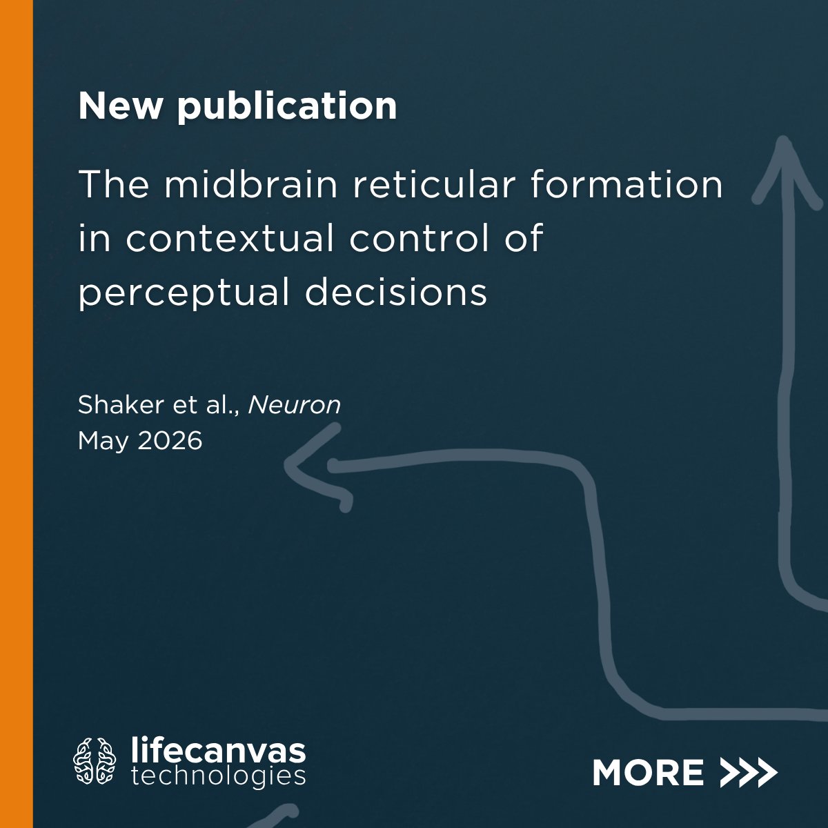
A new study from @SteinmetzNeuro’s lab dissects the neural circuitry of context-dependent responses – like deciding what to do during a fire drill vs a random fire alarm – honing in the role of a region called the midbrain reticular formation (MRF). (1/3) 🧵
1
1
3
175
By using anterograde viral tracing combined with Clear #tissueclearing and #SmartSPIM imaging to map axonal projections from the MRF throughout the brain, they found that MRF neurons connect to decision-making circuits underlying flexible behavior. (2/3)
1
103
Read the study here: cell.com/neuron/abstract/S08… (3/3)
93
This turtle embryo 🐢 was cleared by @MBLScience Embryology course students with SmartBatch & labeled with acetylated tubulin to see innervation. MBL scientists Paul Maier Carsten Wolff imaged the embryo on a #MegaSPIM light sheet microscope #FluorescenceFriday
3
8
515
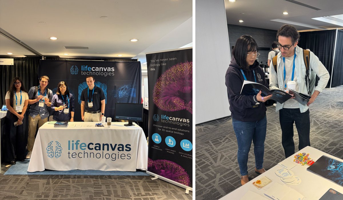
Hello from the last day of @CAN_ACN in Montreal! 🇨🇦 We've had a jam-packed week, including checking out the Neural Circuits & Behavior satellite event hosted by Mark Cembrowski & @Franklandlab.
64
LifeCanvas Technologies retweeted

May 20
Love microscopy and want to work in a dynamic imaging CoRE? 🔬 We’re hiring! Join our team to support cutting-edge microscopy, train users, and help drive exciting research projects. Apply now 📧

1
2
165
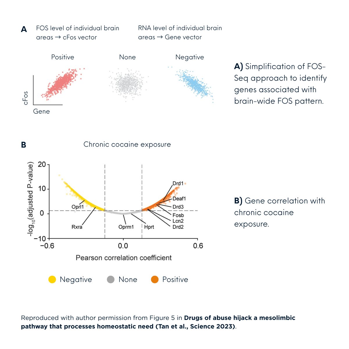
How do you use c-FOS and other neural activity markers in your research? What if that data were brain-wide and spatially resolved?
Check out these studies to see how scientists are using LifeCanvas whole-brain c-FOS mapping to drive their investigations. 🧵 (1/5)
#neuroscience
1
101
📝 # 2: Drugs of abuse hijack a mesolimbic pathway that processes homeostatic need (Bowen Tan et al., Science 2023). Read it here: science.org/doi/10.1126/scie… (4/5)
1
53
By correlating whole 🧠 neural activity data w/ whole 🧠 in situ hybridization data, @EricJNestler's lab IDed genes affected by repeated use of cocaine & morphine. They then delineated a shared reward pathway by which these drugs interfere w/ homeostatic need fulfillment. (5/5)
48