We create hardware and software for light sheet and multiphoton microscopy, fiber photometry, brain mapping, stereology, and image analysis.
Joined July 2009
- Tweets 2,678
- Following 711
- Followers 1,002
- Likes 1,067
862 Photos and videos


Jun 12
We're delighted to see this work highlighted by @TheBioPhotonics.
HySIL and SCOPE represent exciting advances in optical design, helping deliver high-resolution 3D tissue imaging while reducing the cost and complexity traditionally associated with advanced microscopy.
We're proud to have collaborated on bringing these advances to researchers through the SLICE™ Light Sheet Microscope.
📖photonics.com/Articles/Hybri…
#BioPhotonics #Microscopy #Optics #LightSheetMicroscopy #slicelightsheetmicroscope #mbfbioscience #3Dimaging
1
29
Jun 11
Great to see this work featured by @physorg_com.
The article highlights the Nature Biotechnology publication from Dr. Raju Tomer and collaborators introducing HySIL and SCOPE, innovative optical technologies designed to make high-resolution 3D tissue imaging more accessible and scalable.
These advances have also been incorporated into the SLICE™ Light Sheet Microscope, helping bring powerful 3D imaging capabilities to researchers across neuroscience, developmental biology, cancer research, and pathology.
Read more: phys.org/news/2026-06-3d-mic…
#LightSheetMicroscopy #3DImaging #Neuroscience #Microscopy #SLICElightsheetmicroscope #lightsheetimaging
38
MBF Bioscience retweeted

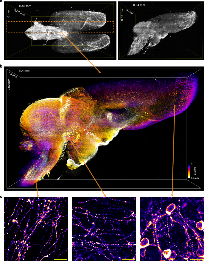
Hybrid solid−liquid optics enable scalable, high-resolution light-sheet microscopy across diverse immersion media go.nature.com/4xdDkQR
15
84
4,093
Jun 9
🔬 Celebrating a breakthrough from Dr. Raju Tomer's lab @Columbia, just published in @NatureBiotech!
SCOPE technology SLICE microscopy = affordable, high-resolution 3D imaging for all.
Dr. Tomer's team developed SCOPE, which transforms inexpensive air objectives into submicron-resolution tools. Combined with SLICE (which we commercialized from his pLSM invention), researchers can image centimeter-scale samples at nanoscale detail.
The research team—spanning Columbia, NIH, Lehigh, NYU Langone, and MBF—demonstrated:
• Whole mouse & salamander brains
• Human tissue 3D histopathology
• Brain organoids with microglia
• Multi-immersion compatibility
This collaboration shows what's possible when academic innovation meets commercial accessibility. Dr. Tomer invented the platform; we help make it available to labs worldwide.
Huge congratulations to Dr. Tomer, the biologist collaborators, and the entire research team! This is the kind of science that expands what's possible for everyone. 🎉
Read the full paper: nature.com/articles/s41587-0…
#Microscopy #Neuroscience #LightSheet #ScientificImaging #Innovation #SLICElightsheetmicroscope
2
9
2,494
Jun 8
Attending C. elegans 2026 (MAPSS DevCell)?
Stop by our booth to see the latest in WormLab for C. elegans tracking, imaging, and behavioral analysis. Whether you're looking to improve imaging workflows or streamline behavioral studies, we'd be happy to show you what's new and discuss your research needs.
Looking forward to connecting with the C. elegans community!
uwmadison.eventsair.com/maps…
.
.
#Celegans #Celegans2026 #WormLab #BehaviorAnalysis #Imaging #Neuroscience #MAPSS #DevCell2026
50
May 20
We’re having a great time at the Canadian Association for Neuroscience Meeting in Montréal!
We’re here today and tomorrow. If you’re attending, stop by our booth to see live demos of:
🔬 SLICE™ light sheet microscope for accessible, high-performance 3D imaging
🧠 FP3002 for flexible fiber photometry experiments
🧬 Software solutions for neuron tracing, stereology, brain mapping, multiphoton imaging, and more
We’d love to hear about your research and imaging workflows and connect with more of the neuroscience community!
can-acn.org/meeting-2026/
@CAN_ACN #CAN2026 #Neuroscience #LightSheetMicroscopy #FiberPhotometry #BrainMapping #Stereology #Scienceinnovation #MBFBioscience


50
May 19
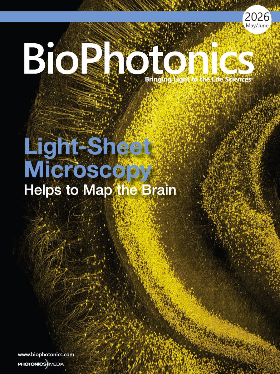
We’re excited to share that our article, “Light-Sheet Microscope Design Puts Whole-Brain Imaging in Reach for Scientists,” is featured as the cover article in the latest issue of @TheBioPhotonics magazine!
The article highlights how the SLICE Light Sheet Microscope system combines projected light-sheet microscopy (pLSM), advanced optics for cleared tissue, and simplified system design to make high-performance whole-brain fluorescence imaging more accessible to researchers.
From subcellular-resolution imaging to large-scale brain mapping and connectomics, this approach brings advanced 3D imaging capabilities within reach of laboratories around the world.
A huge thank you to everyone involved in the development of this technology, especially Dr. Raju Tomer and collaborators at @Columbia University.
📖 Read more about how accessible light-sheet microscopy is accelerating neuroscience research and whole-brain imaging: photonics.com/Articles/Light…
.
.
#3Dimaging #SLICElightsheetmicroscope #lightsheetimaging #wholebrainimaging #neuroscience
1
1
89
May 15
We’re excited to be heading to Montréal for the Canadian Association for Neuroscience Meeting 2026, taking place May 18–21!
If you’re attending, stop by our booth to see live demos of:
🔬 SLICE™: our award-winning light sheet microscope built to make high-performance 3D imaging more accessible and scalable.
🧠 FP3002 Fiber Photometry System: a flexible platform for recording neural activity in freely moving animals.
We'll also be showcasing solutions for neuron tracing, stereology, brain mapping, multiphoton imaging, and more.
Can’t wait to talk science, see what everyone’s working on, and connect with the neuroscience community!
can-acn.org/meeting-2026/
.
.
@CAN_ACN #CAN2026 #Neuroscience #LightSheetMicroscopy #FiberPhotometry #BrainResearch #Microscopy #3DImaging #MBFBioscience
2
48
May 6
We had a great time at the AQLM course this week showcasing the SLICE light sheet microscope and connecting with passionate researchers exploring the future of 3D imaging. 🔬
Thank you to the AQLM team and everyone who stopped by to see SLICE in action!
@MBLScience #LightSheetMicroscopy #3DImaging #Microscopy

3
65
May 1
Join us at the Weinstein Cardiovascular Development & Regeneration Conference
📍 Hyatt Regency, Jersey City, NJ
📅 May 6–8
We’ll be showcasing 🔬SLICE, our award-winning light-sheet microscope built for fast, scalable, and accessible 3D imaging. If you’re working in vascular biology or large-scale 3D imaging, this is something worth seeing.
We’ll also be demoing Vesselucida 360, our powerful image analysis software for automated vessel reconstruction and advanced morphometric analysis.
Stop by to see live demos, explore our full suite of imaging solutions, and chat about your research. We’d love to connect!
weinstein-conference.oa-even…
#WeinsteinConference #VascularBiology #LightSheetMicroscopy #3DImaging #MBFBioscience
60
Apr 7
New research is reshaping how we understand the adolescent brain, and Neurolucida played a key role. 🔬
A new study published in Science Advances found that during adolescence, a specific spine "hotspot" on primary somatosensory cortical neurons sees a dramatic, experience-dependent surge in dendritic spine density. When this accumulation failed to develop properly in mouse models of schizophrenia, researchers suggest that localized regulation of spine density is critical for maturing cognitive functions.
The study pairs Neurolucida with several imaging techniques, including super-resolution, confocal, and two-photon microscopy – creating precise maps of spine density and meaningful morphometric data.
The research reframes how we think about adolescent brain development and underscores the value of quantitative morphology in understanding the biology behind mental health disorders.
Huge congratulations to Ryo Egashira, Meng-Tsen Ke, Takeshi Imai, and team on this work.
🔗Read the full study: science.org/doi/10.1126/scia…
.
.
#Neuroscience #Neurolucida #BrainResearch #Adolescence #MentalHealth #MBFBioscience #sciencematters
4
12
1,387
Mar 30
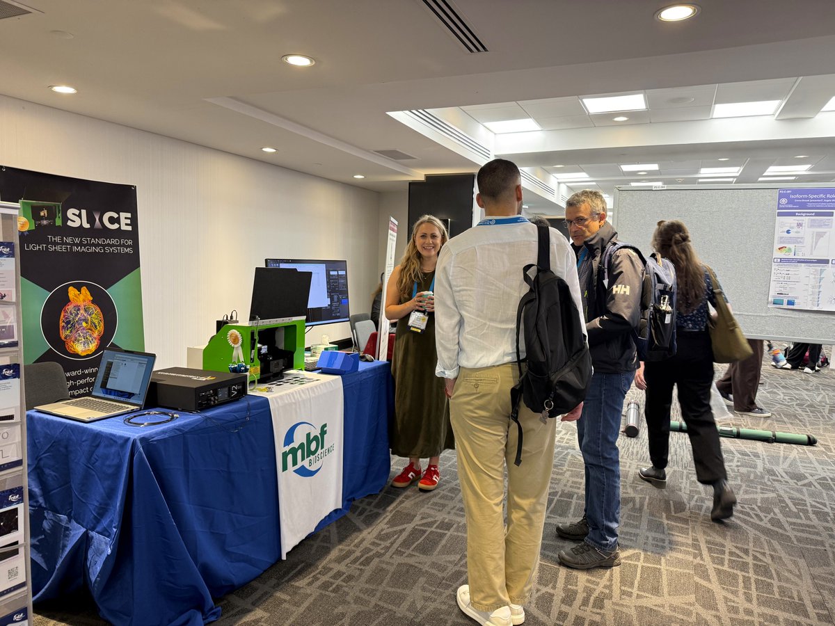
If you're at FOM 2026, come find us at booth 46! 👋
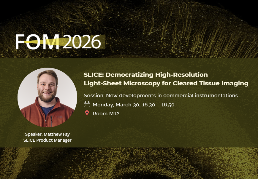
We're here showcasing how SLICE 🔬 is making high-resolution 3D imaging of cleared tissue more accessible, and today our own Matthew Fay is taking the stage to share how.
His talk will highlight how SLICE enables accessible, high-resolution 3D imaging of cleared tissues using advanced projected light-sheet microscopy (pLSM), along with a streamlined workflow for acquisition, visualization, and analysis.
📍 Session: New Developments in Commercial Instrumentation
🗓️ Today, March 30 | 16:30 – 16:50
📌 Room M12
Swing by the session or stop by our booth. We're always happy to geek out about imaging!
#FocusOnMicroscopy #LightSheetMicroscopy #SLICElightsheetmicroscope
#Microscopy #Neuroscience #3DImaging #FluorescenceImaging
#ClearedTissue #MBFBioscience
70
Mar 19
Join us at Booth #46 at FOM 2026 in Stockholm (March 29 – April 1)
We're showcasing SLICE™, our award-winning light-sheet microscope for scalable, accessible, cost-effective high-performance 3D imaging, alongside our full suite of solutions including neuron tracing, stereology, multi-photon imaging, fiber photometry, brain mapping, and more.
📖 Attend Our Lecture: SLICE: Democratizing High-Resolution Light-Sheet Microscopy for Cleared Tissue Imaging
🗓 March 30 | 16:30–16:50 | Room M12
We look forward to seeing you in Stockholm! 🔬
focusonmicroscopy.org/
#FOM2026 #FocusOnMicroscopy #LightSheetMicroscopy #SLICElightsheetmicroscope #Microscopy #Neuroscience #CellBiology #3DImaging #FluorescenceImaging #ClearedTissue
1
74
MBF Bioscience retweeted

Trusted by neuroscientists worldwide, Neurolucida from @MBFBioscience enables precise tracing of neuronal structures from histological specimens & generates quantitative morphometric data.
📚 6,000 citations advancing neuroscience research.
#Neuroscience #NeuroAnatomy
1
2
73
MBF Bioscience retweeted

Breaking the Size Barrier: Sub-Micron 3D Imaging with ClearScope arnastech.com/f/breaking-the…
#microscopy #lightsheet #neuroscience @MBFBioscience
1
1
213
Mar 10
We are excited to share our latest publication on ClearScope, a fully integrated light-sheet theta microscope for sub-micron resolution imaging without lateral size constraints. 🔬
Using light-sheet theta microscopy (LSTM), ClearScope enables high-resolution 3D imaging of large biological specimens including intact mouse brains and human brain tissue sections.
Read the open-access paper:
doi.org/10.3390/jimaging1203…
.
.
@Columbia @nyulangone
#slicelightsheetmicroscope #3Dimaging #microscopy #neuroscience #Connectomics #Microscopy #OpenAccess
1
2
159
Mar 6
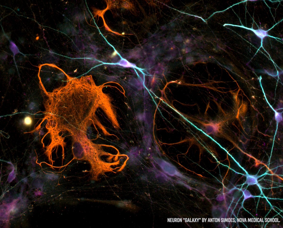
🧠 NeuroArt Image Contest
Neuroscience isn't just about discovery. It's also about beauty.
We invite researchers, students, and imaging enthusiasts to submit their most captivating neuroscience images. Whether it’s neurons, brain tissue, or fluorescent microscopy, we want to see the artistry in your science.
Submit your image today!
🔗 neuroart.com/march-image-con…
.
.
#NeuroArt #MicroscopyArt #NeuroscienceResearch #BrainScience #ScientificImaging #LifeScience
1
1
121