
Let’s make time spent on X worthwhile. Elevate your medical knowledge with high-yield MCQs and AI-powered visuals. #MedTwitter #MedX #Healthcare #USMLE #FOAMed
Joined June 2024
- Tweets 13,073
- Following 1,708
- Followers 13,577
- Likes 29,709
662 Photos and videos


Pinned Tweet

29 Nov 2024
🚑 You’re the Doctor:
A 25-year-old marathon runner collapses at the finish line. Bystanders say he was confused and agitated before collapsing. On arrival:
•BP: 88/60 mmHg
•HR: 145 bpm
•Temp: 41°C (105.8°F)
•RR: 30/min
•Skin: Hot, dry, no sweating.
What’s your next step? 🩺
A. IV fluids and antipyretics 🌡️
B. Rapid sequence intubation 🛏️
C. Ice-water immersion 🧊
D. Administer adrenaline 💉
E. CT brain to rule out stroke 🧠
👇 Comment your answer & reasoning!
#MedTwitter #FOAMed #EmergencyMedicine #MedicalEducation #MedStudentTwitter #DoctorLife #HeatStroke #CriticalCare

182
490
2,747
432,786
May 22
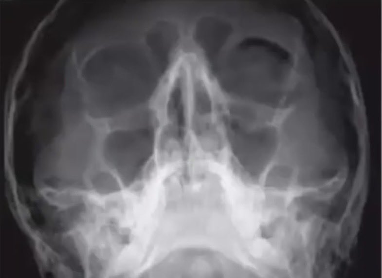
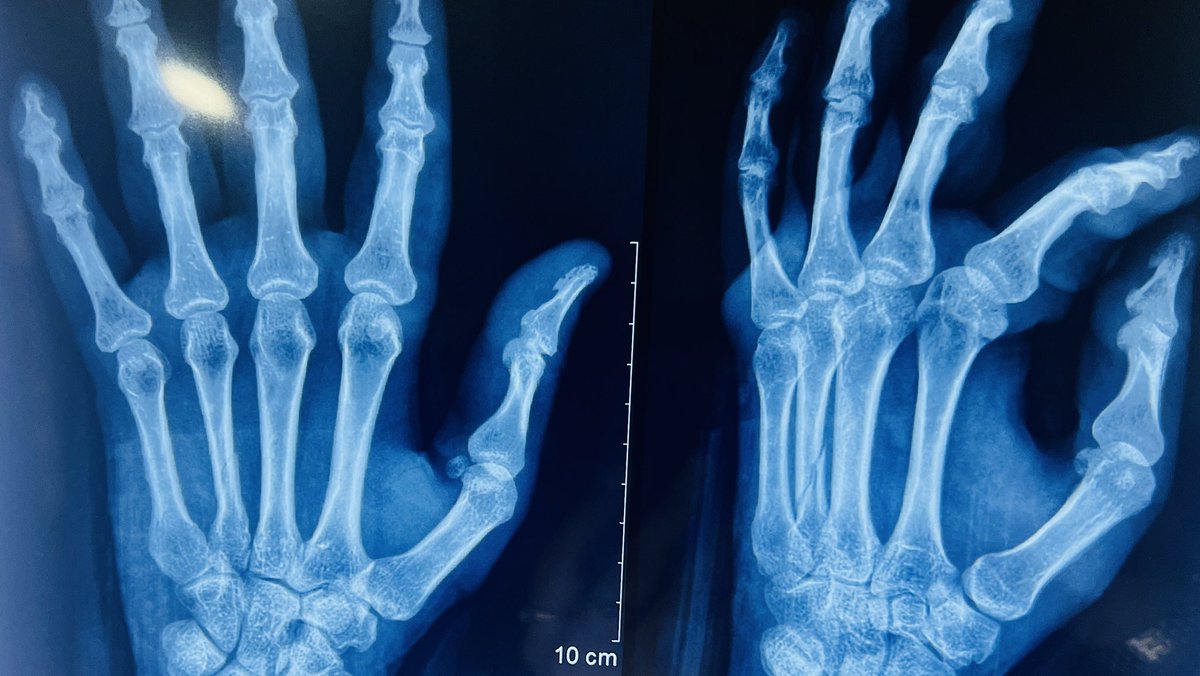
A 28-year-old man presents to the Emergency Department after a blunt facial trauma during a sports injury. He complains of pain, swelling around the right eye, and double vision on upward gaze.
A plain facial radiograph is performed.
What is this radiological sign known as, and what does it most likely indicate?
1
1
2
446
A 62-year-old man presents to the emergency department with progressive dyspnea and chest discomfort. He is hypotensive (BP 85/60 mmHg), tachycardic, and has elevated jugular venous pressure. Heart sounds are muffled. Bedside echocardiography shows a large pericardial effusion with right atrial and right ventricular diastolic collapse.
While preparing for urgent pericardiocentesis, further history reveals recent chest trauma following a road traffic accident.
In which of the following situations is pericardiocentesis contraindicated?
A. Cardiac tamponade with hemodynamic instability
B. Large symptomatic pericardial effusion
C. Suspected aortic dissection causing hemopericardium
D. Malignant pericardial effusion with dyspnea
E. Purulent pericarditis
1
1
5
1,568
Answer C: Pericardiocentesis is contraindicated in Aortic dissection–related hemopericardium, as draining the pericardial blood removes the tamponade effect and can precipitate catastrophic bleeding. It is also contraindicated (or avoided unless absolutely life-saving) in Left ventricular free wall rupture, where needle drainage may worsen hemorrhage; these patients require urgent surgical repair. On echocardiography, diastolic collapse of the right atrium and right ventricle is a hallmark sign of cardiac tamponade.
1
179
A 68-year-old female with a history of hypertension and chronic kidney disease presents with confusion and dyspnea. She is found to have sepsis secondary to pneumonia. Vitals show BP 85/50 mmHg, HR 120/min, RR 28/min. ECG shows nonspecific ST depressions. High-sensitivity troponin is elevated with a rising trend. There is no chest pain.
Which of the following is NOT an example of Type 2 Myocardial Infarction (MI)?
A. Severe anemia causing myocardial oxygen supply-demand mismatch
B. Sustained tachyarrhythmia leading to increased myocardial oxygen demand
C. Septic shock with hypotension causing myocardial hypoperfusion
D. Acute plaque rupture with thrombus formation in a coronary artery
3
1
12
790
Answer: D. Acute plaque rupture with thrombus formation in a coronary artery
Type 2 MI is due to oxygen supply–demand mismatch (e.g., sepsis, anemia, hypoxia, tachyarrhythmia) without a primary coronary event. Plaque rupture with thrombus represents an acute atherothrombotic event, which defines Type 1 MI, not Type 2.
1
133
80-year-old diabetic woman with a history of atrial fibrillation is transferred to your emergency
department (ED) from the local nursing home with a note from the facility stating that she was
complaining of abdominal pain and vomited once. Her vital signs in the ED are blood pressure (BP)
105/75 mm Hg, heart rate (HR) 95 beats/minute, respiratory rate (RR) 18 breaths/minute, and temperature
100.1°F . The patient appears very uncomfortable and has not stopped moaning in pain since arriving to
the ED. You are surprised to find that her abdomen is soft and nontender on palpation. Which of the
following diagnostic tests is most likely to reveal the cause of her symptoms?
a. Capsule endoscopy
b. Colonoscopy
c. Computed tomography (CT) angiography of the abdomen
d. Ultrasound
e. Abdominal radiograph
2
1
10
644
3 Aug 2025
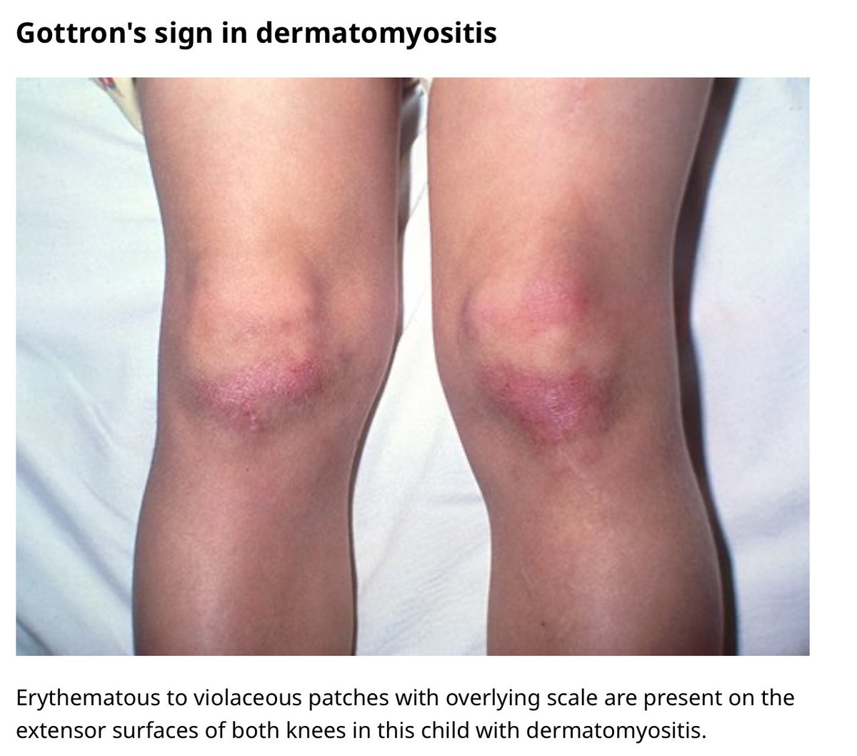
🌟 Gottron’s sign over extensor surfaces of knees 🌟
1
3
27
2,070
Daily Dose of Medicine retweeted

9 May 2025
C-Biopsy of Artery
◦ Temporal artery biopsy (TAB) is currently the gold standard method for the diagnosis of giant cell arteritis (GCA), with a specificity of 98% to 100% when histopathology demonstrates active necrotizing arteritis, characterized by a predominance of mononuclear cell infiltrates or a granulomatous process with multinucleated giant cells. Other features include narrowing and occlusion of the arterial lumen, intimal proliferation, rupture of the internal elastic layer, and lumen thrombosis.
◦ However TAB has a sensitivity of 77% and its false-negative rate ranges from 9% to 61%.
◦ False negatives may be due to the timing of biopsy, the length of the specimen, or the presence of segmental “skipping lesions” (observed in 12%‒28% of cases).
◦ It is also estimated that 25% of TAB for GCA are reported as “healed arteritis”, defined as fibrosis, attenuation and/or neovascularization of the media, irregular intimal proliferation, multifocal to complete loss of internal elastic lamina, adventitial fibrosis, and absence of ongoing chronic medial inflammation. The presence of “healed arteritis” is more often associated with prior history of polymyalgia rheumatica and longer corticosteroid use before TAB.
📸 Histopathological findings in biopsy-proven giant cell arteritis:
(A) Temporal artery with narrowed lumen, atrophic media, interrupted elastic lamina, and granulomatous inflammation consistent with the diagnosis of giant cell arteritis (H&E, x50).
(B) Typical area of granulomatous inflammation with multiple giant cells (black arrows) in the interrupted elastic lamina (H&E, x400).
(C) Similar findings in another temporal artery biopsy with interrupted elastic lamina (black arrow), granulomatous inflammation, and giant cell formation (H&E, x200).
(D) A clearer area of interrupted elastic lamina (red arrowhead) after special staining (Elastin, x200).
*Source: Alkatan, Hind M et al. “Giant cell temporal arteritis: a clinicopathological study with emphasis on unnecessary biopsy.” Frontiers in Ophthalmology vol. 3 1327420. 2023
🔗doi.org/10.3389/fopht.2023.1…

3
15
997
9 May 2025
⭐ Gold standard Diagnostic test for Giant cell arteritis ?
A-ultrasound
B-Angiography
C-Biopsy of Artery
D-Ct head
E-Explain the answer
16
4
51
7,423
5 Apr 2025
A 72-year-old female presents with fever, stiff neck, and headache. You strongly suspect bacterial
meningitis. Which of the following is the most appropriate treatment strategy?
A. Dexamethasone
B. Vancomycin and ceftriaxone
C. Ampicillin
D. A and B
E. A, B, and C
58
28
312
53,022
10 Apr 2025
Answer E: Vancomycin, ceftriaxone are effective against major pathogens in bacterial meningitis (Strep pneumoniae, Neisseria meningitidis, and H influenza). Patients at extremes of age, Listeria
monocytogenes becomes more prevalent and requires specific therapy with ampicillin.
1
6
653
6 Mar 2025
A woman presents with a long history of dysphagia for both liquids & solids. Chest X-ray reveals a dilated lower oesophagus with a fluid level behind the heart
choice of treatment?
A. Atenolol
b-Nifedipine
c-Ramipril
d-Propranolol
e-Doxazosin
21
21
194
21,078
6 Mar 2025
acanthosis nigricans is a sign for insulin resistance in body
1
26
1,789
4 Feb 2025
A 45-year-old male with fatigue, weight gain, and cold intolerance. On examination, his skin is dry, and he has a slow heart rate.
TSH: Elevated
Free T4: Low
❓ likely diagnosis?
A) Hyperthyroidism
B) Hypothyroidism
C) Addison’s disease
D) Cushing’s syndrome
25
6
61
5,319
28 Jan 2025
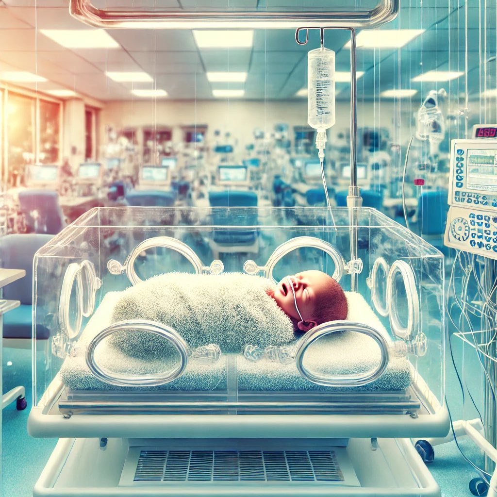
Persistence of fetal circulatory pattern of right to left shunt through PDA and foramen ovale after birth is due to high pulmonary vascular resistance occur in which of the following.
a. PPHN (Persistent Pulmonary Hypertension of the Newborn)
b. Sepsis
c. TTN
d. CHD
e. Pneumothorax
5
8
34
2,495
Daily Dose of Medicine retweeted
16 Jan 2025
Hypoxic –ischemic encephalopathy(HIE) is an important cause of permanent damage to CNS tissue that may result in neonatal death or manifest as cerebral palsy. Which of the following is the characteristic of stage 1.
a. Baby dull and lethargic
b. Weak primitive reflexes
c. Contricted pupils
d. Puplis unequal in size
e. Normal reactive pupils
10
8
27
2,679
Daily Dose of Medicine retweeted
12 Jan 2025
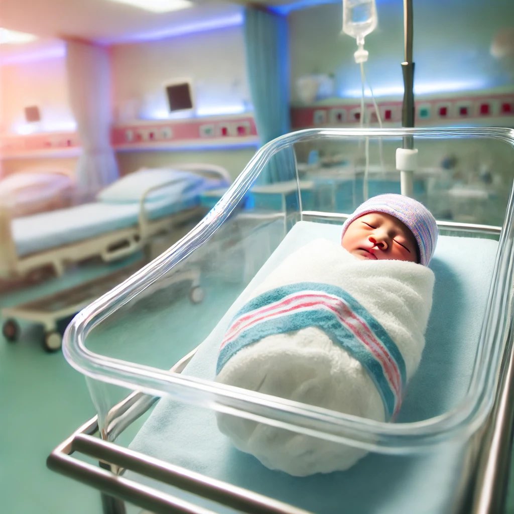
A term baby weighing 3.6kg delivered by c-section has tachypnea and acrocyanosis. Apgar scores were 7 and 8 at 1 and 5 minutes, respectively. Vitals and systemic examination are normal with the exception of a respiratory rate of 84b/m and slight subcostal retractions. What diagnosis you will consider in this baby.
a. Congental diaghpramatic hernia
b. Pneumothorax
c. Cyanotic congenital heart disease
d. Transient tachypnea of newborn
e. Sepsis
8
4
14
2,266