
Musculoskeletal Radiology and Intervention. McGill Alumnus. Cycling Passionate
Joined August 2009
- Tweets 1,652
- Following 673
- Followers 251
- Likes 3,814
Photos and videos


319
5,247
17,452
556,408
Miguel Vega retweeted

8 Dec 2024
🚨 Lateral Cutaneous Nerve Compression
⭐️The lateral cutaneous nerve typically passes under the inguinal ligament, but in 13% of cases, it traverses the ligament, increasing the risk of compression and leading to meralgia paresthetica.
#mskrad #radres #nerveimaging
2
12
69
5,758
Miguel Vega retweeted

26 Nov 2024
Rectus femoris muscle overload: what do we expect to find in ultrasound?
Rectus femoris muscle injuries are very common in sports activities, and ultrasound is a highly sensitive tool for diagnosing them.
Based on the ultrasound criteria described in the previous publication, both in static images and in dynamic studies, to diagnose rectus femoris muscle overload, the following must be observed:
A) Static ultrasound:
A1.- In the axial image, an increase in “tension” of the indirect tendon is observed, characterized by an almost perpendicular orientation instead of its normal coma morphology.
A2.- In the longitudinal plane, the pennation angle increases compared to the contralateral muscle without alteration of the typical fibrillar pattern.
B) Dynamic ultrasound:
B1.- A lesser muscle deformity is seen on sonocompression with the transducer associated with a greater muscle tone on “sonopalpation”.
B2.- There is little muscle mobility during isometric contraction, significantly less than the opposite side and with minimal increase in its anteroposterior diameter.
1
12
76
6,156
Miguel Vega retweeted

23 Sep 2024
🌟 ASAS researchers recommend using axSpA as the overarching term for axial spondyloarthritis. They prefer radiographic-axSpA over ankylosing spondylitis. These changes aim to standardize terminology and improve clarity in diagnosis and treatment.
ard.bmj.com/content/83/5/547…
#ASAS
1
38
92
5,757
Miguel Vega retweeted

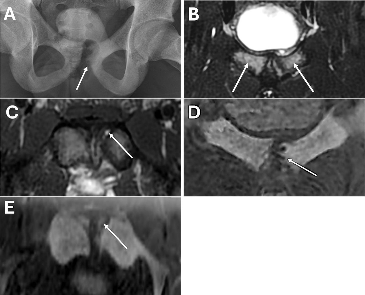
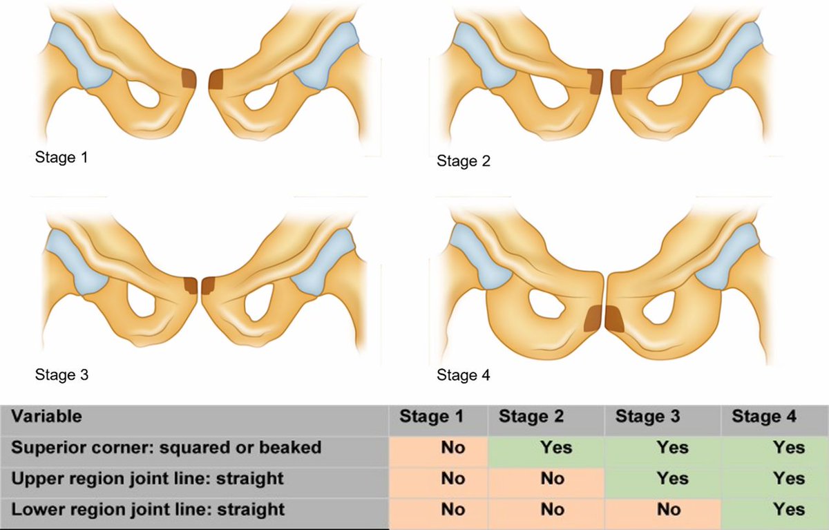
⚠️ Pubic apophysitis - an important cause of groin pain in young athletes
NEW article in the #ImagesinSEM series ✅ using a case of a 17-year-old male right-footed footballer ⚽️
Find out more ➡️ bit.ly/47i3p4F
1
31
90
11,081
Miguel Vega retweeted
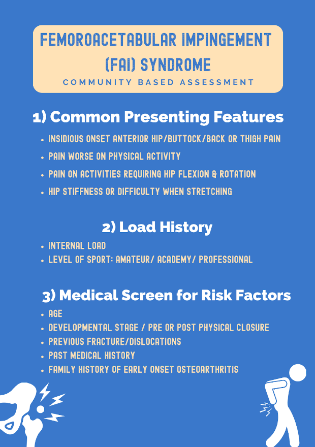
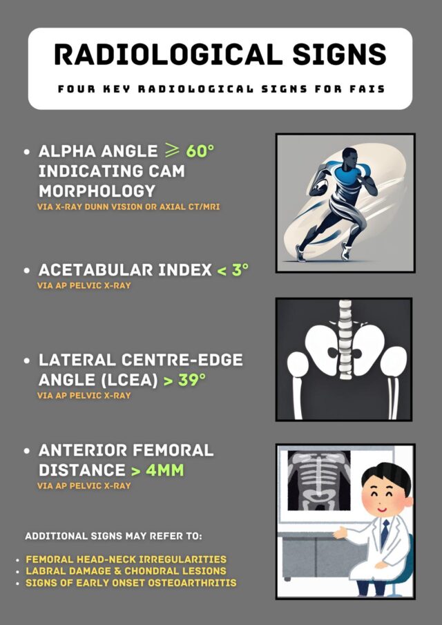
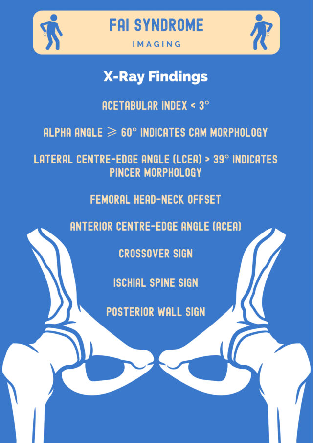
⚠️ Femoroacetabular Impingement (#FAI) Syndrome ⚠️
Dive into key clinical tests, common imaging findings, and treatment options in our latest #MSKPlaybook blog post ✅
Perfect for clinicians looking to enhance their practice! 🏃♂️
READ ➡️ bit.ly/4dmTRYm
25
72
8,318

Miguel Vega retweeted

5 Aug 2024
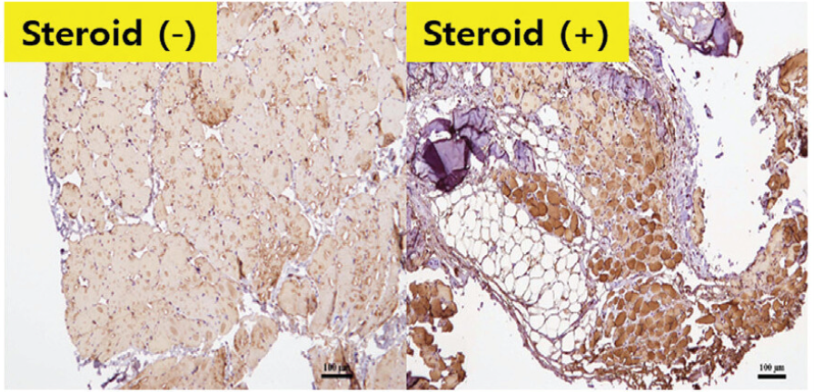
Frequent corticosteroid injection in shoulders with rotator cuff tear leads to upregulation of proteins and genes leading to adipogenesis and muscle atrophy, and downregulation of myogenic and inflammatory substances.
See more on this topic here! journals.sagepub.com/doi/ful…
19
63
7,681
Miguel Vega retweeted

5 Aug 2024
Check out the latest BackTable Brief: Anatomy and setup for genicular nerve ablation with Dr John Smirniotopoulos! #medEd @SDhandMD @JacobFlemingMD @drprestongs @Georgetown_IR open.spotify.com/episode/6I2…
1
5
15
964
Miguel Vega retweeted

28 Jul 2024
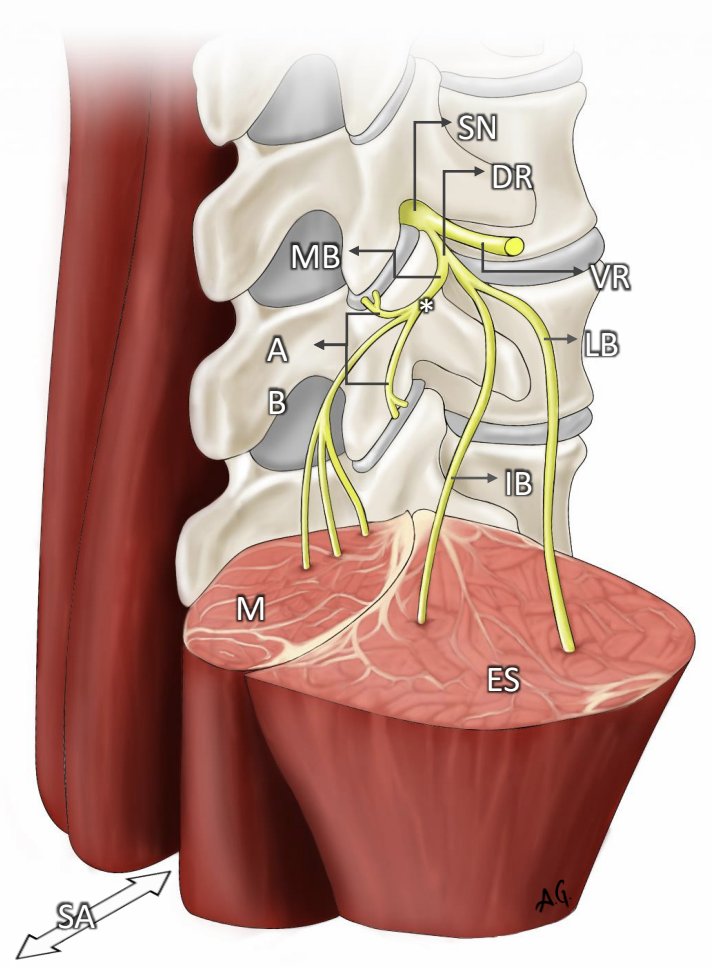
Radiofrequency denervation of the lumbar facets is the most evidenced procedure for treating facet mediated low back pain. But is it helpful or harmful?🧵
bit.ly/3Su3A6K
bit.ly/3SrQuH1
@Ryan_S_DSouzaMD @VinnyFrancioMD @ZackMcCormickMD @Sympathy4TheDr
12
22
99
27,568
Miguel Vega retweeted

28 Jul 2024
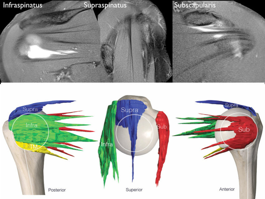
Rotator cuff muscles/tendons
☑️supraspinatus👉🏼a single tendon within a bipennate muscle
☑️subscapularis👉🏼four tendons that span the insertion
☑️infraspinatus👉🏼an oblique head (effective head depressor) & a transverse head (effective external rotator)
jisakos.com/article/S2059-77…
156
768
68,692
Miguel Vega retweeted
26 Jul 2024
Tendinopathy
Main management strategies👇🏼
✅exercise-based interventions
✅extracorporeal shock-wave therapy
✅injection-based therapy
✅surgery
Prevention 👉🏾 exercise regimens to improve strength & coordination of muscle-tendon unit
#tendon
nature.com/articles/s41572-0…

2
95
328
37,806
Miguel Vega retweeted

25 Jul 2024
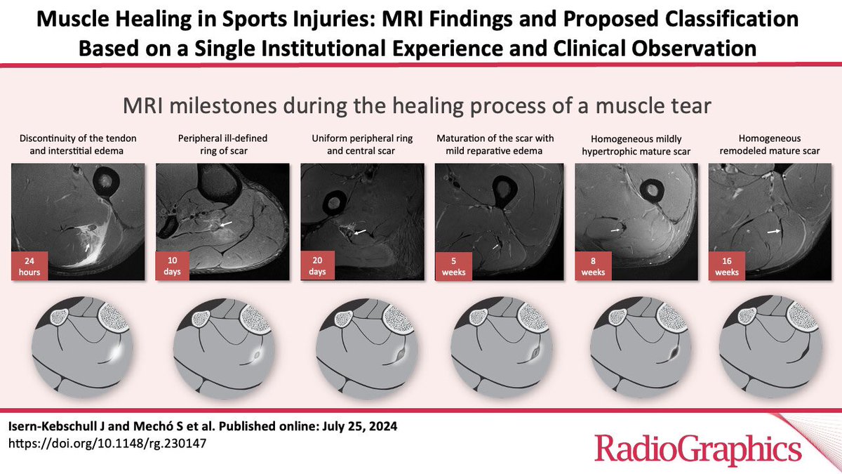
Muscle Healing in Sports Injuries: MRI Findings and Proposed Classification Based on a Single Institutional Experience and Clinical Observation | RadioGraphics pubs.rsna.org/doi/10.1148/rg…
2
27
81
8,122
Miguel Vega retweeted
25 Jul 2024
Thanks to @mechomeca @Alunaalcala @gadolinio61 @akassarjian @carlespedret @gilrodasfont et al
Muscle Healing in Sports Injuries: MRI Findings and Proposed Classification Based on a Single Institutional Experience and Clinical Observation | RadioGraphics pubs.rsna.org/eprint/ZTNVVBA…
9
18
44
5,935
Miguel Vega retweeted

1 Jul 2024
Review
Imaging of calcific tendinopathy: natural history, migration patterns, pitfalls, and management: a review
bit.ly/4bIjrpO
#Radiology
8
13
733
Miguel Vega retweeted

2 Jul 2024
Take a look at this new infographic:
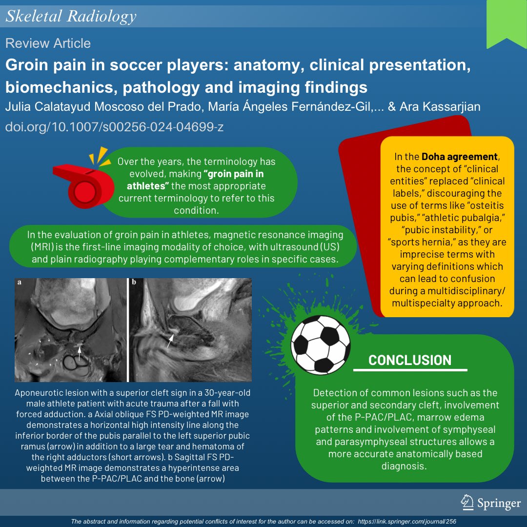
🟢 Groin pain in soccer players: anatomy, clinical presentation, biomechanics, pathology and imaging findings
Read more: rdcu.be/dK0cm
#SkeletalRadiology #MSKrad #radres #orthopedics #sportsmedicine
23
48
5,440
Miguel Vega retweeted

26 Jun 2024
Symptomatic lunotriquetral coalition type 1 of Minaar (abnormal congenital fibrous, cartilaginous, or osseous union secondary to incomplete segmentation of the tarsal or carpal bones). Can become symptomatic after trauma or, more commonly, after microtrauma in sports

1
26
80
5,826
Miguel Vega retweeted

21 Jun 2024
This study provides 12-month follow-up with Knee injury & Osteoarthritis Outcome Score & EuroQol five-dimension five-level index outcomes for saline, single & multiple platelet-rich plasma injections for patients with knee osteoarthritis.
#BJJ #Surgery
ow.ly/zZBa50SkIZY

ALT The Bone & Joint Journal. Abstract extract for article titled 'The effectiveness of leucocyte-poor platelet-rich plasma injections on symptomatic early osteoarthritis of the knee: the PEAK randomized controlled trial'.
4
43
101
86,902
Miguel Vega retweeted

19 Jun 2024
Very honored to work with an exceptional group on this new SR. pdf.sciencedirectassets.com/…
2
25
154
18,112
Miguel Vega retweeted
16 Jun 2024
Trigger finger: ultrasound diagnosis and treatment
Patient with stiffness in the first finger of the right hand, especially in the morning, which is accompanied by a sensation of clicking or crunching when moving the finger, associated with sensitivity and a lump in the palm at the level of the metacarpophalangeal joint.
An ultrasound study shows a diffuse increase in the thickness of the flexor tendon, observing the tendon snapping at the level of pulley A1 in a dynamic study when the tendon slides.
Initially, infiltration of 1 cc of trigon and 1 cc of 2% mepivacaine into the tendon sheath is performed under ultrasound control to subsequently proceed to fenestration of the pulley until the tendon is released.
Sometimes, it can be associated with physiotherapy to unload the muscles in the forearm, performing proximal peritendinous infiltration of the first finger's extensor muscle to prevent the pathology's recurrence.
3
32
134
8,959