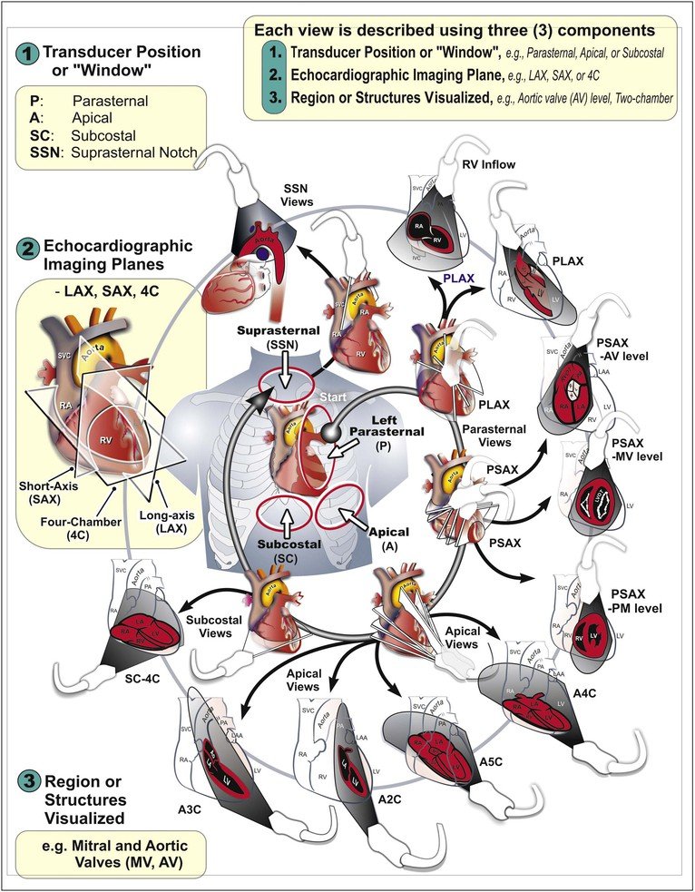


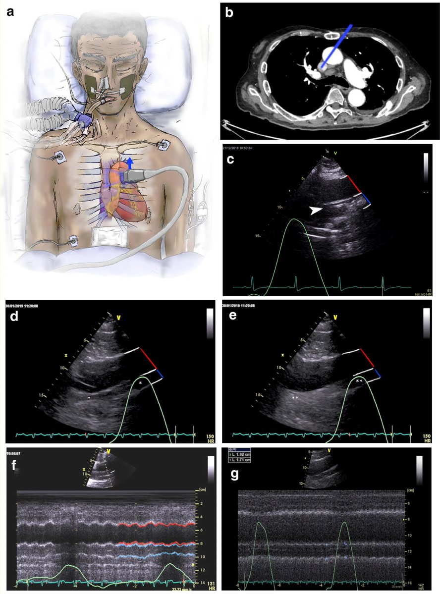
ALT a Diagram of probe position required for identification of the superior vena cava (SVC). b Chest computerized tomography showing orientation of the image plane required for identification of the SVC. c Visualization of a central venous catheter positioned in the SVC (white arrow) with the aorta indicated by a red line, the SVC by a blue line, and the vessel walls highlighted with white lines. d Visualization of the SVC and aorta without agitated saline contrast injection with the SVC and aorta indicated as in (c) and the SVC indicated by an asterisk. e Identical view of the SVC and aorta as in (d) following injection of agitated contrast injection resulting in opacification of the SVC. f M-mode visualization of the SVC demonstrating significant respirophasic diameter variation with the SVC outlined in blue and the aorta in red. g M-mode visualization of the SVC demonstrating minimal respirophasic diameter variation.