
Visual survey of surgical pathology with more than 14,000 high-quality images of benign and malignant neoplasms & related entities. Created by: @DharamRamnani
Joined July 2010
- Tweets 1,661
- Following 1,201
- Followers 14,594
- Likes 7,774
446 Photos and videos


Pinned Tweet

17 Oct 2024
Please consider giving your pathology images a forever home on WebPathology. Now you can load them directly from the site using Submit Images form. Give it a try. Webpathology.com: A Collection of Surgical Pathology Images #PathTwitter #PathX
2
9
33
6,632
May 23
I refrain from posting anything non-pathology related on this account....however, I couldn't resist sharing this with you. If you are ever in Richmond, Virginia, pay them a visit...their single malts and old-fashioned are to die for.....
1
311
Apr 8
Update in #HemePath - Intranodal Palisaded Myofibroblastoma; 15 images; check it out on the website under Hematopathology ➡️Lymph Node➡️Spindle & Vascular Proliferations #pathresidents #pathology
6
46
2,100
Apr 5
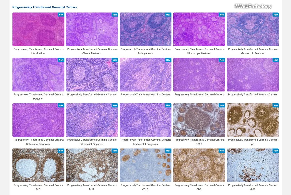
Update in #HemePath - Progressively transformed germinal centers. 20 images. Check it out on the website under Lymphadenopathies - II.
18
74
3,368
WebPathology retweeted

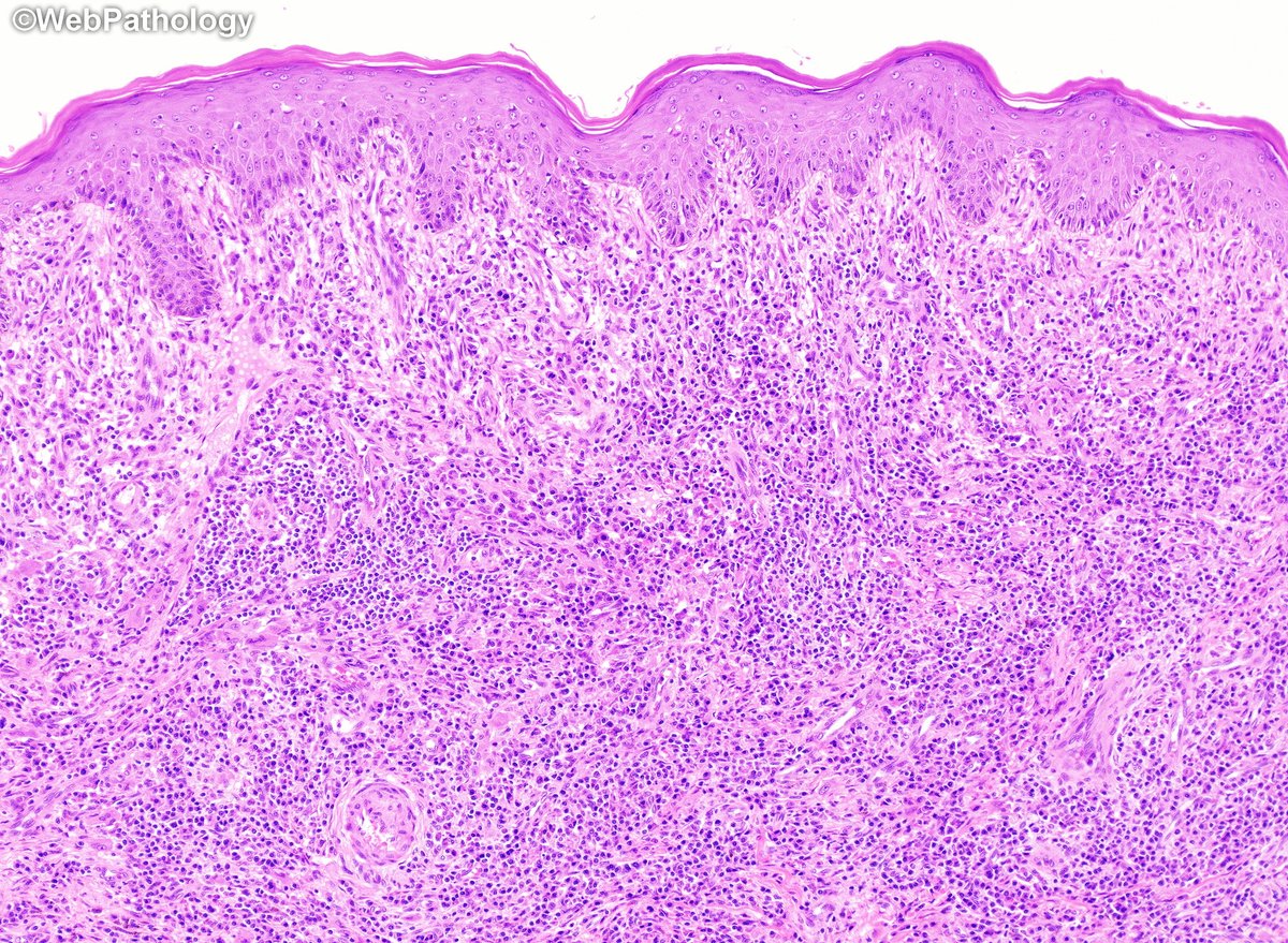
Rosai-Dorfman disease in skin -somebody please call the police - the emperipolice! An S100 protein stain highlights the nuclei (arrow) and cytoplasm of abnormal histiocytes but not the engulfed inflammatory cells in the cytoplasm (emperipolesis - engulfment without destruction).


2
48
157
6,016
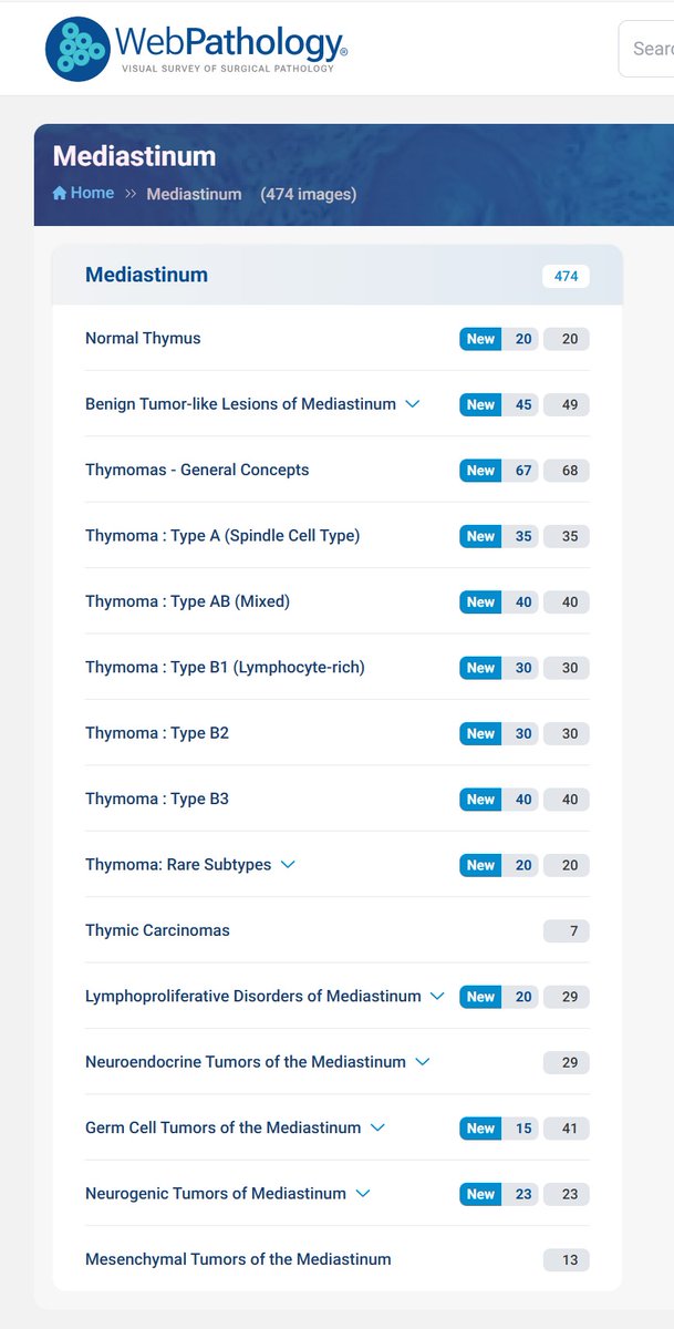
Mar 16
I’m updating mediastinum section and looking for images of T-cell lymphoblastic leukemia/lymphoma. Anyone have a case that they would like to contribute? Thanks. #hemepath
1
2
784
Mar 11
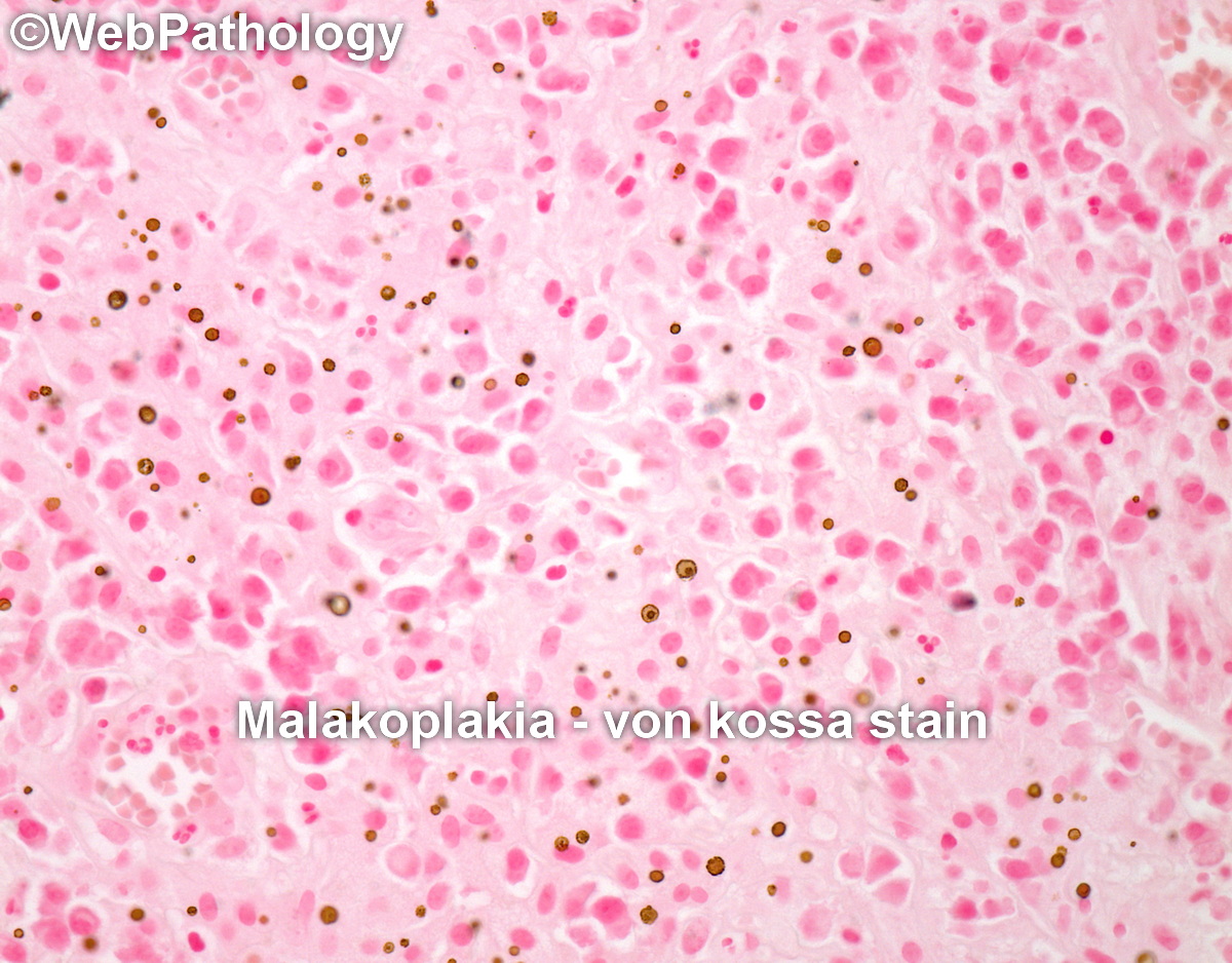
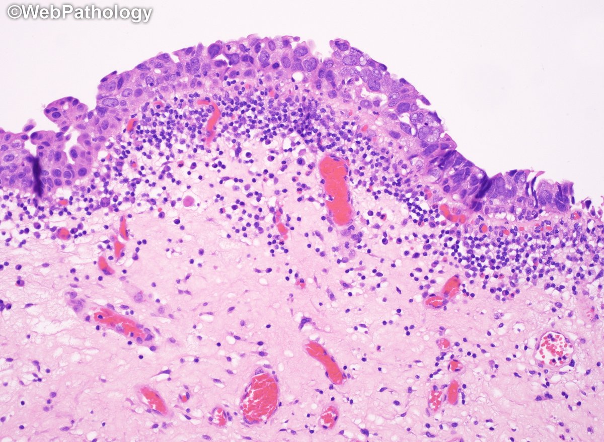
Adult female with history of high-grade uroth. CA. Surveillance cystoscopy showed multiple yellow-tan plaques near trigone. What's your diagnosis? #gupath for #pathresidents Diagnosis & additional images in the comment below.
5
11
43
2,366
Mar 11
Malakoplakia of bladder. Once you start looking for Michaelis-Guttmann bodies, they're everywhere.
7
493
Mar 5
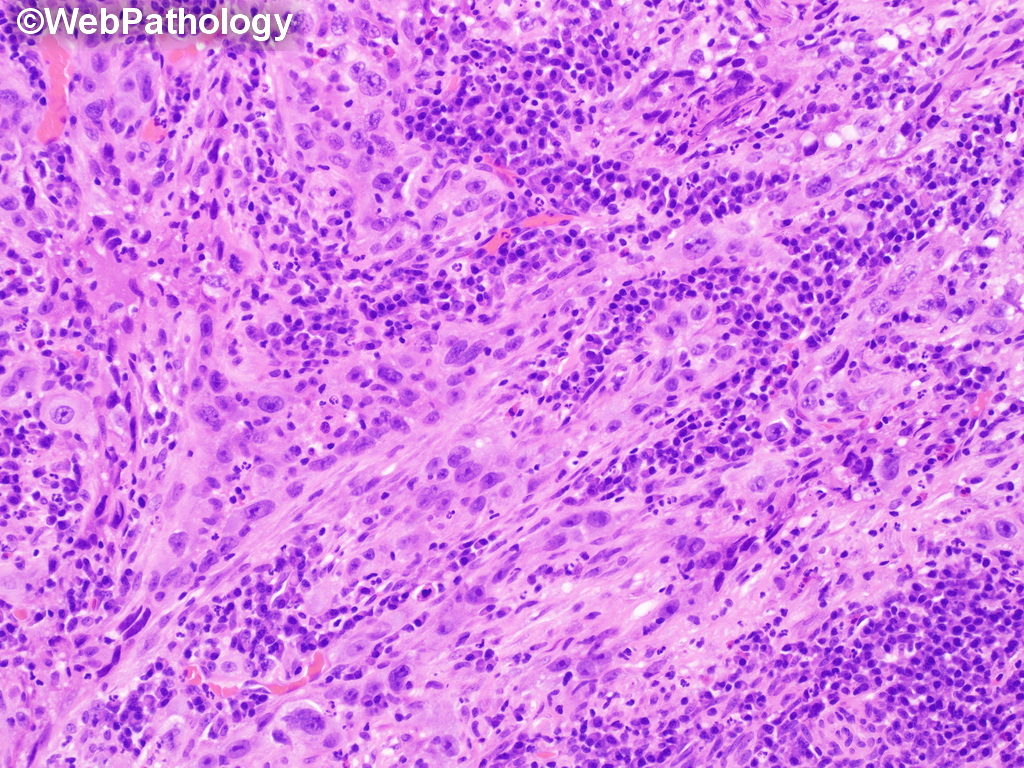
This bladder tumor had a little bit of everything....adult male with gross hematuria and a large bladder mass. #gupath
3
9
33
2,358
Mar 9
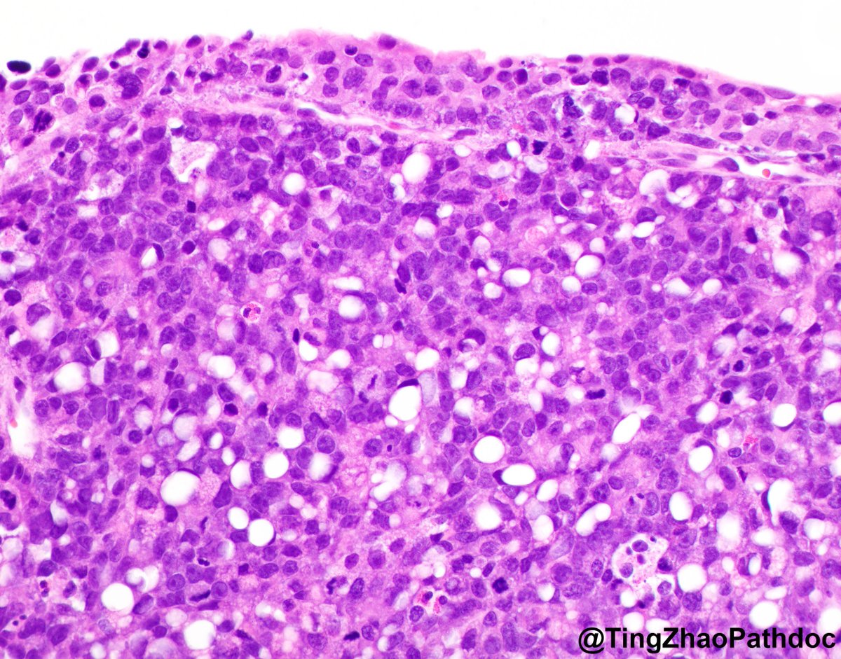
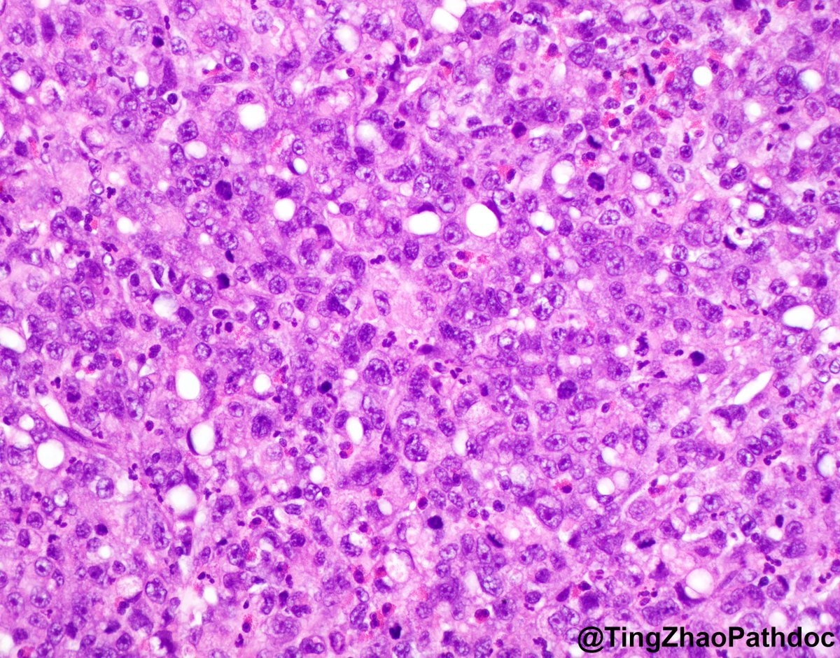
This bladder tumor had small cell neuroendocrine CA (90%) combined with conventional inv. high-grade Uroth. CA (10%), areas of CIS (flat and micropapillary) and adenoCIS. Here are additional images with immunos. #GUpath

3
242
WebPathology retweeted

Macroscopic examination of a prostate specimen, with a focus of malignancy at the extreme superior, dark yellow in color. Necropsy finding

2
9
764
WebPathology retweeted

Feb 14

11
13
61
4,983
WebPathology retweeted

Public service announcement of the day: remember that ALK alterations are not only seen in lung adenocarcinomas.
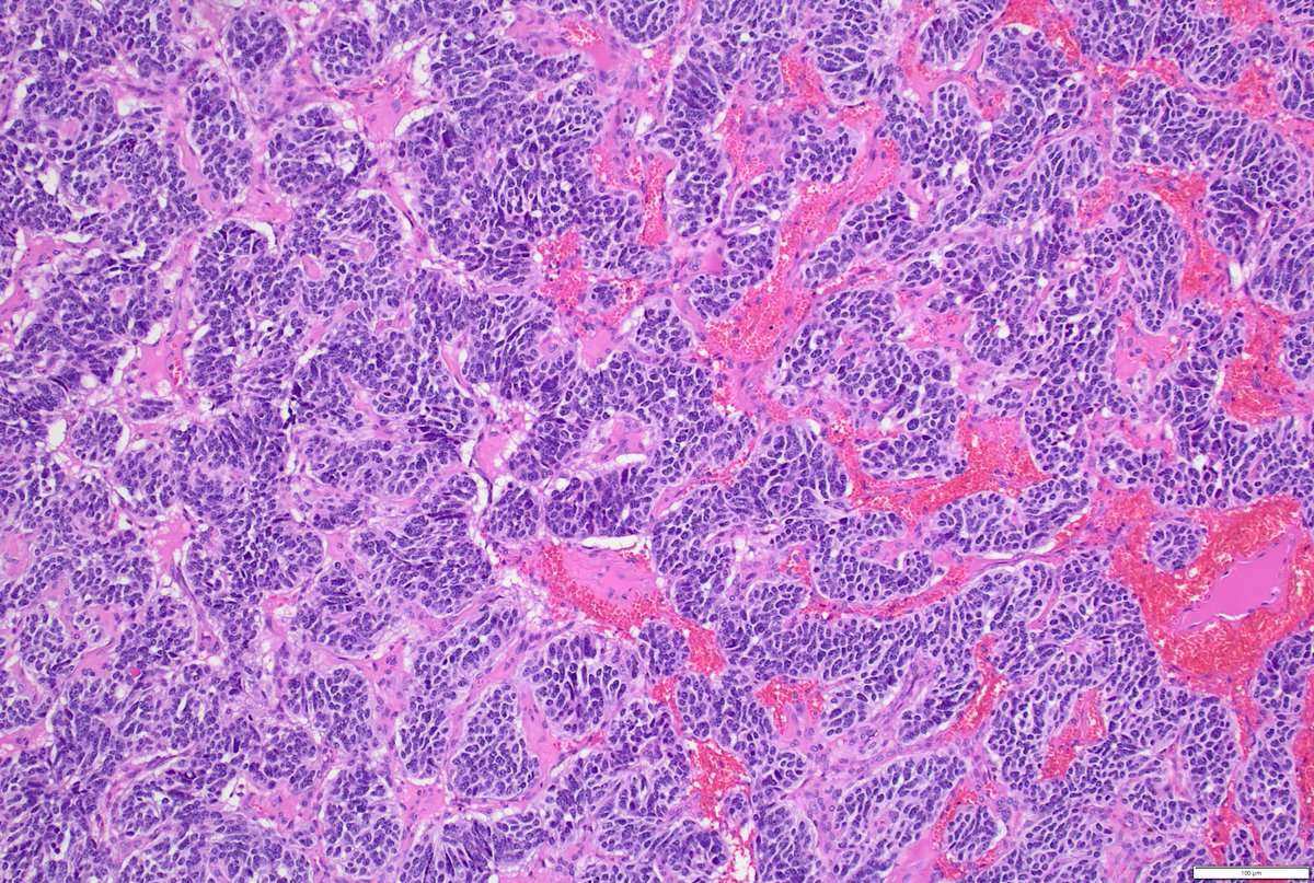
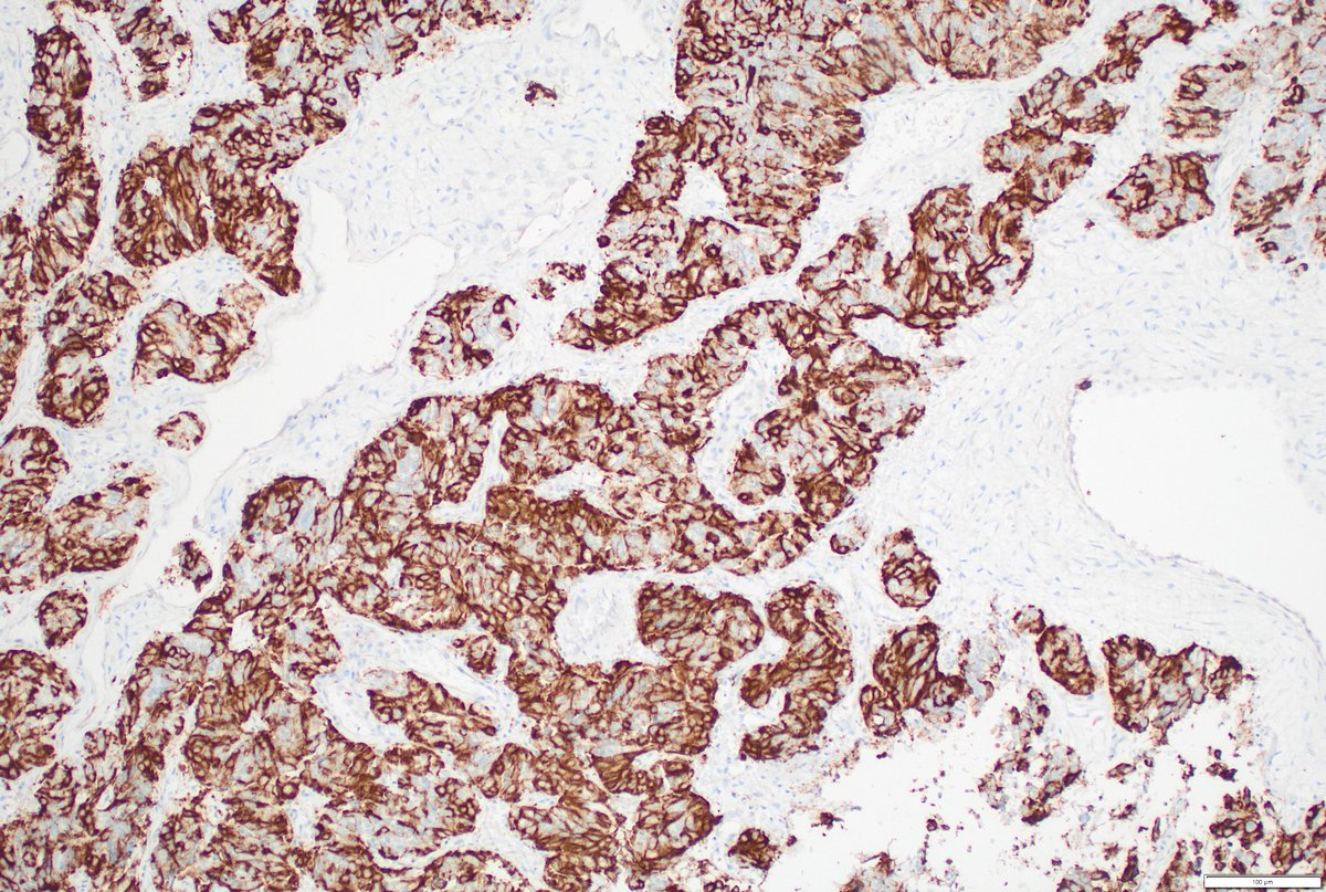
Here is an example of a carcinoid tumor with ALK fusion (extremely rare, except here at #Moffitt). We are certainly writing this one up. The IHC is ALK D5F3.
Pulmonary carcinoid tumors very rarely harbor ALK rearrangements, in contrast to non–small cell lung cancers where ALK fusions represent a well‑established oncogenic driver. The literature documents only isolated case reports, underscoring the exceptional nature of this finding. This scarcity suggests that ALK testing in carcinoid tumors is generally low yield; however, when detected, these fusions may offer meaningful therapeutic implications, particularly given the success of ALK inhibitors in other thoracic malignancies.
#lung #pathology #pulmpath #thoracicpath #MedEd #PathTwitter
17
63
2,057
WebPathology retweeted
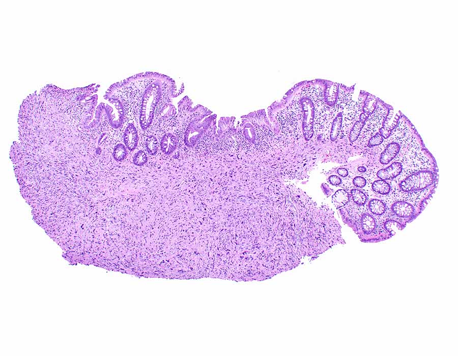
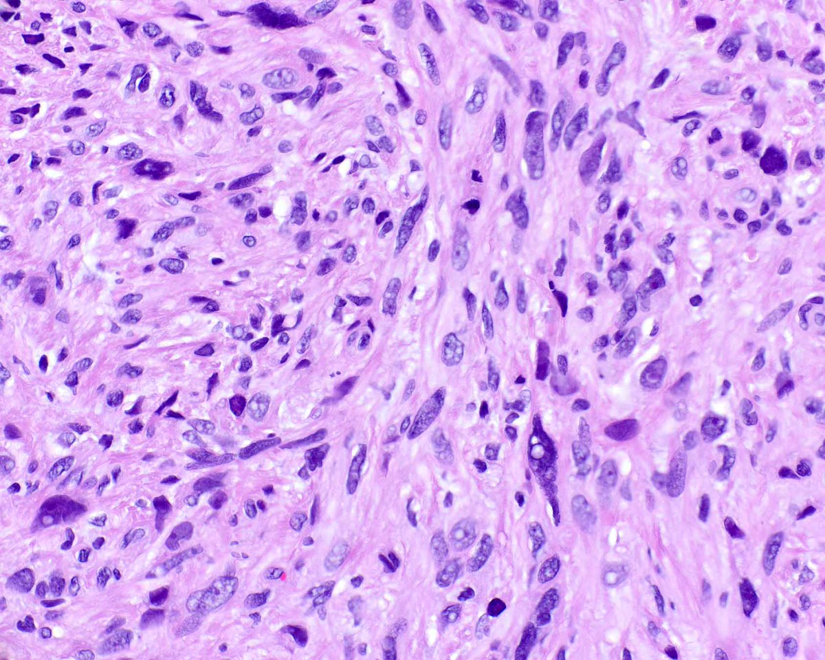
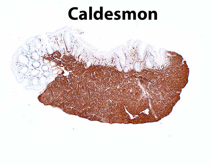
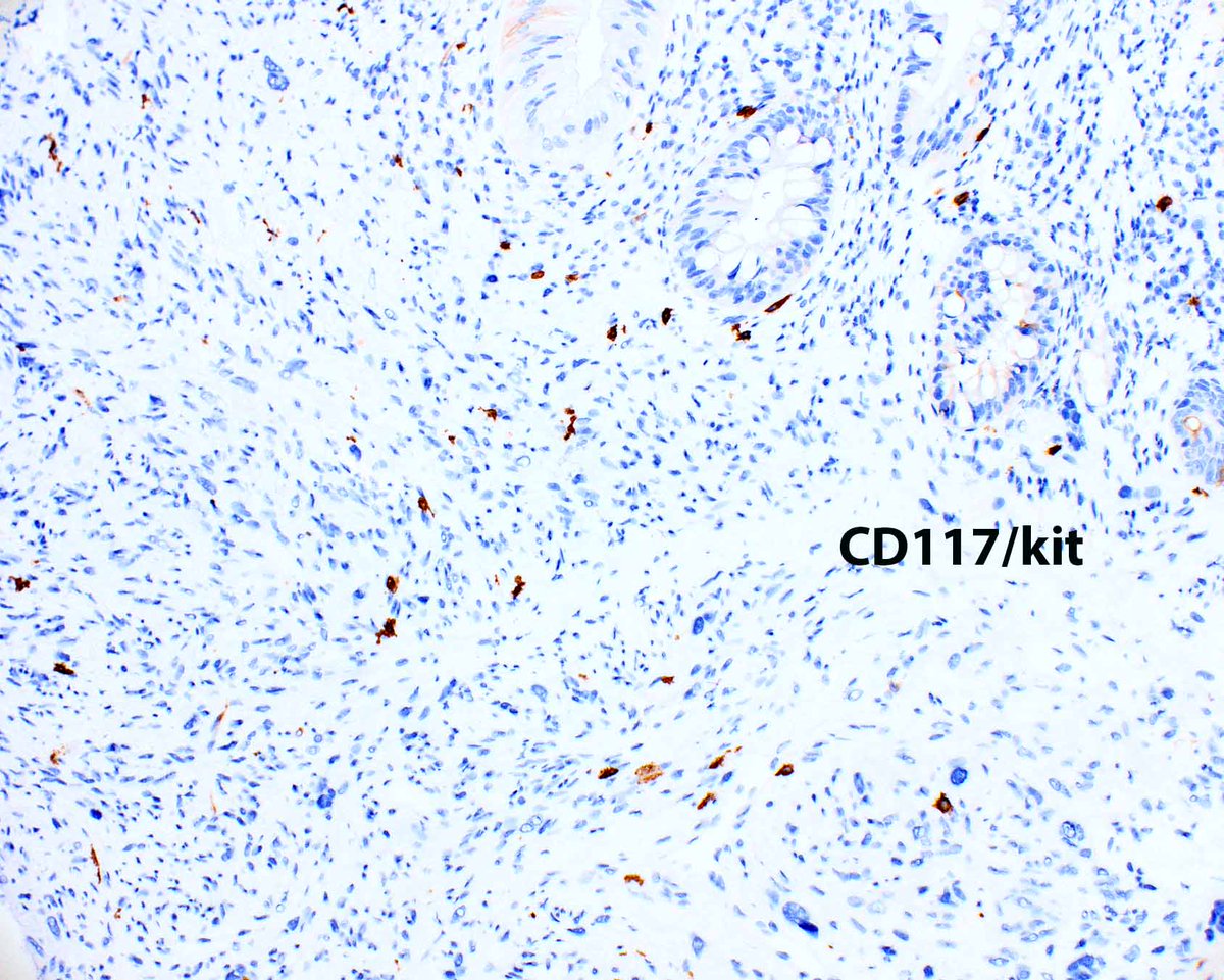
This metastatic leiomyosarcoma reached the colonic mucosa.
2
32
134
3,911
WebPathology retweeted

Jan 28
Pseudomyxoma peritonei, in the form of a high-grade mucinous peritoneal neoplasia with signet ring cells (G3). History of appendiceal mucinous neoplasm. CDX2 and Alcian Blue are shown here. #PathX #pathresidents #PathTwitter


13
35
1,291
Jan 27
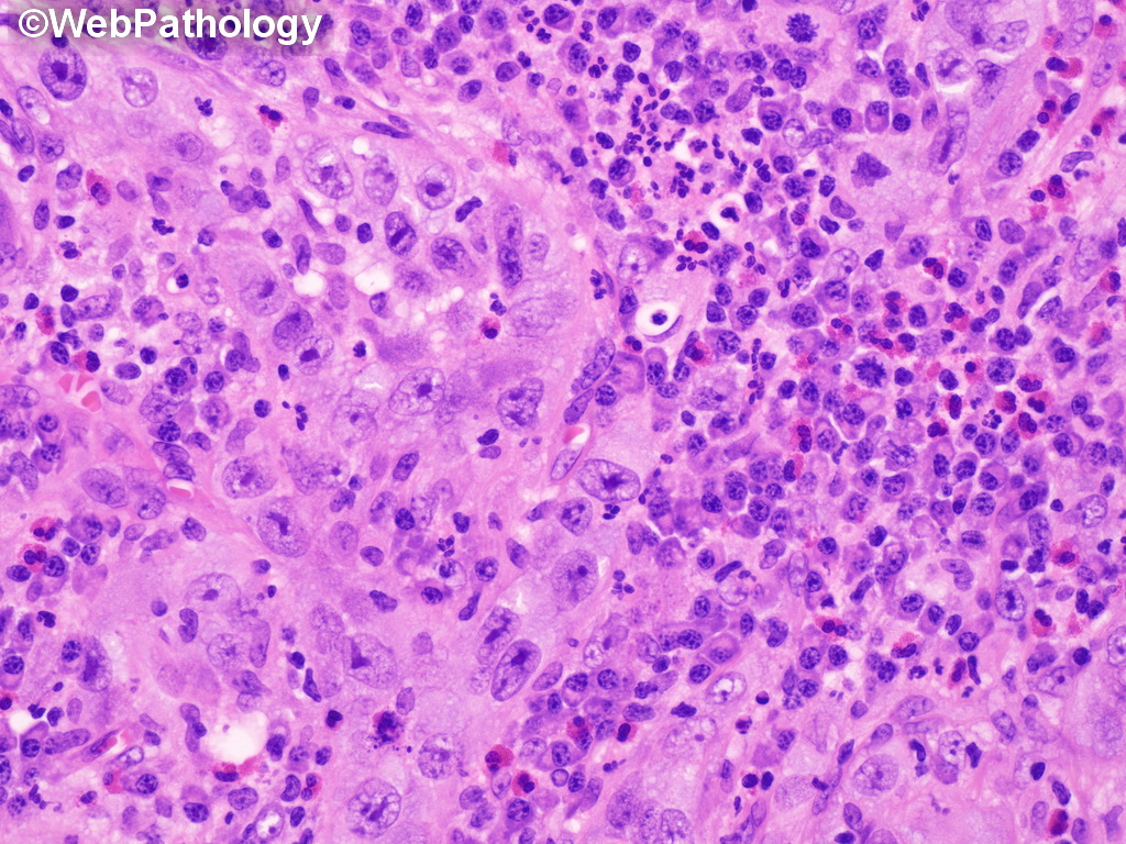
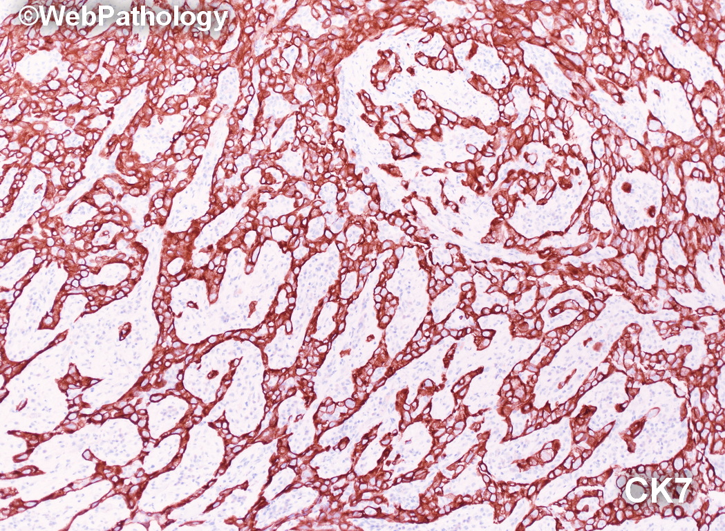
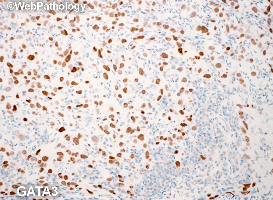
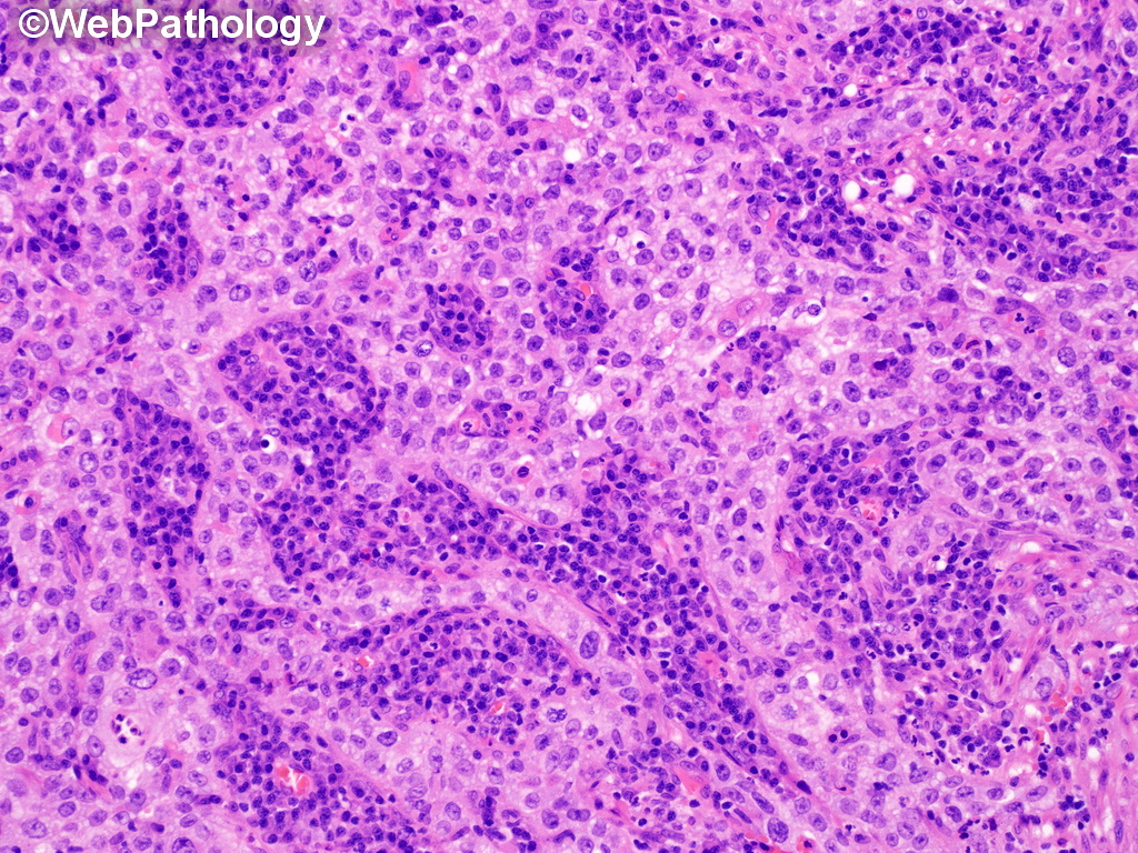
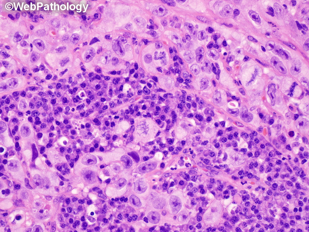
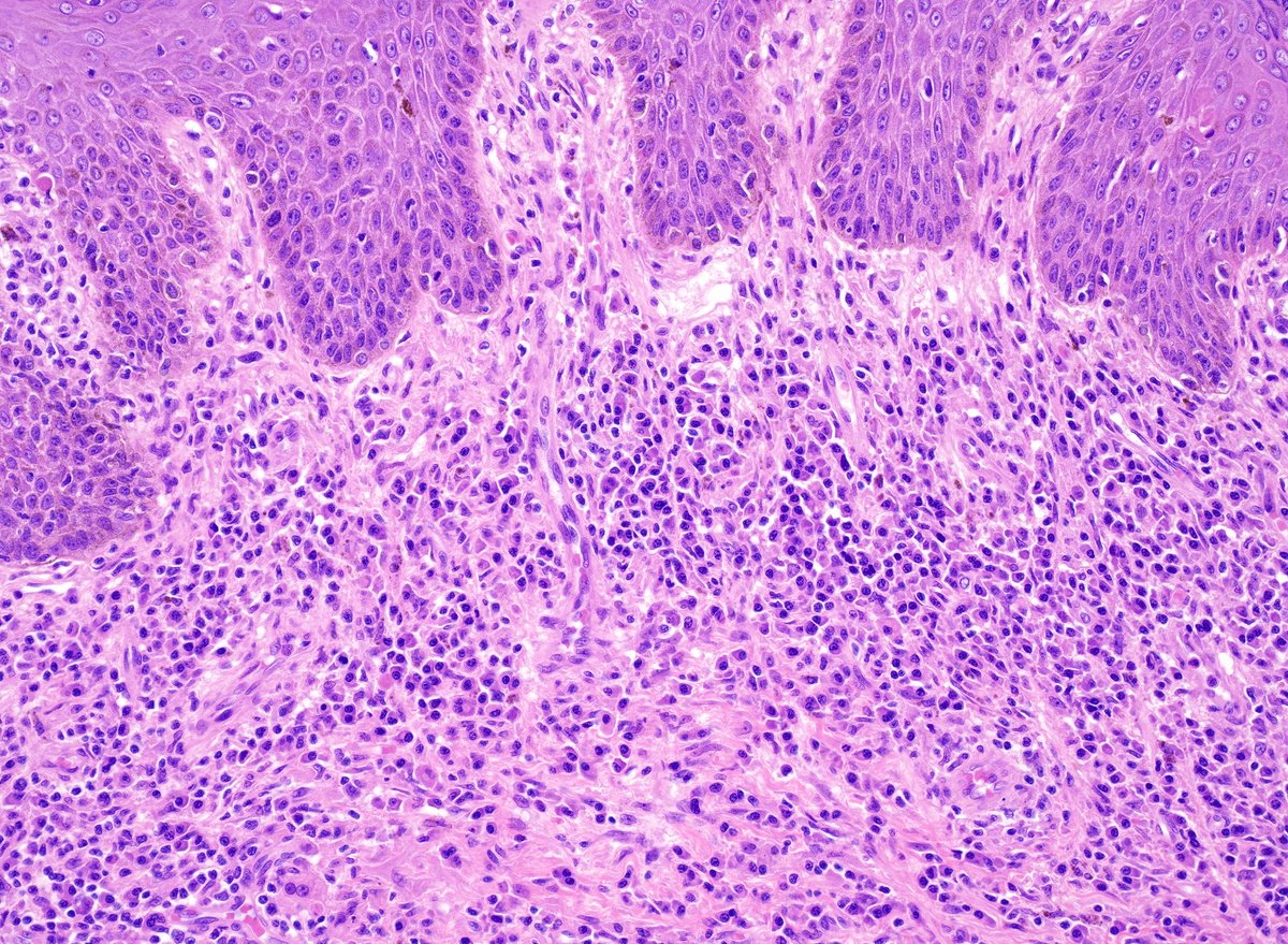
Additional images: Lymphoepithelioma-like Urothelial Carcinoma. Sheets, clusters of undiff. cells with syncytial appearance surrounded by lymphoplasmacytic infiltrate. Resembles nasopharyngeal lymphoepithelioma but lacks association with EBV or HPV. Pos for cytokeratins, GATA3.
12
36
1,603
Jan 26
Bladder mass in an elderly male. What is your diagnosis? Additional images to follow. #gupath
2
20
65
4,680


Jan 22
Adult male with a large ulcerated and indurated lesion on foreskin. What would you do next? Additional images and diagnosis will be posted later. #gupath

2
6
27
2,123
Jan 19
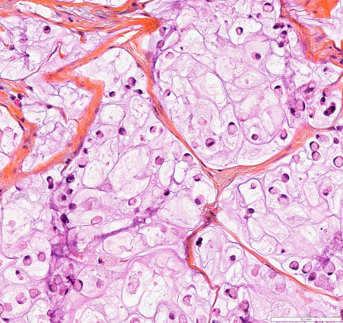
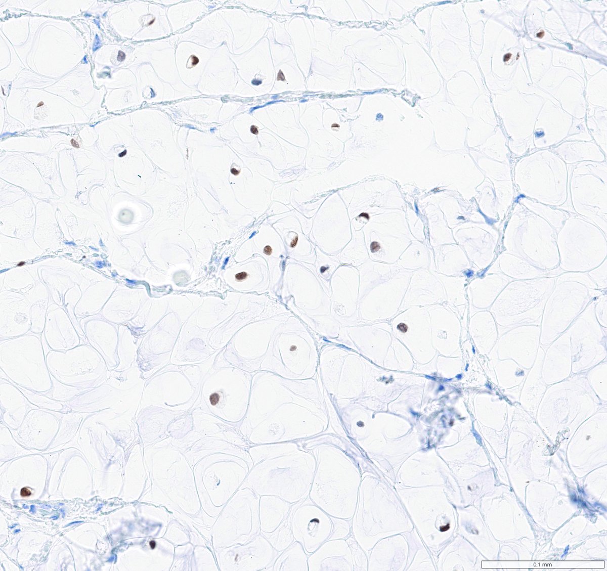
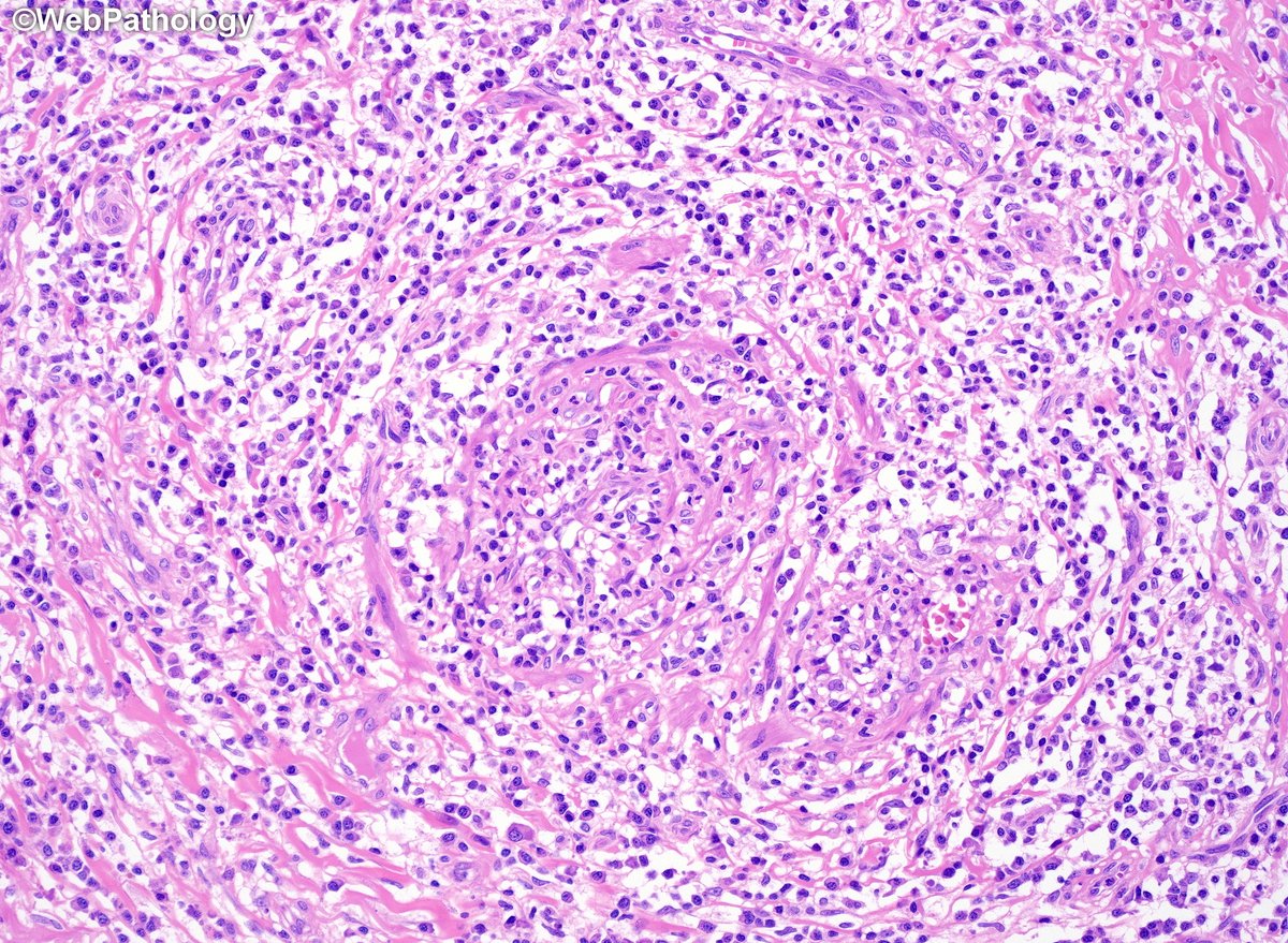
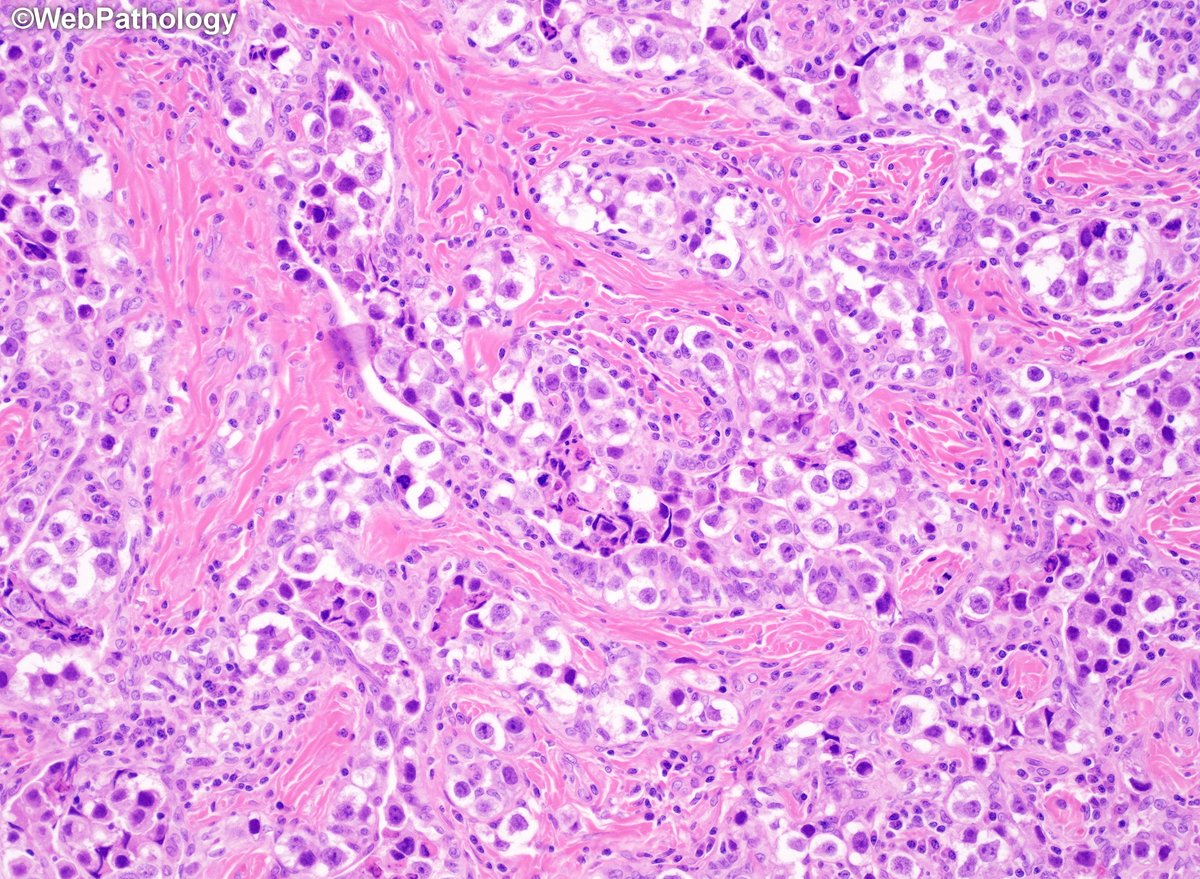
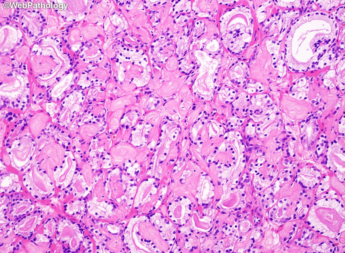
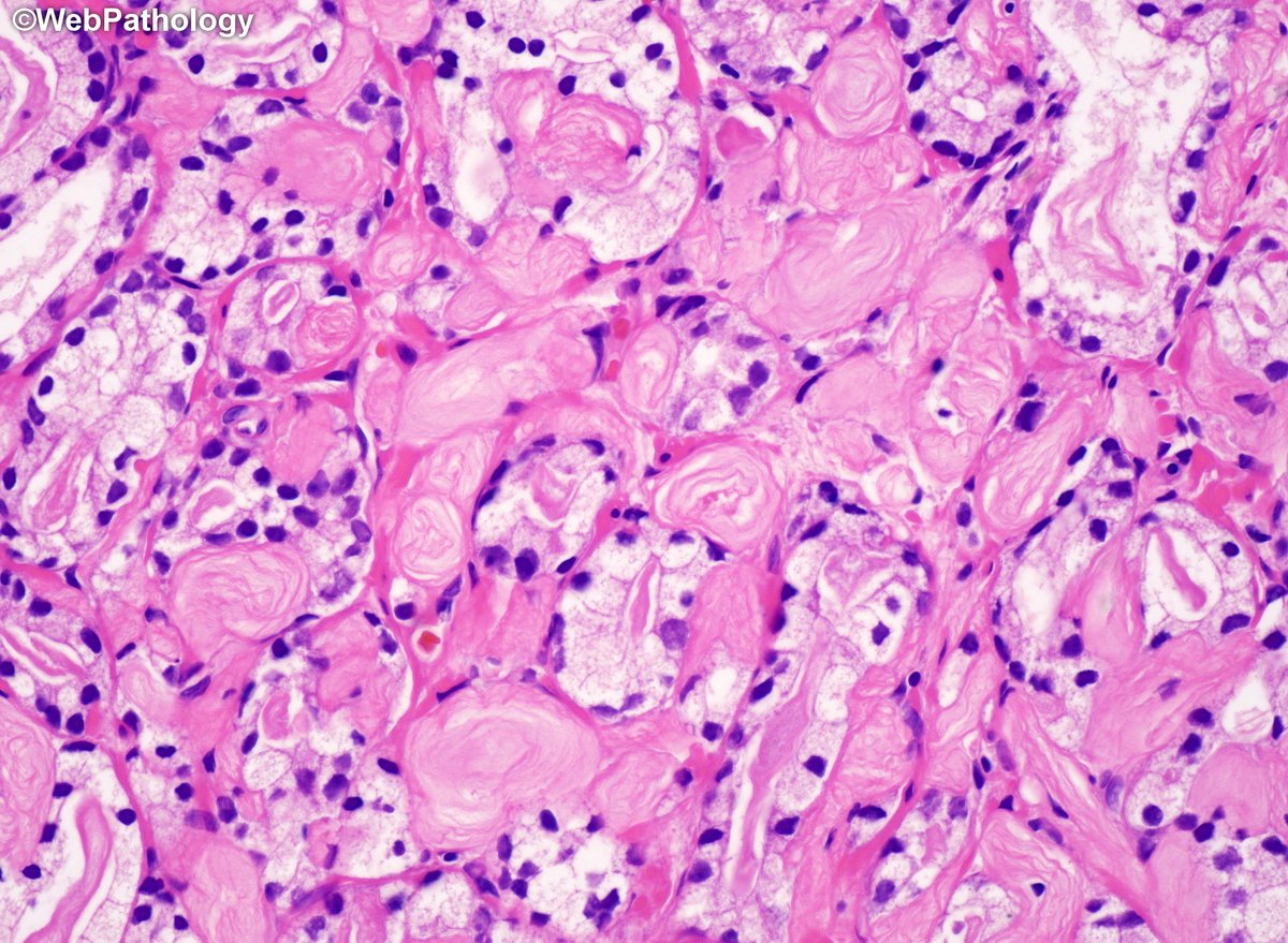
Collagenous micronodules in #ProstateCancer. A specific but infrequent finding, usually in mucin-producing CaP. Not seen with benign prostate, nodular hyperpl or high-grade PIN. #GUPath
25
62
2,780