
Cardiac anesthesiologist | Intensivist • Founder, Africa’s first Hypoxia Lab • Scalable ICU systems in Africa • Global Health • Medical Simulationist
Joined November 2012
- Tweets 1,338
- Following 393
- Followers 871
- Likes 146
17 Photos and videos


Cornelius Sendagire, MD retweeted

🫀 Septic Shock Is Not Just About Blood Pressure: It Is About Ventriculo-Arterial Coupling
For years, septic shock resuscitation has focused on restoring MAP, increasing cardiac output, and normalizing lactate.
But what if the real problem is not flow alone?
What if the heart and arterial system are no longer working together?
This is the concept of ventriculo-arterial coupling (VAC): the dynamic interaction between ventricular contractility (Ees) and arterial load (Ea). When coupling is preserved, the cardiovascular system operates efficiently, maximizing stroke work while minimizing myocardial energy expenditure.
Why VAC Matters in Septic Shock
Sepsis causes profound vasoplegia, myocardial depression, and alterations in vascular tone.
As a result, many patients develop ventriculo-arterial uncoupling, where ventricular contractility and arterial load become mismatched. This leads to:
✅ Reduced cardiovascular efficiency
✅ Increased myocardial energetic cost
✅ Impaired tissue perfusion despite apparently adequate cardiac output
✅ Variable responses to fluids, vasopressors, and inotropes
In other words:
Two patients may have the same MAP and cardiac output but completely different cardiovascular efficiency and energetic burden.
The Norepinephrine Paradox
One of the most interesting concepts highlighted by Pinsky and Guarracino is that increasing blood pressure does not always improve cardiovascular performance.
In some septic shock patients with depressed contractility:
🔹 Norepinephrine increases arterial elastance (Ea)
🔹 MAP rises
🔹 Left ventricular afterload increases
🔹 Stroke volume may fall
🔹 VAC worsens
🔹 Cardiac output may remain unchanged or even decrease
The monitor shows a better blood pressure.
The ventricle may actually be working less efficiently.
Why Some Patients Respond and Others Do Not
The review provides a physiological explanation for the heterogeneity we see every day in the ICU.
Patients with preserved contractile reserve may tolerate increased afterload and maintain efficient coupling.
Patients with septic cardiomyopathy may not.
This may explain why identical norepinephrine doses can produce dramatically different hemodynamic responses among seemingly similar septic shock patients.
Beyond Left Ventricular Function
The same principles apply to the right ventricle.
In septic patients with ARDS:
🔹 Pulmonary vascular resistance rises
🔹 RV afterload increases
🔹 RV-pulmonary artery coupling deteriorates
🔹 Venous congestion develops
🔹 Organ perfusion worsens despite acceptable systemic pressures
This reminds us that shock physiology extends far beyond MAP alone.
Clinical Takeaway
Perhaps the next evolution of septic shock management is not simply asking: "Did cardiovascular efficiency improve?"
Reference 📚
Pinsky MR, Guarracino F. Pathophysiological implications of ventriculoarterial coupling in septic shock. Intensive Care Medicine Experimental. 2023;11:87.
doi.org/10.1186/s40635-023-0…

ALT
72
192
6,754
Cornelius Sendagire, MD retweeted
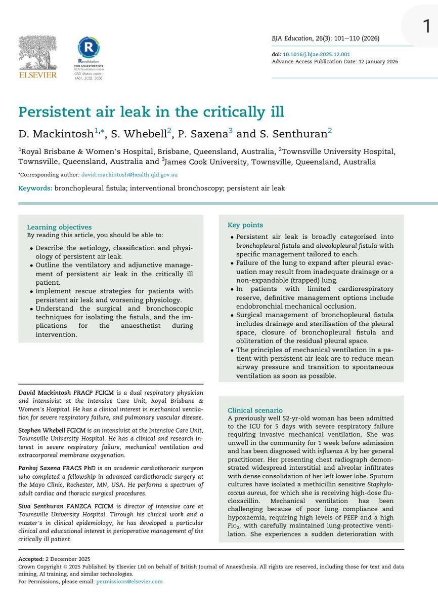
🫁 Persistent Air Leak in the ICU: Are We Treating the Fistula or Feeding It?
Few complications are as frustrating in critical care as a persistent air leak (PAL).
The patient remains hypoxemic, the chest drain keeps bubbling, the pneumothorax refuses to resolve, and every ventilator adjustment feels like a compromise.
A recent review in BJA Education provides a physiology-based framework for approaching this challenging problem.
The first key concept is understanding that not all persistent air leaks are the same.
🔹 Bronchopleural fistula (BPF): communication between a proximal airway and pleural space.
🔹 Alveolopleural fistula (APF): communication originating from distal airways or alveoli, often associated with pneumonia, ARDS, trauma, or diffuse lung injury.
The article emphasizes a principle that every intensivist should remember:
Air leak flow is largely driven by mean airway pressure (Pmean/Pmaw).
The higher the mean airway pressure, the greater the airflow through the fistula and the harder it becomes for the defect to heal.
This creates a therapeutic dilemma:
We need sufficient airway pressure to maintain oxygenation, but excessive pressure may perpetuate the air leak.
The recommended ventilatory strategy therefore focuses on:
✅ Lowest feasible PEEP
✅ Lowest feasible tidal volume and inspiratory pressure
✅ Short inspiratory time
✅ Lower respiratory rate when tolerated
✅ Permissive hypercapnia when appropriate
✅ Early transition to spontaneous breathing and extubation whenever possible
Interestingly, the authors challenge a common reflex in ICU practice:
Routine chest tube suction may not always help.
Although suction can maintain lung expansion in large leaks, excessive suction may increase the pressure gradient across the fistula and potentially delay healing. When suction is required, the lowest effective level should be used.
For refractory cases, modern management extends beyond conventional drainage:
🔹 Endobronchial valves
🔹 Bronchoscopic mechanical occlusion
🔹 Lung or lobar isolation with bronchial blockers or double-lumen tubes
🔹 VV-ECMO allowing ultra-protective ventilation or even temporary apnea to facilitate fistula healing
Perhaps the most important message is that persistent air leak is not merely a chest drain problem.
It is fundamentally a problem of respiratory physiology, pleural mechanics, and multidisciplinary decision-making, requiring close collaboration between intensivists, respiratory physicians, interventional pulmonologists, thoracic surgeons, and anesthesiologists.
Reference 📚
Mackintosh D, Whebell S, Saxena P, Senthuran S. Persistent air leak in the critically ill. BJA Education. 2026;26(3):101–110. DOI: 10.1016/j.bjae.2025.12.001.
ALT
30
96
3,777
Cornelius Sendagire, MD retweeted

May 26
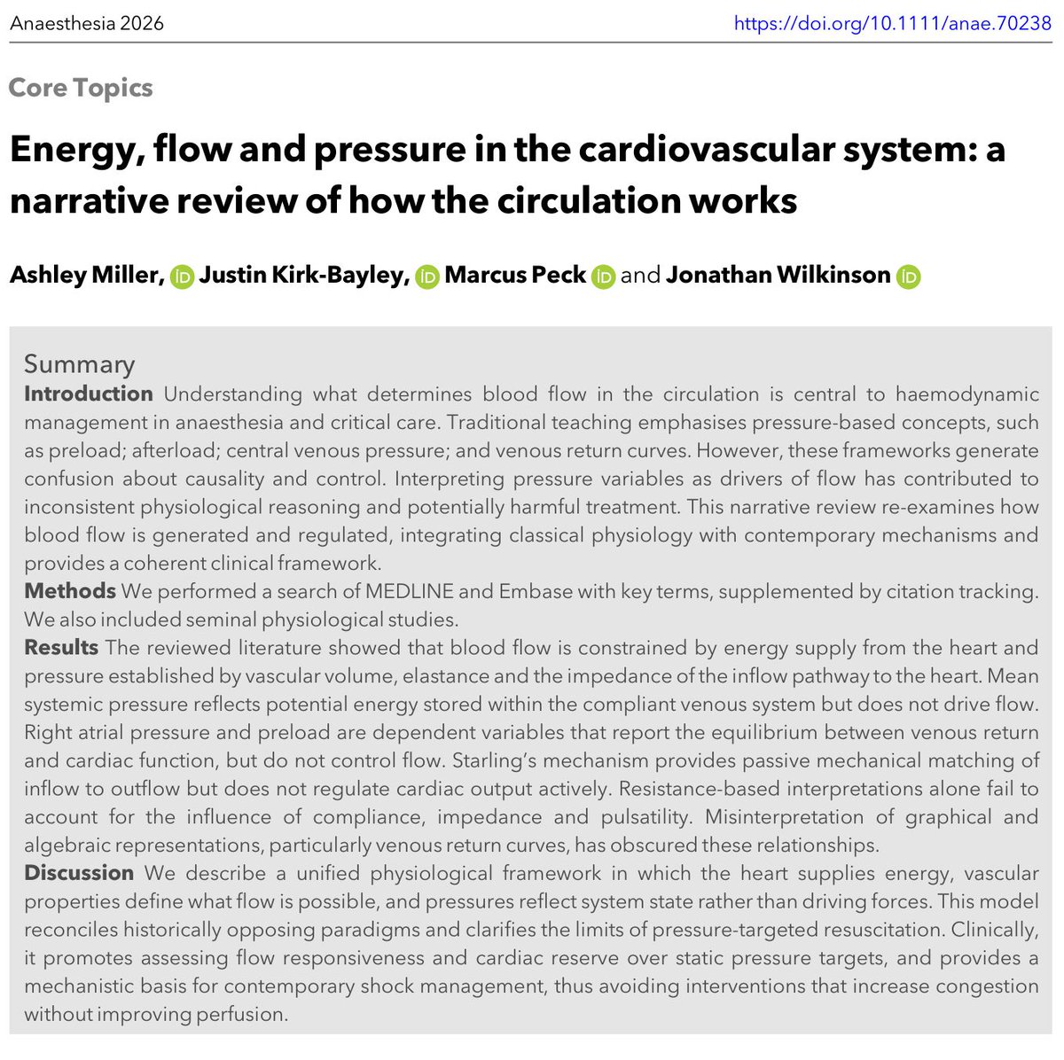
Haemodynamic equations are useful. But they also mislead.
Take:
CO = HR × SV
CO ≈ (MAP − RAP) / SVR
Both are mathematically true. But they can make the variables they contain look like the controllers of output.
Often they are not.
In the intact circulation, these equations describe the resolved state of the system. They do not, by themselves, tell you what is supplying energy, what is constraining flow, or what is actually limiting output.
That is one of the central themes of our review:
Energy, flow and pressure in the cardiovascular system: a narrative review of how the circulation works.
doi.org/10.1111/anae.70238
10
95
295
18,597
Cornelius Sendagire, MD retweeted
🫀📈 One of the most controversial debates in critical care may be shifting again.
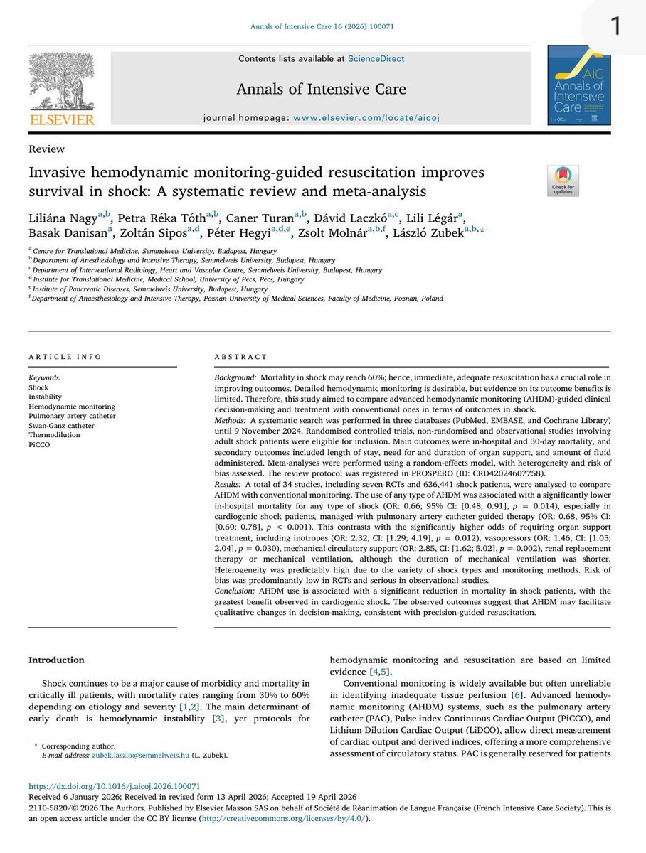
This new 2026 systematic review and meta analysis in Annals of Intensive Care challenges that narrative directly.
The study analyzed:
📊 34 studies
📊 636,441 shock patients
📊 PAC, PiCCO and advanced hemodynamic monitoring guided resuscitation strategies
Main finding: ✅ significant reduction in in hospital mortality with advanced hemodynamic monitoring guided management (OR 0.66)
The strongest signal appeared in:
⚠️ cardiogenic shock particularly with pulmonary artery catheter guided therapy.
One of the most interesting physiological observations:
Patients monitored with advanced hemodynamic systems received:
• more vasopressors
• more inotropes
• more mechanical circulatory support
• more RRT
Yet mortality was LOWER.
That is extremely important.
This suggests the benefit may not come from the device itself, but from:
🧠 earlier recognition of instability
🧠 physiology informed escalation
🧠 more precise therapeutic targeting
In other words: better decision making.
The paper strongly supports a concept many intensivists intuitively recognize at bedside:
Not all shock is “vasoplegia plus fluids.”
Different hemodynamic phenotypes require:
• different vasoactive strategies
• different fluid approaches
• different escalation timing
• different mechanical support thresholds
Advanced monitoring may allow clinicians to move away from: “one size fits all resuscitation.”
Another important nuance:
The mortality benefit was strongest in cardiogenic shock.
The evidence in septic shock remains less definitive, although trends still favored advanced monitoring.
This may reflect an important reality: cardiogenic shock is fundamentally a hemodynamic disease.
One particularly valuable message from this paper:
The authors emphasize that modern AHDM is not simply “placing a Swan Ganz catheter.”
It is:
📌 integrating dynamic physiology
📌 interpreting perfusion targets
📌 understanding ventricular interactions
📌 identifying fluid responsiveness limitations
📌 tailoring escalation
Technology without physiology remains insufficient.
Interesting practical point:
The analysis did NOT show major increases in serious complications related to advanced monitoring devices.
That matters because procedural fear has been one of the strongest arguments against invasive monitoring.
My personal takeaway:
Critical care may be entering a new era where: precision hemodynamics returns to the center of shock resuscitation.
Not because catheters are fashionable again because modern shock management increasingly requires individualized physiology rather than protocolized averages.
📖 Reference
Nagy, L., Tóth, P. R., Turan, C., et al. (2026). Annals of Intensive Care, 16, 100071. doi.org/10.1016/j.aicoj.2026…
ALT
3
57
182
8,721
Cornelius Sendagire, MD retweeted
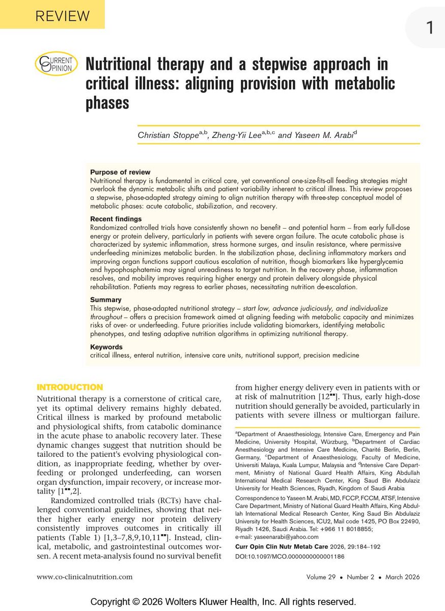
🍽️ ICU nutrition: we’ve been doing it wrong for years?
More calories
More protein
Earlier feeding
Sounds logical
But physiology disagrees
⚠️ The key update
High-quality RCTs now show:
❌ Early full-dose nutrition
→ NO benefit
→ Potential harm
Especially in:
• Shock
• Multiorgan failure
• High metabolic stress
👉 This is not neutral
👉 This is dangerous practice
🧠 Critical illness is not static
It evolves through metabolic phases:
1. Acute catabolic phase
2. Stabilization phase
3. Recovery phase
👉 Feeding must follow physiology
Not protocols
🔥 Phase 1: Acute catabolic
• High inflammation
• Insulin resistance
• Endogenous substrate mobilization
👉 The body is NOT ready for full nutrition
💡 Strategy:
✔️ Permissive underfeeding
✔️ Low protein
Why?
Because early overload leads to:
• Hyperglycemia
• Hepatic dysfunction
• Renal stress
• Impaired autophagy
👉 You are feeding dysfunction, not recovery
⚖️ Phase 2: Stabilization
• Inflammation decreasing
• Organ function improving
👉 Now metabolism starts to tolerate nutrition
💡 Strategy:
✔️ Slow escalation
✔️ Daily reassessment
⚠️ Red flags:
• Hypophosphatemia
• Hyperglycemia
→ Patient is NOT ready
💪 Phase 3: Recovery
• Anabolism returns
• Mobility improves
👉 NOW nutrition matters most
💡 Strategy:
✔️ Higher calories
✔️ Higher protein
✔️ Combine with rehab
👉 This is where you rebuild muscle and function
📊 The real takeaway
Nutrition is NOT:
❌ A fixed prescription
❌ A calorie target
It is:
✔️ A dynamic therapy
✔️ A metabolic intervention
🎯 The new principle
“Start low
Advance judiciously
Individualize throughout”
👉 Precision ICU nutrition
⚠️ Final thought
Overfeeding early harms
Underfeeding late harms
👉 Timing is everything
📚 Stoppe C et al. Curr Opin Clin Nutr Metab Care 2026
DOI: 10.1097/MCO.0000000000001186
ALT
44
117
5,857
Cornelius Sendagire, MD retweeted

In healthcare, the simplest actions save the most lives!
Congrats to our Theatre team for their outstanding Hand Hygiene Performance!
Their dedication to international safety standards helps prevent infections and keeps our patients safe.
#ActionsSaveLives #HandHygiene


2
7
29
2,249
Cornelius Sendagire, MD retweeted
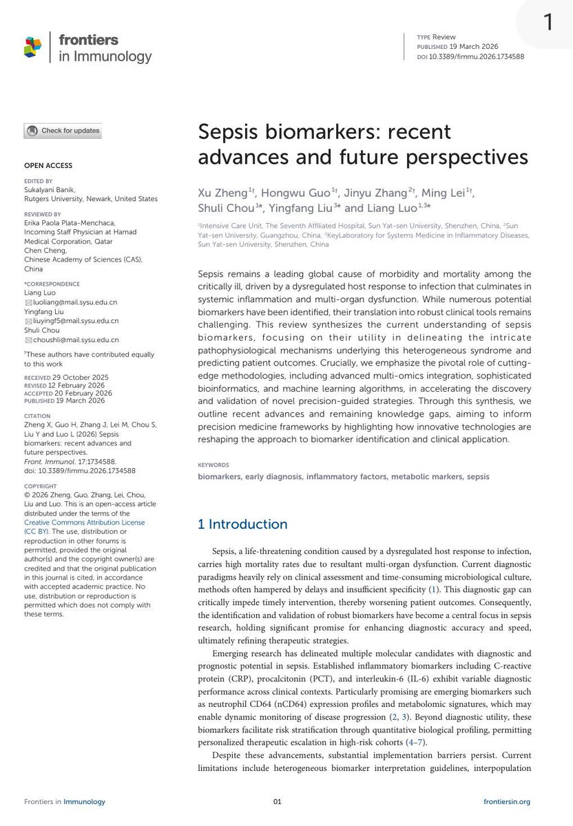
🧬 Sepsis biomarkers: are we finally moving beyond CRP and PCT?
Sepsis is not just infection.
It is a dysregulated host response leading to multi-organ dysfunction.
And yet…
👉 Our diagnostic tools are still slow
👉 Our biomarkers are still imperfect
🧠 The current reality
We rely on:
• CRP → sensitive but non-specific
• PCT → better for bacterial infection, useful for antibiotic guidance
• Lactate → cornerstone for perfusion and prognosis
➡️ But none of them truly capture the complex biology of sepsis
📊 What is changing?
New biomarkers are emerging across multiple domains:
🔥 Inflammatory markers
• IL-6 → correlates with severity and mortality
• IL-10 → reflects immunosuppression
• TNF-α → early hyperinflammatory signal
⚙️ Immune cell markers
• CD64 → helps differentiate bacterial infection
• Presepsin → early diagnostic and prognostic potential
🧪 Metabolic markers
• Lactate kinetics still critical
• Metabolomics → early prediction of shock progression
🧬 The real revolution: multi-omics AI
👉 Transcriptomics
👉 Proteomics
👉 Metabolomics
Combined with:
🤖 Machine learning
➡️ Allowing:
• Sepsis phenotyping (endotypes)
• Hyperinflammatory vs immunosuppressed states
• Personalized risk stratification
🚨 Critical insight
Sepsis is:
❌ Not one disease
❌ Not one pathway
❌ Not one biomarker
➡️ It is a dynamic, evolving biological network
⚠️ Why biomarkers still fail in real life
• Heterogeneity between patients
• Variable kinetics over time
• Lack of standardized thresholds
• Overreliance without clinical context
➡️ Biomarkers alone will NEVER replace clinical reasoning
🔥 Take-home message
The future is not:
👉 “Which biomarker is best?”
The future is:
➡️ Integrated biomarker panels physiology .... AI?
📚 Zheng X. et al. (2026)
Frontiers in Immunology
doi.org/10.3389/fimmu.2026.1…
ALT
1
54
174
8,313
Cornelius Sendagire, MD retweeted

Apr 30
Congratulations to Prof Doruk Ozgediz for the UCSF Exceptional Physician Award! Many Ugandan babies and paediatric surgeons have witnessed his exceptional dedication to surgical (and anaesthesia too!) care. Well deserved! @UCSF_CHESA @ASOU_Official @KidsOperating @stellahalyce

7
20
514
Cornelius Sendagire, MD retweeted
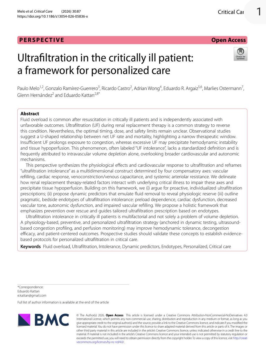
💧 Ultrafiltration in the ICU is not fluid removal…It is hemodynamic stress testing.
🚨 New perspective in critical care:
👉 Ultrafiltration (UF) is a double-edged sword
👉 The relationship with outcomes is U-shaped
📉 Too slow → persistent congestion
📈 Too fast → hypoperfusion & organ injury
🎯 The safe zone is narrow and patient-specific
🧠 We have been thinking about UF incorrectly
❌ “UF intolerance = hypovolemia”
👉 This is wrong
🚀 UF intolerance is MULTIDIMENSIONAL
Defined by failure of 4 physiological axes:
▪️ Vascular refilling
▪️ Cardiac response
▪️ Venous tone / capacitance
▪️ Arteriolar resistance
⚡ Key concept
👉 UF is not just removing fluid
👉 It is testing physiological reserve
🔥 Why patients crash during UF
Not just volume…
🧩 5 clinical endotypes
1. Preload dependence
2. Cardiac dysfunction
3. Vasoplegia (↓ vascular tone)
4. Autonomic dysfunction
5. Low vascular refill
👉 Different mechanism = different treatment
⚠️ Same hypotension ≠ same problem
Giving fluids to all = mistake
Stopping UF always = mistake
🧬 Practical bedside shift
Before UF:
👉 Don’t ask “how much fluid to remove?”
👉 Ask “can this patient tolerate removal?”
🛠️ New tools proposed
▪️ Passive leg lowering (reverse PLR)
▪️ UF challenge (mini fluid removal test)
▪️ Perfusion markers (CRT, PI)
▪️ Multimodal POCUS (LUS VExUS cardiac)
🔥 Major clinical implication
👉 UF should be:
✔️ Dynamic
✔️ Personalized
✔️ Preventive (not reactive)
🚀 Paradigm shift
We mastered fluid resuscitation with physiology…
👉 Now it’s time to master fluid removal the same way
⚠️ Take-home
UF is not dialysis mechanics
👉 It is cardiovascular physiology under stress
📚 Melo et al. Critical Care 2026
doi.org/10.1186/s13054-026-0…
ALT
2
45
126
7,956
Cornelius Sendagire, MD retweeted

STATEMENT FROM ALEXANDRA MEDICAL CENTER
723
262
1,319
212,427
Cornelius Sendagire, MD retweeted
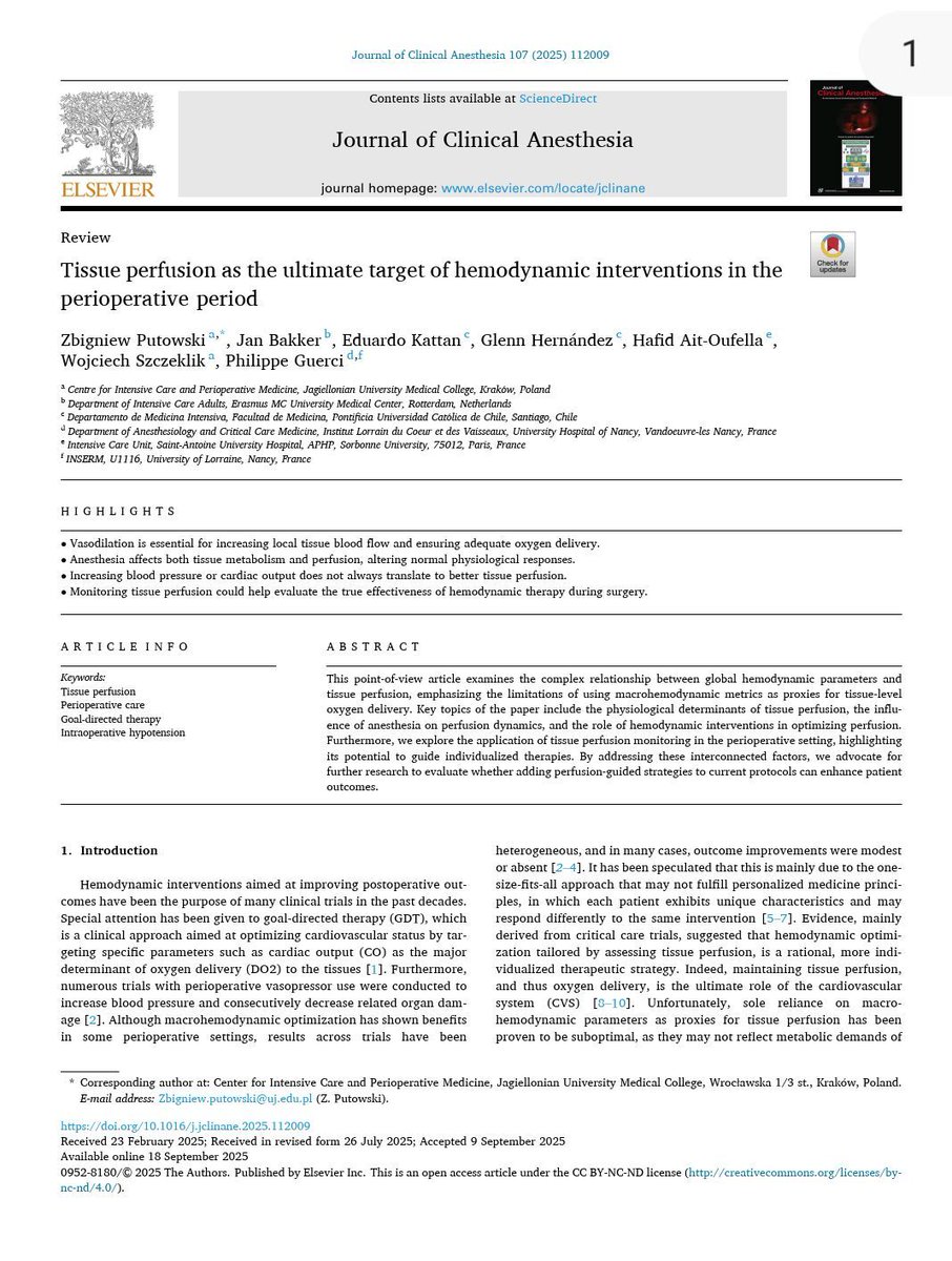
🫀 Are we treating numbers… or perfusion?
A compelling perspective from perioperative physiology challenges a core assumption in anesthesia and critical care:
👉 Optimizing MAP or CO does NOT guarantee adequate tissue perfusion.
🔬 Physiological reality: macro ≠ micro
▪️ Tissue perfusion is primarily driven by metabolic demand and local vasoregulation, not just systemic pressure
▪️ Arteriolar vasodilation and capillary recruitment are the real determinants of oxygen delivery
▪️ Increasing MAP via vasopressors may restore pressure but impair flow, especially at the microcirculatory level
⚠️ Anesthesia changes the rules
▪️ ↓ VO₂ (around 25%) alters the DO₂-VO₂ relationship
▪️ Volatile agents → vasodilation uncoupling of metabolism and perfusion
▪️ Blunted autonomic responses → dependence on external hemodynamic manipulation
➡️ The result: hemodynamic coherence is frequently lost intraoperatively
📉 Why classic strategies fall short
▪️ Goal-directed therapy often targets stroke volume maximization, not perfusion
▪️ Fluid responsiveness ≠ fluid requirement
▪️ Vasopressors may ↑ MAP but ↓ CO and regional oxygenation (brain, kidney)
💡 This explains why large RCTs show heterogeneous or neutral outcomes
🧠 What should we do differently?
👉 Shift the target:
From: MAP, CO, SVR
To:
✔️ Tissue perfusion
✔️ Microcirculatory flow
✔️ Organ oxygenation
But here’s the limitation:
⚠️ We still lack a reliable, practical intraoperative perfusion monitor
🎯 Clinical takeaway
Hemodynamic success should not be defined by “normalized numbers,”
but by restored tissue perfusion.
Until we measure it properly,
we risk treating physiology we assume, not physiology that exists.
📚 Putowski Z et al. (2025).
Journal of Clinical Anesthesia, 107, 112009
doi.org/10.1016/j.jclinane.2…
ALT
2
22
64
3,493
Cornelius Sendagire, MD retweeted
⚠️ We’ve been explaining calcium in hyperkalemia WRONG for decades.
“Membrane stabilization”?
➡️ Probably not 🤔
🧠 What we were taught:
👉 Calcium restores resting membrane potential (RMP)
👉 That’s why it “stabilizes” the myocardium
Sounds clean.
But… it may be wrong physiology.
🔬 What this study shows:
▪️ Hyperkalemia →
→ ↑ RMP
→ ↓ sodium channel availability
→ severe conduction slowing → QRS widening → sine wave ECG
✔️ Calcium DOES improve ECG
✔️ Calcium DOES restore conduction
❌ But it does NOT restore RMP
⚡ So what is calcium really doing?
👉 It restores conduction velocity, not membrane potential
Mechanism:
▪️ ↑ L-type Ca²⁺ current
▪️ Enables Ca²⁺-dependent propagation
▪️ Bypasses impaired sodium channel conduction
➡️ The heart switches from Na⁺-dependent conduction → Ca²⁺-supported conduction
📉 Key physiological shift
Hyperkalemia:
▪️ Sodium channels ↓
▪️ Conduction fails
Calcium:
▪️ Doesn’t fix the membrane
▪️ Keeps the signal moving anyway
💡 Clinical implication:
👉 Calcium is NOT a “cosmetic ECG fix”
👉 It is a conduction rescue therapy
🎯 Use it when: ▪️ QRS widening
▪️ Conduction delay
▪️ Pre-arrest patterns
⚠️ NOT just for: ▪️ Isolated peaked T waves
🔥 Take-home
We should stop saying:
❌ “Calcium stabilizes the membrane”
And start saying:
✔️ “Calcium restores cardiac conduction during hyperkalemia”
📚 Piktel JS et al. (2024)
Critical Care Medicine
DOI: 10.1097/CCM.0000000000006376

ALT
5
146
517
40,461
Cornelius Sendagire, MD retweeted
🩺 Arterial line ≠ just a number on the monitor
If you’re only looking at MAP…
you’re missing most of the physiology.
🧠 Invasive BP is a real-time hemodynamic language
Every component tells a different story:
▪️ MAP → organ perfusion
▪️ DAP → vascular tone
▪️ SAP → LV afterload
▪️ Pulse Pressure (PP) → stroke volume surrogate
➡️ It’s not one number.
It’s a dynamic physiological system
⚠️ First rule, often ignored:
👉 If the waveform is wrong → everything is wrong
Before interpreting:
✔️ Check damping
✔️ Perform fast flush test
✔️ Look for:
Rapid upstroke
Dicrotic notch
Smooth diastolic decay
➡️ Bad waveform = bad decisions
📉 MAP alone is NOT enough
We target MAP ≥65 mmHg…
but:
▪️ Duration of hypotension matters
▪️ Individual physiology matters
▪️ CVP matters
👉 Think instead:
🎯 Perfusion pressure = MAP − CVP (MPP)
➡️ A “normal MAP” can still mean hypoperfusion
🔥 DAP = your vasopressor trigger
Low DAP = low vascular tone
▪️ Septic shock → ↓ DAP
▪️ Early signal before MAP collapses
👉 Use it to:
✔️ Start norepinephrine earlier
✔️ Avoid delayed vasopressor therapy
➡️ It’s one of the most underused variables in ICU
⚡ Pulse Pressure = hidden CO monitor
PP reflects:
▪️ Stroke volume
▪️ Arterial stiffness
👉 Dynamic changes = key:
✔️ PLR
✔️ Fluid challenge
✔️ Ventilator cycles (PPV)
➡️ You can track CO trends without a CO monitor
🧬 Next-level physiology (very underrated):
New indices:
▪️ DSI = HR / DAP
→ identifies vasoplegia early
▪️ VNERi = DAP / (HR × NE dose)
→ detects norepinephrine resistance
👉 These may define who needs vasopressin early
💡 Mindset shift
Don’t ask:
❌ “What’s the MAP?”
Ask:
✔️ “What is the physiology behind this waveform?”
🧠 Take-home
Arterial line monitoring is not passive.
It’s:
▪️ Diagnostic
▪️ Therapeutic
▪️ Predictive
➡️ If you read it correctly…
it becomes a precision resuscitation tool
📚 Bertrand M et al. (2025)Annals of Intensive Care
DOI: 10.1186/s13613-025-01608-y

ALT
158
516
47,290
Cornelius Sendagire, MD retweeted

22 Oct 2025
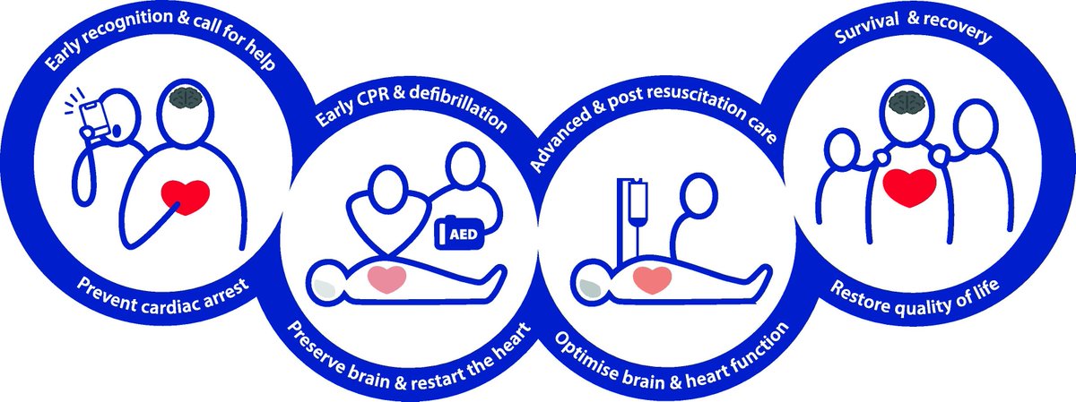
The 2025 European Resuscitation Council Guidelines have been released
All 11 plus the executive summary included below
CCR Journal Watch
criticalcarereviews.com/late…
6
81
194
15,356
Cornelius Sendagire, MD retweeted

21 Oct 2025
Hello @UEDCLTD
No power in Bugolobi, Can you please look into it....Tuffa 😢.
3
2
5
2,588
Cornelius Sendagire, MD retweeted

18 Oct 2025
I was priviledged to make the presentation. Thanks for the invitation @Nabukenya8 @AnesthesiaUg
4
5
395
Cornelius Sendagire, MD retweeted

16 Oct 2025
CHEST X-RAY 36: What is the classic pattern of shadowing on this chest X-ray? What does it signify? Answer buff.ly/3PA4Cfl

16
27
285
29,937
Cornelius Sendagire, MD retweeted
16 Oct 2025
🧩 Part 3 – Why you usually can’t move one curve without the other
1️⃣
So far, we’ve treated the cardiac and venous return curves as two lines that meet.
In theory, you can move one without the other – and sometimes that’s true.
But in physiology, they almost always move together – because they share the same inlet.
4
49
192
31,892
Cornelius Sendagire, MD retweeted

17 Oct 2025

1
55
360
26,302
Cornelius Sendagire, MD retweeted
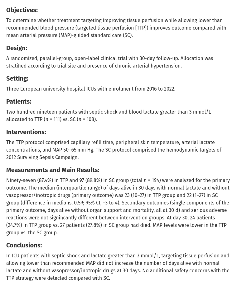
17 Oct 2025
Interesting trial result, ahead of ANDROMEDA-SHOCK 2 in a fortnight
Targeted Tissue Perfusion vs Macrocirculatory-Guided Standard Care in Patients With Septic Shock: - The TARTARE-2S RCT
CCR Journal Watch
criticalcarereviews.com/late…
28
81
5,874