
GS resident at al jala hospital
Joined July 2014
- Tweets 2,401
- Following 972
- Followers 228
- Likes 7,308
104 Photos and videos


Pinned Tweet

28 Feb 2021
الحمد الله حتي يبلغ الحمد منتهاه
الحمد الله حتي ترضي وبعد الرضا
الحمد الله حمدا كثيرا مباركا فيه🙏🙏🙏🙏🙏
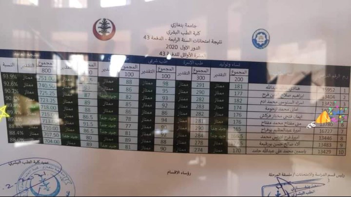
الحمد الله نجحت من سنة رابعة بشري
🎉🎉🎉🎉🎉🎉🎉🎉🎉🎉
23
24


Mona Elsaity retweeted

Surgical removal of a herniated disc.💥
114
310
3,998
4,208,918


Mona Elsaity retweeted

May 6
To win you have to be control your emotion.

44
845
4,807
76,509


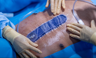
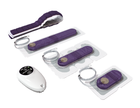
Help protect and optimize the healing environment of closed surgical incisions with Solventum™️ Prevena™️ Therapy. Visit Prevena.com to learn more. #PaidAdvertising
9
33
5,175


Mona Elsaity retweeted

Apr 3
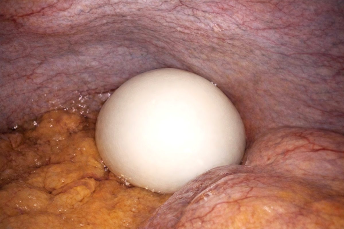
Found on diagnostic laparoscopy in a patient with abdominal discomfort. What’s the diagnosis?
68
22
272
67,308
Mona Elsaity retweeted

x.com/i/status/2040144733885…
That is a classic "spot diagnosis" from a laparoscopic view. This smooth, white, egg-like structure found free-floating in the peritoneal cavity is a Peritoneal Loose Body, also commonly known as a "Peritoneal Mouse."
Here is a high-yield summary of the pathology for your records:
Pathophysiology
These are generally thought to result from torsion and infarction of an appendix epiploica (the small, fat-filled sacs along the colon).
The pedicle of the appendix epiploica twists, cutting off its blood supply.
It undergoes aseptic necrosis and eventually detaches.
Over time, the detached tissue becomes saponified and calcified.
As it rolls around the peritoneal cavity, it acquires layers of albuminous serum, giving it that smooth, "boiled egg" appearance.
Clinical Presentation
Asymptomatic: Most are incidental findings during laparoscopy or imaging (CT/MRI).
Size: They typically range from 1 to 5 cm, though "giant" peritoneal loose bodies (over 5 cm) are occasionally reported.
Mobility: On sequential imaging, they may change position, which is a hallmark diagnostic feature.
Differential Diagnosis
While the appearance is quite distinct, in a clinical or exam setting, one might consider:
Leiomyoma: A subserosal fibroid that has become parasitic.
Gallstone: A dropped gallstone post-cholecystectomy (though these are usually darker and more irregular).
Teratoma: Though usually attached and more complex in structure.
Calcified Mesenteric Lymph Node.
Management
No treatment is necessary if found incidentally, as they are benign.
If they are large enough to cause extrinsic compression (e.g., on the bladder or bowel), surgical removal via laparoscopy is indicated.

Peritoneal loose body
Peritoneal mous
3
11
1,731
Mona Elsaity retweeted

B. Propylthiouracil
Thyroid Troubles: Hyperthyroidism and Thyrotoxicosis
manualofmedicine.com/topics/…
1
16
2,384
Mona Elsaity retweeted

Biliary tract cancers are best understood through anatomy.
Intrahepatic, perihilar, and distal tumors behave differently — and so does their management.
Clear explanation in this video:
youtu.be/S-zLnK5to1U
12
57
3,698
Mona Elsaity retweeted

Mar 28
عارفين من وجهة نظرى عشان تبقى جراح قوى
لازم يكون عندك
١- بيزكس محترمة
٢- معلومات محترمة
٣- شفت كتير
٤- شجاعة الجراح
٥ - تكون شغال based on حاجات صح
3
5
241
52,433
Mona Elsaity retweeted

This painful fistula needed surgery
147
406
6,921
13,545,881
Mona Elsaity retweeted

Mar 26
This is how a urinary catheter is placed through the penis
12
91
1,379
258,582