
Research Professional with @UAFcfos. #aquaculture, #ecophysiology, and a lot of #seaweed. 🌿🌊
- Tweets 6,608
- Following 1,306
- Followers 2,255
- Likes 12,745


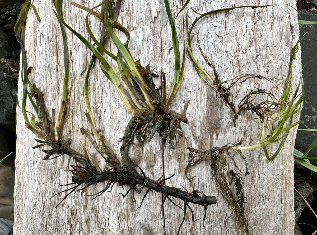


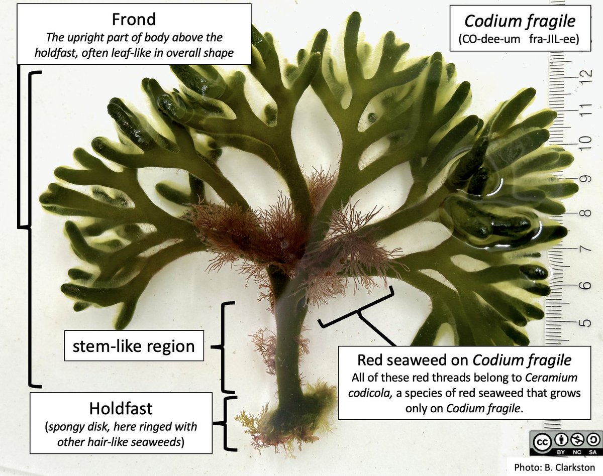
ALT A mature Codium fragile (CO-dee-um fra-JIL-ee) with labelled parts. Holdfast: (spongy disk, here ringed with other hair-like seaweeds); Stem-like region; Frond: The upright part of body above the holdfast, often leaf-like in overall shape. This specimen of Codium is also home to a filamentous red seaweed, Ceramium codicola, that resembles long thin red threads emerging from the Codium body. Ceramium codicola grows only on Codium fragile.
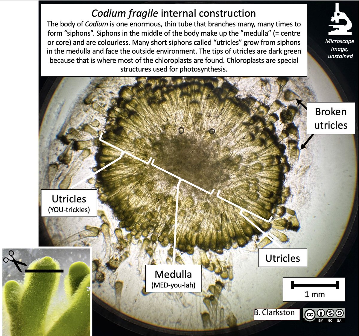
ALT A cross-section of Codium fragile showing the body’s internal construction. A “cross-section” cut reveals the different tissue layers — think of slicing a sandwich and seeing the bread, tomato, lettuce, etc. The insert photo and cartoon scissors shows where the cross-section cut was made. The body of Codium is one enormous, thin tube that branches many, many times to form “siphons”. Siphons in the middle of the body make up the “medulla” (= centre or core) and are colourless. Many short siphons called “utricles” grow from siphons in the medulla and face the outside environment. The tips of utricles are dark green because that is where most of the chloroplasts are found. Chloroplasts are special structures used for photosynthesis. The labelled parts are: Utricles (YOU-trickles), Medulla (MED-you-lah), Broken utricles. The image has been taken with a microscope and the colours are natural. Scale bar = 1mm.
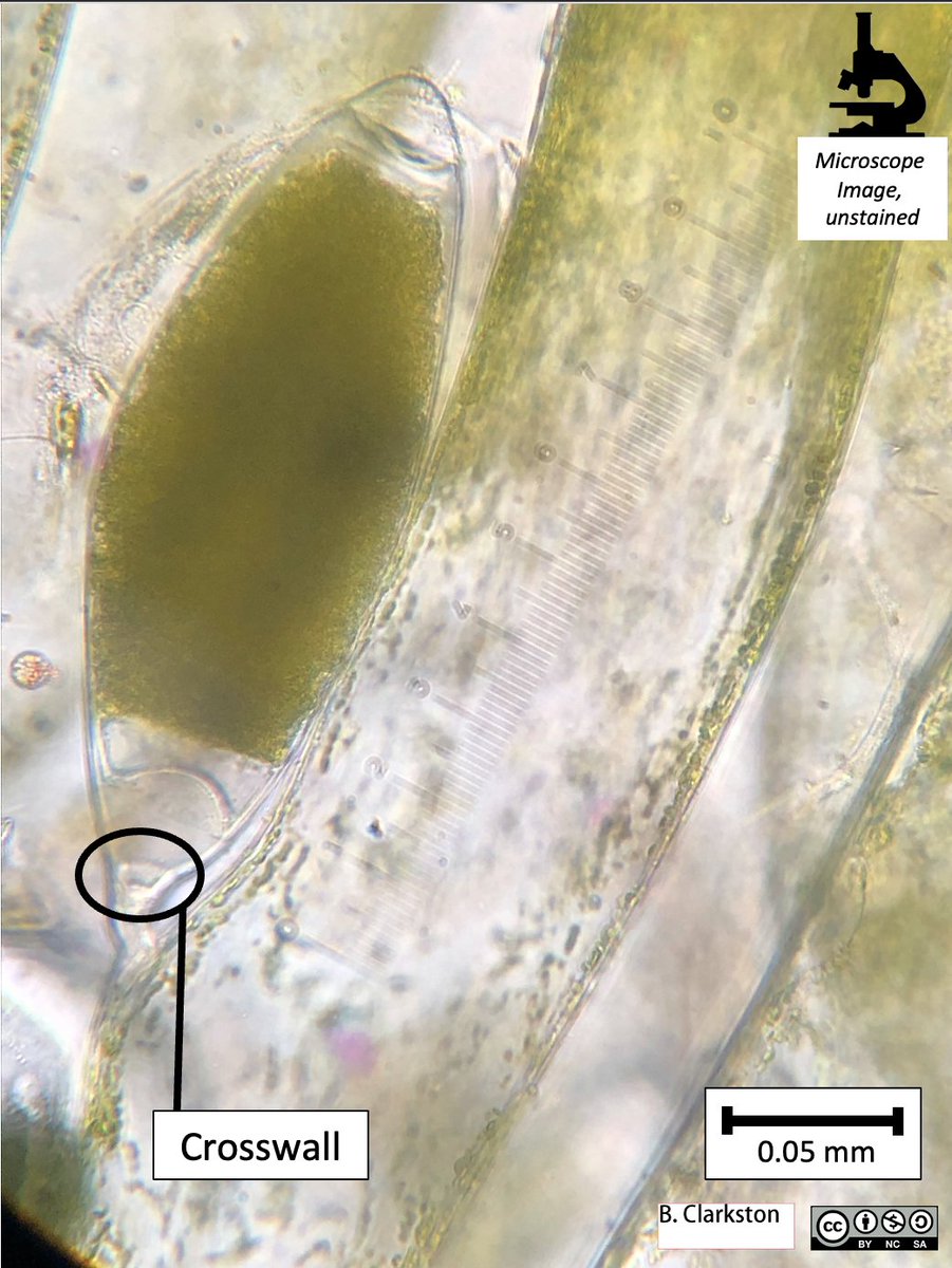
ALT A close-up at higher magnification of a Codium fragile gametangium. The gametangium is very densely-coloured, a sign that it is mature and will soon release gametes into the water. The thick crosswall separating the internal contents of the gametangium from the rest of the body is clearly visible. The labelled parts are: crosswall. The image has been taken with a microscope and the colours are natural. Scale bar = 0.05mm.
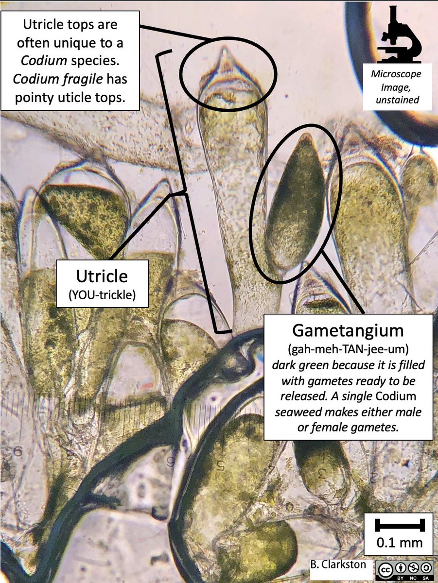
ALT A cross-section of Codium fragile showing utricles and gametangia (singular: gametangium). The gametangia are darker green because there are many gametes densely packed inside, ready to be released into the water when the top of the gametangium ruptures. A single Codium fragile seaweed makes either male or female gametes inside its gametangia. The tops of utricles are often unique to each species of Codium. Codium fragile has pointy utricle tops. The labelled parts are: Utricle (YOU-trickle) and Gametangium (gah-meh-TAN-gee-um). The image has been taken with a microscope and the colours are natural. Scale bar = 0.1mm.


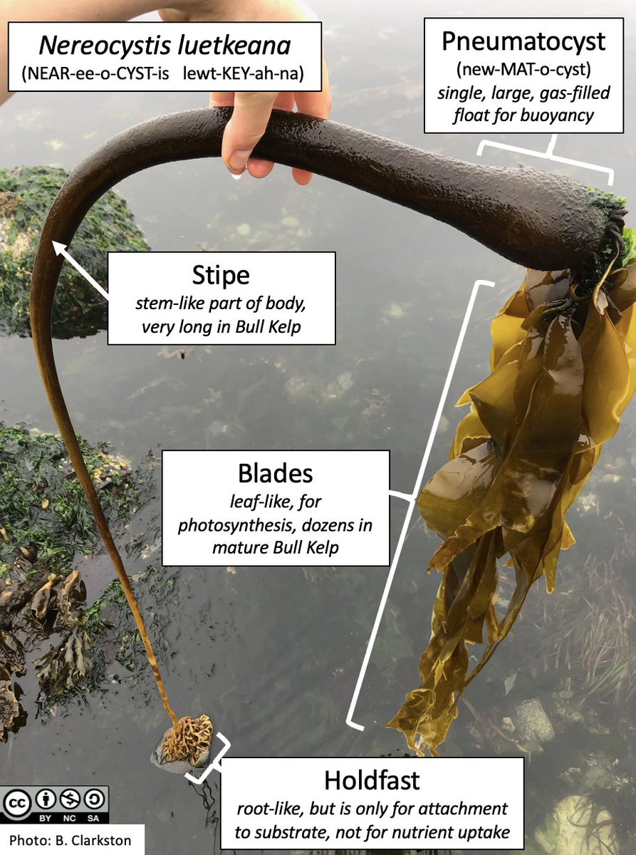
ALT A mature Nereocystis luetkeana (NEAR-ee-o-CYST-is lewt-KEY-ah-na) with labelled parts. Holdfast: root-like, but is only for attachment to substrate, not nutrient uptake; Stipe: stem-like part of body near base, very long in Nereocystis; Blade: leaf-like, primary region of photosynthesis, dozens in mature individuals; Pneumatocyst (new-MAT-o-cyst): single, large, gas-filled float for buoyancy.
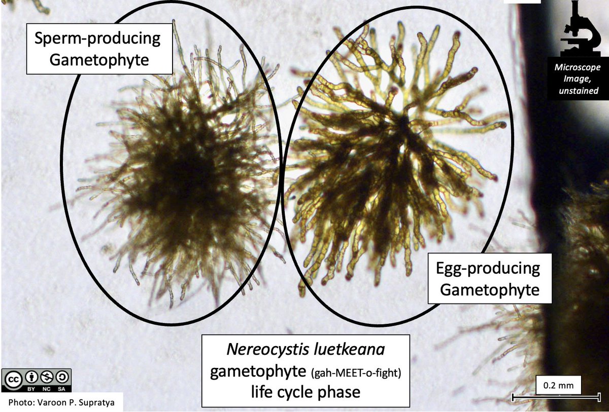
ALT A collection of Nereocystis leutkeana individuals in the gametophyte (gah-MEET-o-fight) life cycle phase. The egg-and sperm-producing gametophytes can be distinguished by their different cell sizes: egg-producing gametophytes produce larger body cells. The cells have not been stained in this image, the colour is natural. The image has been taken with a microscope; all individuals pictured here are less than a millimeter in size. The labelled parts are: Sperm-producing gametophyte, Egg-producing gametophyte.

ALT A collection of Nereocystis leutkeana individuals in the gametophyte (gah-MEET-o-fight) life cycle phase. An egg-producing structure, the oogonium (oo-GO-knee-um) is visible. One very young individual in the sporophyte life cycle stage is also visible. The sporophyte is formed when two gametes fuse, one sperm from a male gametophyte and one egg from a female gametophyte. The cells have not been stained, the colour is natural. The image has been taken with a microscope; all individuals pictured here are less than a millimeter in size. The labelled parts are: Gametophyte filaments, Oogonium, Young sporophyte.