
Jun 15
⚡️NEWS FLASH: Welcome to Alabama, ITU AbsorbTech! The Wisconsin-based provider of launderable, reusable oil absorbents and shop towels recently broke ground on a 50,000-square-foot processing facility in McCalla. It’s the company’s sixth processing site and first in Alabama.

1
2
405

Accidents happen. Be prepared with absorbents and spill cleanup materials to keep leaks and spills out of storm drains and local waterways. Quick action can help prevent stormwater pollution and protect our environment.
#ContractorStormwaterSolutions #CMRSWC

ALT Graphic titled “Accidents Happen!” showing oil or liquid spills on pavement with an arrow pointing to the spill. The text encourages using absorbents to prevent spills from entering nearby storm drains and promotes spill prevention practices for construction and home improvement projects.
1
11

Jun 10
🩹♻️ El tèxtil sanitari, a la resta!
⚫️Amb el canvi de contenidors, ara el tèxtil sanitari ha d'anar al contenidor de la fracció resta 👇
Bolquers, compreses i tampons, bastonets per netejar les orelles, corones absorbents de lactància, tovalloles humides, fil dental o petits residus de cures domèstiques.
Tota la informació sobre les fraccions:
web.girona.cat/netejairesidu…

2
1,267

Jun 9
Industrial Cannabis isn’t waste — it’s the future of the circular economy!
328 different technologies/applications were identified, including:
Textile fibers (for clothes and fabrics)
Bioplastics
Biofuels
Functional foods and supplements
Absorbents (like natural filters)
Natural cosmetics
pubmed.ncbi.nlm.nih.gov/4165…

8
23
65
1,011

Jun 7
Discover our comprehensive Anaesthesia range at #Euroanaesthesia26 on stand 5.67, including anaesthetic face masks, breathing systems, breathing system accessories, CO₂ absorbents and more.
You'll also have the opportunity to see Versi-flo™, our modular Nasal High Flow system, designed for simple, flexible use wherever Nasal High Flow therapy is required across the hospital.
Explore more:
📲 intersurgical.com/products/a…
📧 info@intersurgical.com
#Intersurgical #RespiratoryCare #EA26 #ESAICcongress
1
3
178

Jun 7
The mass shaving scene is real and takes place in Mina, Saudi Arabia, during Hajj (men shaving their heads in large tents).
As for the factory scenes that convert human hair into panels and products (insulation or absorbents), they are real from specialized factories in other countries, but **not in Saudi Arabia**. There are no Saudi factories of this size or form.
The video combines two separate scenes just for sensationalism x.com/mlqbnmmd3/status/20632…
 معلومات تفيد ®️
1
1,503

For research mining & algorithmic tag purposes-
Source: uploaded PDF pages after the Volume II title page.
Continuing from the uploaded PDF’s **List of Engravings** pages.
## LIST OF ENGRAVINGS.
### VOLUME I. continued
**Fig. 35** Longitudinal section of the root of ditto, (id.) . . . 96
**36** Capsule of bulb of ditto laid open, (id.) . . . 96
**37** Muscle in a state of relaxation . . . 101
**38** The same muscle contracted . . . 101
**39** Diagram illustrating the action of oblique muscles . . . 101
**40** Semi-penniform muscle . . . 101
**41** Penniform muscle . . . 101
**42** Complex muscle . . . 101
**43** Tendon of muscle . . . 101
**44** Trapezius muscle . . . 101
**45** Muscular structure of the Ear-drum, (Home) . . . 105
**46** Orbicular muscle of the Eye-lids, (Albinus) . . . 105
**47** Muscular structure of the Iris, (Home) . . . 105
**48** Muscular fibres of a sucking disk . . . 105
**49** Longitudinal muscular fibres of a blood vessel . . . 106
**50** Transverse muscular fibres of ditto . . . 106
**51** Muscular fibres of the human stomach, (Cooper) . . . 106
**52** Muscular fibres of the heart, (id.) . . . 106
**53** Magnified view of a Sponge, (Grant) . . . 114
**54** Spicula in the texture of a Sponge, (id.) . . . 114
**55** Gemmule of a Sponge, (id.) . . . 114
**56** *Lobularia Alcyonium pelagica*, (Deterville) . . . 122
**57** Detached polype of ditto, (id.) . . . 122
**58** *Zoanthus*, (*Actinia sociata*,) (Ellis) . . . 122
**59** *Hydra viridis*, (Trembley) . . . 122
**60** *Sertularia pelagica*, (Deterville) . . . 124
**61** *Tubipora musica*, (Ellis) . . . 125
**62** Section and polypes of ditto, magnified, (id.) . . . 125
**63** *Flustra carbasea*, (id.) . . . 125
**64** Cells of ditto, magnified, (id.) . . . 125
**65** *Corallium rubrum*, (id.) . . . 125
**66** Polypes of ditto, magnified, (id.) . . . 125
**67** Section of *Gorgonia Briareus*, (id.) . . . 125
**68** *Isis hippuris*, (id.) . . . 125
**69** Polype of *Flustra carbasea*, (Grant) . . . 129
**70** Tentaculum of ditto, magnified, (id.) . . . 129
**71** *Pennatula phosphorea*, (Ellis) . . . 131
**72** Magnified view of the polypes of ditto, (id.) . . . 131
**73 to 76** Mode of progression of the *Hydra viridis*, (Trembley) . . . 133
**77** *Vorticella cyathina*, (Muller) . . . 136
**78** *Proteus diffluens*, (id.) . . . 139
**79** *Volvox globator*, (id.) . . . 139
**80** *Brachionus urceolaris*, (id.) . . . 140
---
**81** *Medusa Pulmo*, (Macri) . . . 142
**82** *Beroe ovatus*, (Bruguiere) . . . 144
**83** *Beroe pileus*, (id.) . . . 144
**84** *Velella limbosa*, (Guérin) . . . 144
**85** *Physalia atlantica*, (id.) . . . 144
**86** *Actinia rufa*, (original) . . . 146
**87** Ditto expanded, (original) . . . 146
**88** *Asterias serrulata*, (Bruguiere) . . . 147
**89** *Asterias regularis*, (id.) . . . 147
**90** *Echinus Ananchites ovata*, (id.) . . . 147
**91** *Clypeaster rosaceus*, (id.) . . . 147
**92** *Ophiura lacertosa*, (id.) . . . 147
**93** *Euryale muricatum*, (id.) . . . 147
**94** *Pentacrinus europæus*, (Thomson) . . . 147
**95** Ambulacra, and feet of *Asterias*, viewed from the under side, (Reaumur) . . . 148
**96** Ditto, viewed from the upper side, (id.) . . . 148
**97** Vesicles appended to the feet of the *Asterias* . . . 148
**98** Polygonal pieces composing the test of the *Echinus* . . . 150
**99** Structure of a detached piece of ditto . . . 150
**100** Spine of the *Cidaris*, (Carus) . . . 150
**101** Shell of *Unio batava*, (Goldfuss) . . . 159
**102** Adductor muscle of Oyster, (Hunterian Museum) . . . 160
**103** Shell of *Pholas candida*, with abductor muscle, (Osler) . . . 161
**104** Foot of *Cardium edule*, (Reaumur) . . . 162
**105** *Planorbus cornutus*, (Cuvier) . . . 166
**106** Magnified view of the striæ on the surface of Mother of Pearl, (Herschel) . . . 169
**107** Directions of the fibres in the component strata of shells . . . 170
**108** Shell of *Achatina zebra*, (De Blainville) . . . 176
**109** Longitudinal section of ditto, (id.) . . . 176
**110** Shell of *Pterocerus scorpio*, at an early stage of growth, (id.) . . . 178
**111** Shell of the same when completely formed, (id.) . . . 178
**112** Shell of *Cyprea exanthema* at an early period of growth, (id.) . . . 178
**113** Shell of the same animal, when completed, (id.) . . . 178
**114** Transverse section of the shell of the *Cyprea exanthema*, (Hunterian Museum) . . . 179
**115** Shell of *Conus* . . . 181
**116** Longitudinal section of the same, (original) . . . 181
**117** Transverse section of the same, (Bruguiere) . . . 181
**118** Inner surface of the Epiphragma of the *Helix pomatia*, (De Blainville) . . . 183
---
**119** Outer surface of the same, (id.) . . . 183
**120** *Clio borealis*, (Cuvier) . . . 186
**121** *Sepia loligo*, (De Blainville) . . . 187
**122** Suckers of the same, (id.) . . . 187
**123** Suckers of the Octopus, (original) . . . 187
**124** Shell of *Spirula australis*, (De Blainville) . . . 191
**125** Longitudinal section of the same, (id.) . . . 191
**126** Shell of *Nautilus pompilius*, (id.) . . . 191
**127** Longitudinal section of the same, (id.) . . . 191
**128** *Pontobdella muricata*, (Bruguiere) . . . 195
**129** *Nereis*, (id.) . . . 195
**130** *Erpobdella vulgaris*, Lam. *Hirudo hyalina* . . . 195
**131** Diagram illustrating the rings and muscles of *Annelida*, (original) . . . 195
**132** *Gordius aquaticus* . . . 198
**133** *Serpula opercularia* . . . 198
**134** *Terebella conchilega*, (De Blainville) . . . 198
**135** *Arenicola piscatorum*, or *Lumbricus marinus* . . . 198
**136** *Aranea diadema*, (Roesel) . . . 202
**137** Divisions of the limb of a Crustaceous animal . . . 205
**138** Mandible and palpus of *Mysis Fabricii*, (Bruguiere) . . . 205
**139 to 141** Feet-jaws belonging to the first, second, and third pairs, (id.) . . . 205
**142** True foot, belonging to the first pair, (id.) . . . 205
**143** *Julus terrestris* . . . 213
**144** Muscles of the trunk of the *Melolontha vulgaris*, (Straus Durckheim) . . . 214
**145** Eggs of *Bombyx mori* . . . 217
**146** Larva of the same . . . 217
**147** Pupa of the same . . . 217
**148** Imago of the same . . . 217
**148*** A Caterpillar of the *Phalæna striaria*, (Hubner) . . . 224
**B** The same in a rigid position, (Lyonet) . . . 224
**149** *Calosoma Sycophanta*, (Kirby and Spence) . . . 227
**150** Analysis of skeleton of the same, (Carus) . . . 228
**151** Hind view of the segment of the head in the same, (id.) . . . 228
**152** Suckers on the foot of the *Musca vomitoria*, expanded; magnified view, (Bauer) . . . 235
**153** Cushions on the foot of the *Cimex lutea*, magnified, (id.) . . . 235
**154** Suckers on the under side of the foot of a male *Dytiscus marginalis*, (id.) . . . 235
**155** Cushions and sucker of the *Acridium biguttulum*, Latr., (id.) . . . 235
---
**156** *Dytiscus marginalis*, upper side, (Roesel) . . . 237
**157** Lower side of the same insect, (id.) . . . 237
**158** *Notonecta glauca*, (Roesel) . . . 238
**158*** Fore leg of *Gryllotalpa*, (Kidd) . . . 242
**159** Wing of *Gryllus nasutus*. Orthoptera . . . 246
**160** Wing of *Libellula grandis*. Neuroptera . . . 246
**161** Wing of *Ichneumon persuasorius*. Hymenoptera . . . 246
**162** Wing of *Tipula oleracea*. Diptera . . . 246
**163** Sting of *Anthophora retusa*, (original) . . . 248
**164** Separate scales of the wing of *Hesperia Sloanus*, (original) . . . 250
**165** Arrangement of the scales in the wing of the same . . . 250
**172** Longitudinal section of the thigh-bone to show the cancellated structure, (Cheselden) . . . 262
**173** Longitudinal section of the humerus, (id.) . . . 262
**174** Ossification of the parietal bone, (id.) . . . 265
**175** Early stage of ossification of the bones of the skull, (Cloquet) . . . 265
**176** The same in the adult, showing the sutures . . . 265
**177** Dorsal vertebra, human . . . 271
**178** Junction of vertebræ forming the spinal column . . . 271
**179** Longitudinal section of the same, showing the spinal canal . . . 271
**180** Elements of structure of a vertebra, (Carus) . . . 274
**181** Skeleton of Hog, (Pander and D’Alton) . . . 279
**182** Sternum, clavicle, and scapula; human . . . 279
**184** Skeleton of *Cyprinus carpio*, (Bonnaterre) . . . 286
**185** Diagram illustrating the progressive motion of Fishes . . . 287
**186** Front view of the vertebra of a Cod, (*Gadus morrhua*) . . . 288
**187** Side view of the same . . . 288
**188** Vertical and longitudinal section of a part of the spinal column in the same . . . 288
**189** A similar section, showing the gradation of structure . . . 288
**190** Similar section in the *Squalus centrina*, (Carus) . . . 288
**191** Bones of the shoulder of the *Lophius piscatorius*, (id.) . . . 293
**192** Pectoral fin of the *Raia clavata*, (id.) . . . 293
**193** Belt of bones of the shoulder of a Ray, (id.) . . . 294
**194** Muscular system of *Cyprinus alburnus*, (id.) . . . 295
**195** Air bladder of *Cyprinus carpio*, (Blasius) . . . 298
**196** Eggs of the Frog . . . 303
**197** Side view of Tadpole magnified, (Rusconi) . . . 303
**198** Upper view of the same, (id.) . . . 303
**199** Adult Frog . . . 303
**200** Skeleton of Frog, (Cheselden) . . . 306
**201** Skeleton of the Viper . . . 310
Next
Continuing the **List of Engravings** from the uploaded PDF.
## LIST OF ENGRAVINGS.
### VOLUME I. continued
**202** Ribs and spine of *Boa constrictor*, (Home) . . . 312
**203** Bones of the foot of the same, (Mayer) . . . 311
**204** Muscles moving the claw of the same, (id.) . . . 311
**205** Rudimental bones of the foot of the *Tortryx scytale*, (id.) . . . 311
**206** - of the *Tortrix corallinus*, (id.) . . . 311
**207** - of the *Anguis fragilis*, (id.) . . . 311
**208** - of the *Amphisbaena alba*, (id.) . . . 311
**209** - of the *Coluber pullutatus*, (id.) . . . 311
**210** *Chalcides pentadactylus*, (Bonnaterre) . . . 311
**211** Under surface of the foot of the *Lacerta gecko*, magnified four times, (Bauer) . . . 319
**212** Side view of a longitudinal section of the same, (id.) . . . 319
**213** Skeleton of the Tortoise, (Carus) . . . 322
**214** Section of the thigh bone of the same, (id.) . . . 322
**215** Hind view of skull of *Testudo mydas*, (id.) . . . 325
**216** Bones sustaining the fin of the *Delphinus phocena*, (Pander and D’Alton) . . . 336
**217** Fore part of the Skeleton of an Ox with the *ligamentum nuchae*, (original) . . . 346
**218** Skeleton of the Stag, (Cheselden) . . . 350
**218*** a. Longitudinal section of the horn of an Ox, (original) . . . 355
b. Ditto of an Antelope, (original) . . . 355
c. Extremity of the same, (original) . . . 355
**219** Subcutaneous muscles of the Hedge-hog, relaxed, (Carus) . . . 364
**220** The same muscles contracted, and drawn over the body, (Cuvier) . . . 364
**221** Skeleton of the Lion, (Pander and D’Alton) . . . 365
**222** Skeleton of *Draco volans*, (Tiedemann) . . . 379
**223** Skeleton of *Vespertilio Molossus*, (Temminck) . . . 380
**224** Skeleton of the Swan, (Cheselden) . . . 385
**225** Lateral section of the cervical vertebra of the Ostrich, (original) . . . 388
**226** Fibrils of the vane of a feather, magnified, (original) . . . 393
**227** Edges of the fibres, magnified, (original) . . . 393
**228** Feather, showing its structure, (F. Cuvier) . . . 396
**229** Capsule, or Matrix of the feather, (id.) . . . 396
**230** View of the parts enclosed in the Capsule, when laid open, (id.) . . . 396
**231** Section of the stem, while growing, exhibiting the series of conical membranes, (id.) . . . 396
**233** Extensor muscles of the foot and toes of a bird, (Borelli) . . . 405
**234** Position of a bird in roosting, (id.) . . . 405
---
## VOLUME II.
**239** Cyclosis, or partial circulation in the cells of the *Caulinia fragilis*, magnified, (Amici) . . . 42
**240** The same in the jointed hair of the *Tradescantia virginica*, (Slack) . . . 42
**241** Section of the *Hydra viridis*, magnified, (Trembley) . . . 58
**242** *Hydra viridis* seizing a worm, (id.) . . . 59
**243** The same after swallowing a minnow, (id.) . . . 59
**244** A Hydra which has swallowed another of its own species, (id.) . . . 59
**245** Compound Hydra, with seven heads, (id.) . . . 59
**246** *Veretilla lutea*, showing the communicating vessels of the Polypes, (Quoy et Gaimard) . . . 64
**247** Nutrient vessels of the *Tenia solium*, (Chiaje) . . . 64
**248** *Tenia globosa*, or Hydatid of the Hog, (Goeze) . . . 64
**249** Horizontal section of the *Rhizostoma Cuvieri*, Peron, (Eysenhardt) . . . 67
**250** *Geronia Hexaphylla*, Peron, *Medusa proboscidalis*, (Forskal) . . . 67
**251** Vascular net-work in margin of the disk of the *Rhizostoma Cuvieri*, (Eysenhardt) . . . 67
**252** Vertical section of the *Rhizostoma Cuvieri*, (id.) . . . 68
**253** Transverse section of one of the arms of the same, (id.) . . . 68
**254** Transverse section of the extremity of a tentaculum of the same, (id.) . . . 68
**255** *Leucophra patula*, highly magnified, (Ehrenberg) . . . 73
**256** Alimentary canal and ceca of the same, viewed separately, (id.) . . . 73
**257** Vertical section of the *Actinia coriacea*, (Spix) . . . 75
**258** Digestive organs of the *Asterias*, (Tiedemann) . . . 76
**259** Stomachs of the *Nais vermicularis*, (Roesel) . . . 77
**260** Stomachs of the *Hirudo medicinalis*, (original) . . . 78
**261** Mouth of the same, showing the three semicircular teeth, (original) . . . 78
**262** Tooth of the same, detached, (original) . . . 78
**263** *Glossopora tuberculata*; *Hirudo complanata*, Lin., (Johnson) . . . 78
**264** The same seen from the under side, showing the digestive organs, (id.) . . . 78
**265** Diagram showing the arrangement and connexions of the organs of the vital functions in Vertebrata, (original) . . . 81
---
**266** Spiral probosces of *Papilio urticae*, (Griffith) . . . 87
**267** Trophi of *Locusta viridissima*, (Goldfuss) . . . 92
**268** Filaments composing the rostrum, or proboscis, of the *Cimex nigricornis*, (Savigny) . . . 94
**269** Sheath of the proboscis of the same insect, (id.) . . . 94
**270** Toothed cartilage of the *Helix pomatia*, (Cuvier) . . . 95
**271** Mechanism for projecting and retracting the tongue of the Woodpecker, (original) . . . 99
**272** Laminae of Whalebone descending from the palate of the *Balena mysticetus*, (Bonnaterre) . . . 102
**273** Teeth of the *Delphinus phocena*, (Cloquet) . . . 106
**274** Skull of Tiger, (Cuvier) . . . 108
**275** Skull of Antelope, (Pander and D’Alton) . . . 109
**276** Skull of Rat, (id.) . . . 110
**277** Longitudinal section of simple tooth, (Rousseau) . . . 111
**278** Surface of the grinding tooth of a Horse, (Home) . . . 111
**279** Surface of the grinding tooth of a Sheep, (id.) . . . 111
**280** Longitudinal section of the incisor tooth of the Rodentia . . . 111
**281** Vertical section of the grinding tooth of the Elephant, (Home) . . . 114
**282** Grinding tooth of the African Elephant, (id.) . . . 114
**283** Grinding tooth of the Asiatic Elephant, (id.) . . . 114
**284** Succession of teeth in the Crocodile, (Carus) . . . 120
**285** Venomous fang of the *Coluber naia*, (Smith) . . . 121
**286** Transverse section of the same, (id.) . . . 121
**287** The same tooth, at an earlier period of growth, (id.) . . . 121
**288** The same, still less advanced in its growth, (id.) . . . 121
**289** Base of the former, (id.) . . . 121
**290** Base of the latter, (id.) . . . 121
**291** Transverse section of the young fang, about its middle, (id.) . . . 121
**292** A section, similar to the last, of another species of serpent, (id.) . . . 121
**293** *Squalus pristis*. Under side of its snout, (Latham) . . . 122
**294** Interior of the Stomach of a Lobster, (original) . . . 123
**295** Gastric teeth of *Bulla aperta*, (Cuvier) . . . 123
**298** Gizzard of the Swan, (Home) . . . 124
**299** Crop and gizzard of the Parrot, (id.) . . . 131
**300** Crop of the Pigeon, (id.) . . . 131
**301** Human stomach, (id.) . . . 133
**302** Interior of the stomach of the African Ostrich, (id.) . . . 135
**303** Gastric glands of the same, (id.) . . . 135
**304** Gastric glands of the American Ostrich, (id.) . . . 135
**305** Longitudinal section of the gastric glands of the Beaver, (id.) . . . 135
**306** Stomach of Dormouse, (id.) . . . 139
Next
Continuing the **List of Engravings - Volume II** from the uploaded PDF.
## LIST OF ENGRAVINGS.
### VOLUME II. continued
**307** Stomach of *Hyrax capensis*, (Cuvier) . . . 139
**308** Stomach of Porcupine, (id.) . . . 139
**309** Stomach of Kanguroo, (id.) . . . 139
**310** Stomach of *Delphinus phocena*, (id.) . . . 139
**311** Cardiac valve of the Horse, (Gurlt) . . . 140
**312** The four stomachs of a Sheep, (Carus) . . . 141
**313** Inner surface of the honey-comb stomach, (Home) . . . 141
**314** Inner surface of the many-plies stomach of an Ox, (id.) . . . 141
**315** Interior cellular surface of the second stomach of the Camel, (id.) . . . 141
**316** Spiral valve in the intestine of the Shark, (Blasius) . . . 149
**317** Digestive organs of the *Mantis religiosa*, (Marcel de Serres) . . . 153
**318** *Melolontha vulgaris*, (Léon Dufour) . . . 154
**319** *Cicindela campestris*, (id.) . . . 154
**320** Portion of a hepatic vessel of the *Melolontha*, highly magnified, (Straus Durckheim) . . . 155
**321** Alimentary canal of the *Acrida aptera*, (original) . . . 155
**322** Interior of the gizzard of the same, magnified, (original) . . . 155
**323** Row of large teeth in the same, still more magnified, (original) . . . 155
**324** Profile of one of those teeth, still more highly magnified, (original) . . . 155
**325** Base of the same tooth, seen from below, (original) . . . 155
**326** Alimentary canal of the Larva of the *Sphinx ligustri*, (original) . . . 157
**327** - of the Pupa of the same, (original) . . . 157
**328** - of the Imago of the same, (original) . . . 157
**329** - of the *Patella*, (Cuvier) . . . 159
**330** Stomachs of the *Pleurobranchus Peronii*, (id.) . . . 159
**331** Pyloric appendices in the Salmon, (id.) . . . 160
---
**333** Detached Dorsal vessel of *Melolontha vulgaris*, (Straus Durckheim) . . . 172
**334** The same, with its ligamentous and muscular attachments, (id.) . . . 172
**335** Side view of the anterior extremity of the same vessel, (id.) . . . 172
**336** Section of the dorsal vessel, to show its valves, (id.) . . . 172
**337** Circulation in the antenna of the *Semblis viridis*, (Carus) . . . 175
**338** Course of circulation in the same insect, (id.) . . . 175
**339** Dorsal vessel of the Caterpillar of the *Sphinx ligustri*, side view, (original) . . . 177
**340** The same in the Chrysalis, (original) . . . 177
**341** The same in the Moth, (original) . . . 177
**342** The same viewed from above, (original) . . . 177
**343** Magnified lateral view of the anterior extremity of the dorsal vessel, (original) . . . 177
**344** Magnified dorsal view of the same, (original) . . . 177
**345** Structure of the valves of the dorsal vessel, (original) . . . 177
**346** Heart and vessels of the *Aranea domestica*, (Treviranus) . . . 180
**346*** Circulation in the *Planaria nigra*, (Dugés) . . . 181
**347** Course of circulation in the *Erpobdella vulgaris*, (Morren) . . . 183
**348** Vessels in abdominal surface of the same, (id.) . . . 183
**349** Vascular dilatations, or hearts of the *Lumbricus terrestris*, (Morren) . . . 184
**350** Cavities and great vessels of the Heart . . . 187
**351** The Heart laid open to show its Valves . . . 188
**352** Plan of simple circulation . . . 189
**353** Plan of double circulation . . . 191
**354** Branchial circulation in *Maia squinado*, (Audouin) . . . 193
**355** Organs of circulation in the *Loligo sagittata*, (id.) . . . 195
**356** Plan of circulation in Fishes . . . 196
**357** Plan of circulation in Batrachia . . . 197
**359** Plan of double or warm-blooded circulation . . . 199
**360** Heart of the Dugong, (Home) . . . 200
---
**365** Valves of the Veins, (Cloquet) . . . 206
**366** Heart, branchial artery and gills of a fish, (Blasius) . . . 216
**367** Branchial apertures in the *Squalus glaucus*, (Bonnaterre) . . . 216
**368** Branchial apertures in the *Petromyzon marinus*, (id.) . . . 216
**369** Internal structure of the branchiae of the same, (Home) . . . 216
**370** Stigmata in the abdominal surface of the *Dytiscus marginalis*, (Léon Dufour) . . . 222
**371** Stigmata of the *Cerambyx heros*, Fab., magnified, (id.) . . . 222
**372** Longitudinal tracheae of *Carabus auratus*, (id.) . . . 222
**373** Air vesicles and tracheae of the *Scolia hortorum*, Fab., highly magnified, (id.) . . . 222
**374** Respiratory apparatus of the *Scorpio europaeus*, (Treviranus) . . . 225
**375** Internal structure of the lungs of the Turtle, (Bojanus) . . . 229
**377** Air cells of the Ostrich, (Parisian Academicians) . . . 233
**378** Lymphatic Absorbents . . . 250
**379** Passage of Nerves through a ganglion . . . 255
**380** Plexus of Nerves . . . 255
**381** Varieties of forms of antennae of Insects, (Goldfuss) . . . 272
**382** Vertical and longitudinal section of the right nostril in man . . . 283
**383** Vertical transverse section of the same . . . 284
**384** Transverse section of the nostril of a Sheep, (Harwood) . . . 285
**385** Turbinated bones of the Seal, (id.) . . . 285
---
**386** Turbinated bones of the Turkey, (id.) . . . 287
**387** Nerves distributed to the bill of the Duck, (id.) . . . 288
**388** Nasal cavities of the *Perca fluviatilis*, (Cuvier) . . . 291
**389** Nasal cavity of the *Raia batis*, or Skate, (Harwood) . . . 291
**390** Human ear, (Cloquet) . . . 299
**391** Posterior surface of the cavity of the tympanum, (id.) . . . 301
**392** *Ossicula auditus*, or small bones of the tympanum . . . 301
**393** The position of the latter in the tympanum . . . 301
**394** Magnified view of the labyrinth detached from the surrounding parts, (Breschet) . . . 303
**395** Interior structure of the labyrinth, (id.) . . . 304
**396** Membranous labyrinth, with its nerves, (id.) . . . 304
**397** Cretaceous bodies in the labyrinth of the Dog, (id.) . . . 304
**398** Ditto in that of the Hare, (id.) . . . 304
**399** Organ of hearing in the Lobster, (Carus) . . . 308
**400** Groove in the sac of the former, (id.) . . . 308
**401** Organ of hearing in the *Astacus fluviatilis*, (id.) . . . 308
**402** Interior view of the same, (id.) . . . 308
**403** Membranous labyrinth of the *Lophius piscatorius*, (id.) . . . 310
**404** Organ of hearing in the Frog, (Bell) . . . 311
**405** Ear of the Turkey, (Carus) . . . 311
---
**406** Diagram illustrating one mode of obtaining images of objects, (original) . . . 319
**407** Simple Camera Obscura . . . 320
**408** Law of the refraction of a ray of light . . . 321
**409** Convergence of rays to a focus . . . 322
**410** Convergence by a double convex lens . . . 324
**411** Spherical Aberration . . . 324
**412** Variations of focal distance consequent upon variations of divergence of the incident rays . . . 325
**415** Horizontal section of right human eye, magnified, (Home) . . . 326
**416** Straight and oblique muscles of the eye-ball . . . 328
**417** Lacrymal apparatus . . . 330
**418** Eye of *Helix pomatia*, (Muller) . . . 339
**419** Stemmata of Caterpillar, (Marcel de Serres) . . . 342
**420** Eye of the *Scorpio tunensis*, (Muller) . . . 342
**421** Conglomerate eyes of *Julus terrestris*, (Kirby and Spence) . . . 342
**422** External magnified view of the compound eye of the *Melolontha vulgaris*, (Straus Durckheim) . . . 344
**423** Ditto of that of a *Phalena* . . . 344
**424** Section of the compound eye of the *Libellula vulgata*, magnified, (Dugés) . . . 344
Next
2
2
2
1,010

Jun 4
Ashapura Minechem Ltd - Q4 Result and Earnings Call Analysis
1. FY26 was the strongest year in the company’s history: consolidated revenue rose to about ₹5,237 crore (up 91% YoY) and EBITDA to about ₹674 crore, driven largely by Guinea bauxite volumes.
2. Q4 was a very strong topline quarter (₹1,969 crore, up 105% vs Q3), but EBITDA per tonne and margins compressed meaningfully versus earlier quarters due to weaker bauxite prices and higher fuel/freight.
3. Guinea exports were ramped from 3.5 million tonnes to about 8 million tonnes in FY26, with management targeting 10–12 million tonnes in FY27 and 15 million tonnes in FY28, subject to freight and quota outcomes.
4. Management openly acknowledges that EBITDA per tonne in Guinea has fallen “significantly” versus two quarters ago and expects Q1 FY27 margins to be similar to, or slightly worse than, Q4, with improvement only post-quota implementation and freight normalization.
5. The biggest prospective positive is the proposed Guinea bauxite quota system, which management believes will curb output from very large Chinese players, reallocate volumes to mid-size players like Ashapura, and structurally support net bauxite prices and freight.
6. India business (bentonite, white minerals, specialty absorbents, advanced ceramics) faced cost headwinds (fuel, transport, sulphuric acid, mix) but still delivered ~₹998 crore revenue and ₹112 crore EBITDA, and management is doubling down with ~₹150 crore capex into value-added products.
7. Iron ore in Guinea is still in commissioning; management sees a few hundred million tonnes of resource and potential to beneficiate to >60% Fe, but contribution in the next 1–2 years is framed as “meaningful but single-digit margins” and still speculative.
8. Balance sheet is described as healthy; Guinea currently pays no income tax (beyond negligible minimum tax) for at least the next 1–2 years, and the Board has recommended a 100% dividend, but working capital will grow with volume and debt reduction will be gradual.
9. Management tone is confident on the structural opportunity (Guinea bauxite oligopoly, growing aluminium demand, India value-added minerals) but cautious on near-term margins, explicitly flagging Q1 FY27 as still weak and tying a margin recovery to the quota system and freight normalization.
10. Key risks highlighted by management themselves: elevated fuel/freight due to geopolitics, uncertainty on the exact design of Guinea quotas, slower-than-expected iron ore ramp-up, and ongoing cost pressures in Indian operations.
Overall, the business is structurally strengthening (especially in resource and port capacity), but near-term earnings momentum is vulnerable; investors should expect volatility in quarterly margins rather than a straight-line improvement.
#ashapuraminechem #ashapura #concall #earningscall #results
@AethosWealth

1
5
447
May 30
🩹♻️ El tèxtil sanitari, a la resta!
⚫️Amb el canvi de contenidors, ara el tèxtil sanitari ha d'anar al contenidor de la fracció resta 👇
Bolquers, compreses i tampons, bastonets per netejar les orelles, corones absorbents de lactància, tovalloles humides, fil dental o petits residus de cures domèstiques.
Tota la informació sobre les fraccions:
web.girona.cat/netejairesidu…
1
3
3
895

May 29
Hi ha persones que són fil vermell; una conexió profunda que perdura per damunt de temps i distància.
Hi ha persones que venen i se'n van; un aprenentatge.
Hi ha persones-paparra; dependents, absorbents.
(Filosofia de pa sucat amb oli🌸)
6
1
30
374

May 27
Sponsors have taken a fair bit of stick recently, but what an incredible effort from three local companies, Technica, Tech Absorbents & Ramsdens 👏
Sponsoring three stands & injecting much-needed revenue into the club is a hugely generous gesture & a significant boost for Town !
6
1
122
4,188

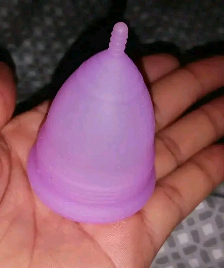
What is a Menstrual Cup?
It's a small, flexible, bell-shaped cup (usually medical-grade silicone) that you insert into the vagina to collect menstrual blood instead of absorbing it like pads or tampons. You empty, rinse, and reuse it. It sits below the cervix and creates a gentle seal.
Why Menstrual Cups are often BETTER than pads:
1. Eco-friendly : One cup replaces thousands of disposable pads/tampons. Lasts 2–10 years. Huge reduction in landfill waste and plastic pollution.
2. Cost-saving : Buy once (around ₦5,000–15,000 depending on brand) and save massively over years. No more monthly pad runs!
3.
Holds more & longer wear: Can hold 3x more than a super tampon/pad. Safe for up to 12 hours (great for night, work, travel). Less frequent changes.
4. More comfortable & discreet: No bulky feeling, no wings, no shifting. Perfect for sports, swimming, or just daily life. Many say you barely feel it.
5.
Healthier for many: Collects blood (doesn't dry out or disrupt vaginal pH like absorbents can). Lower risk of irritation, odor, or leaks when fitted right. No TSS risk like tampons.
6.
Less mess & odor: Blood stays contained inside, less exposure to air.
Note: There's a learning curve (practice makes perfect), and sizing matters (try different brands/sizes). Not everyone loves them, but many who switch never go back.
Consult a doctor if you have specific health concerns.
If you want a plug @HG_Collections sells for #5000


May 21

How do ladies use this and be comfortable?
Ladies do virgins also uses this?

3
2
683

May 18
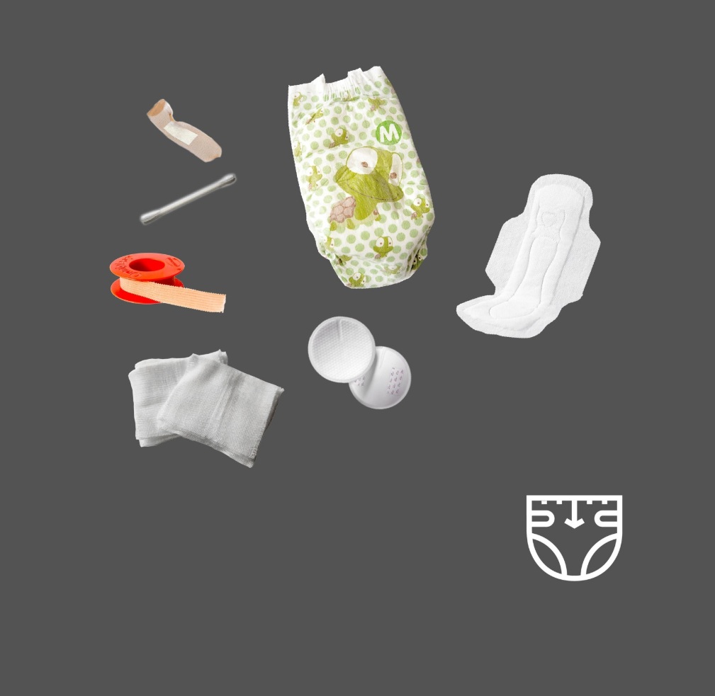
#BreaktheTaboo!
Using clean and hygienic sanitary pads, along with regular stain checks and timely changing of absorbents, helps girls and women manage their periods safely, comfortably, and confidently.
#WaterAidPakistan #PeriodFriendlyWorld #MHDay2026 @WaterAidUK @MHDay28May


1
2
30

Mind you, Stanford Stadium is completely open air. NOT designed with acoustics in mind. It’s a football stadium. For games. Stanford stadium is also below-grade bowl.
One of the most obvious things affecting the way sound travels is the lack of a roof canopy that we see in most stadiums. This means sound escapes straight up into the sky with no roof curves to bounce off back into the floor. This makes it less overwhelmingly loud. Sound is not bounding. It’s just escaping upwards and prevents the reverberation.
Second, Stanford stadium is built below ground level. So the dirt and the soil around the stadium act as acoustic absorbents. Most stadiums have concrete or steel walls, allowing sound waves to hit and vibrate back into the crowd. With Stanford, the waves are hitting earth. And gets dampened.
Third, Stanford Stadium’s strict local city noise ordinance means sound pressure decibels must be controlled. AND. The stadium has a built-in sound system using precision digital beam-steering technology. Where this technology is directly designed to aim audio towards crowds and prevent from spillage to the outside.
Four, atmospheric air pressure. We all saw that it was still daylight despite it being 8pm. Daylight affects sound waves due to thermal refraction. With the sun still out, air closest to the ground is hot. Air on top is cool. This causes sound waves coming from the bottom to travel faster upwards, bending over the crowd and out.
The opposite happens at night when theres no more sun bc the lower air cools down.
And lastly, as to why certain sections were louder than most, why someone from this section couldn’t hear people from the other sections. Same answer. It’s the lack of surfaces for soundwaves to bounce. No bouncing means it’s not travelling back and forth.
Remember, sound travels in concentric circles. Like water ripples. And like water ripples, they vibrate. So if they have surfaces to bounce against, vibration frequency increases. When the vibration frequency of the sound wave increases, the decibel of the sound you hear also increase. If there’s no surfaces to bounce. It escapes.
There. Hope this helps.
47
691
3,297
172,613
May 14
🩹♻️ El tèxtil sanitari, a la resta!
⚫️Amb el canvi de contenidors, ara el tèxtil sanitari ha d'anar al contenidor de la fracció resta 👇
Bolquers, compreses i tampons, bastonets per netejar les orelles, corones absorbents de lactància, tovalloles humides, fil dental o petits residus de cures domèstiques.
Tota la informació sobre les fraccions:
web.girona.cat/netejairesidu…

3
2
382
May 14
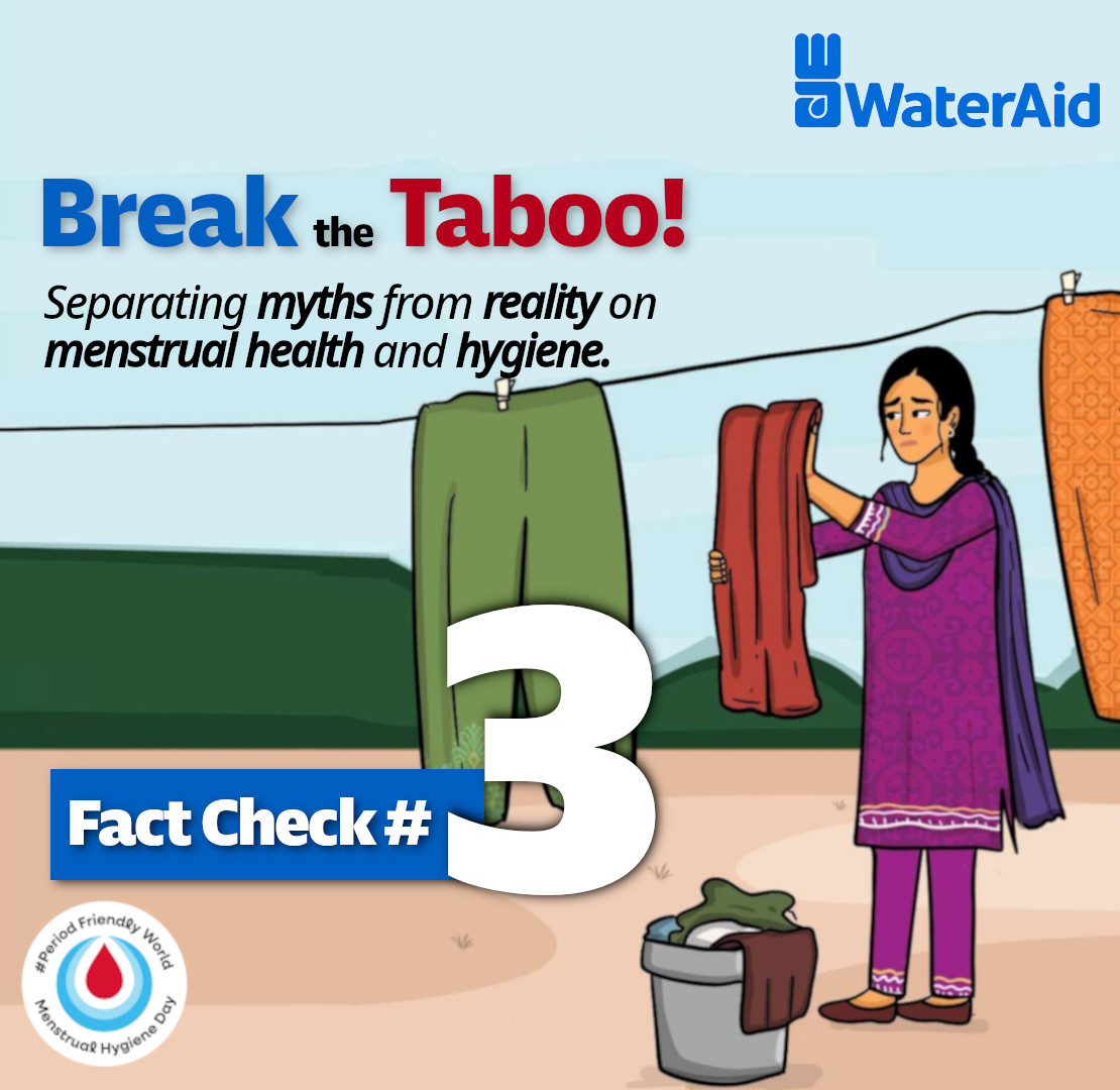
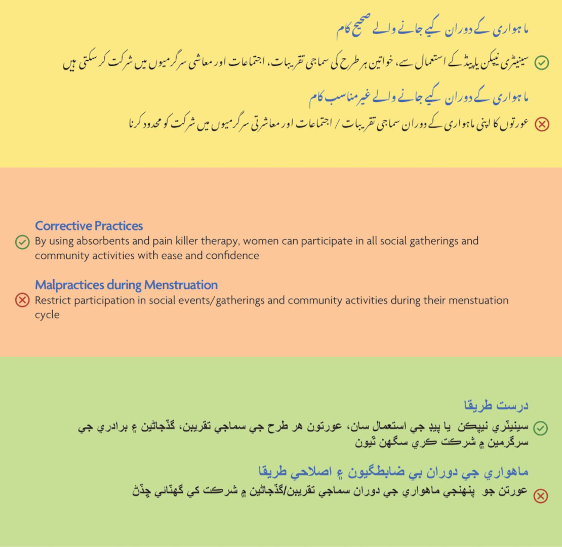
#BreaktheTaboo!
With proper #menstrualhygiene management, access to absorbents & pain management when needed, women & girls can continue participating in social gatherings, education & community activities with comfort & confidence.
#WaterAidPakistan @WaterAidUK #MHDay2026

1
1
2
37

May 11
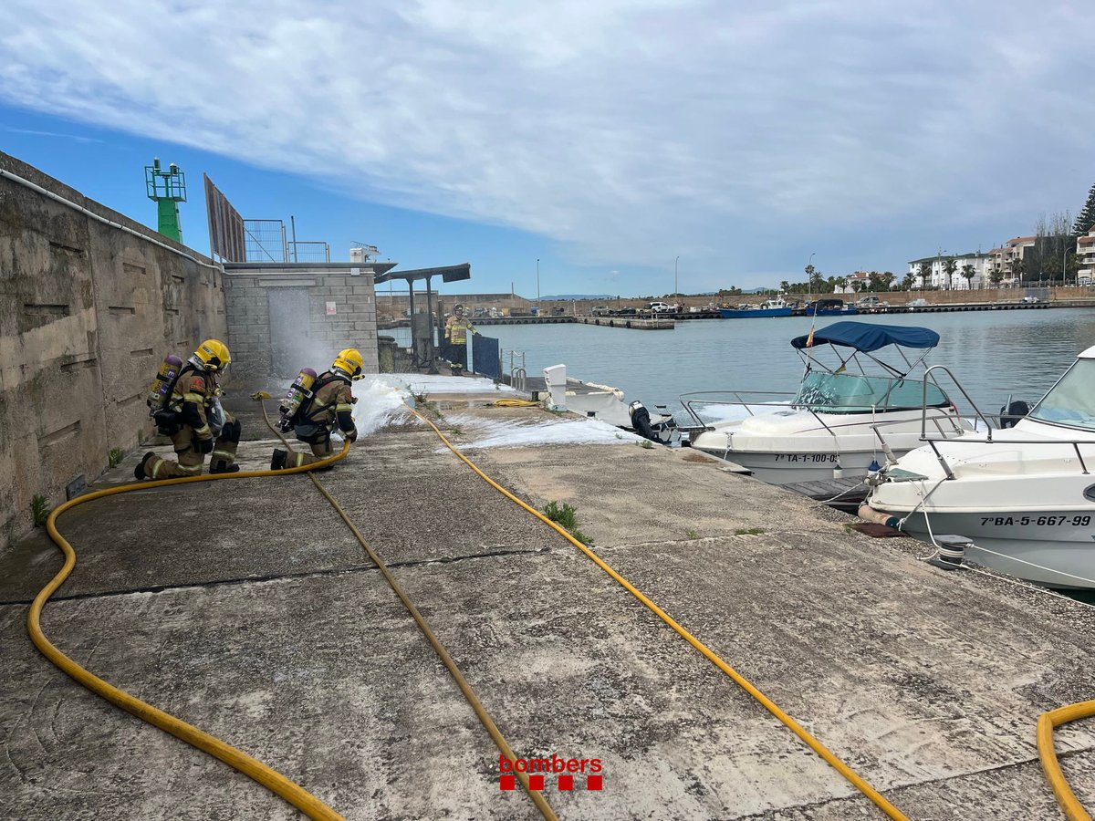
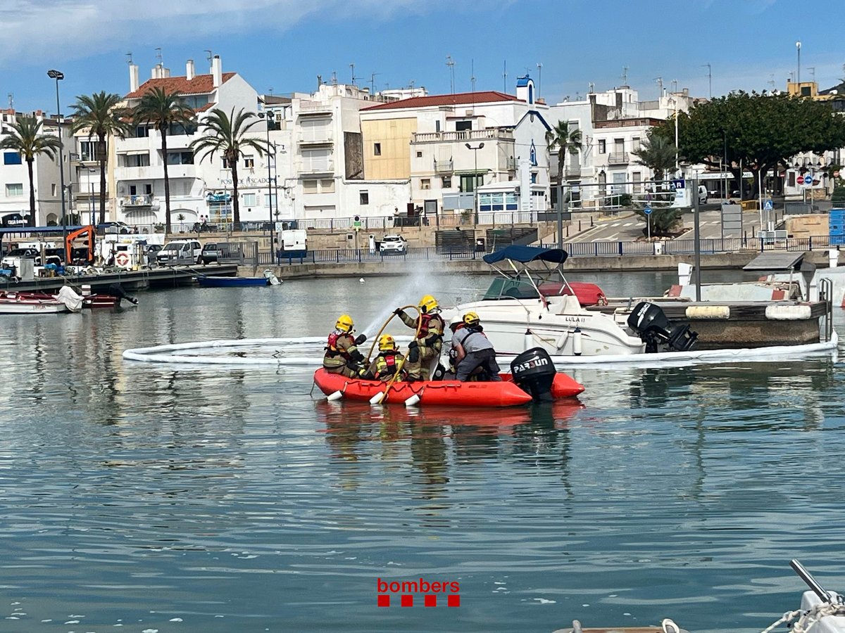
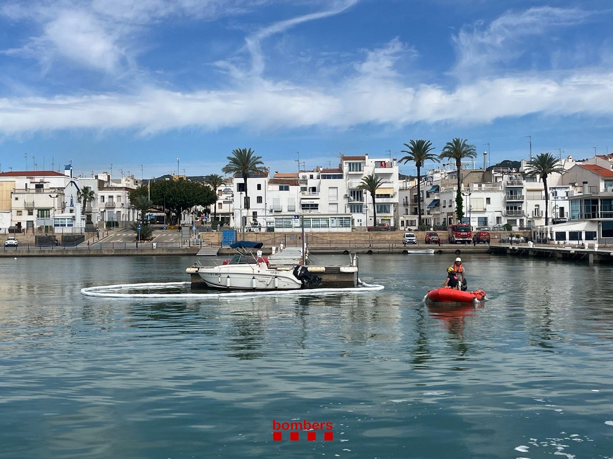
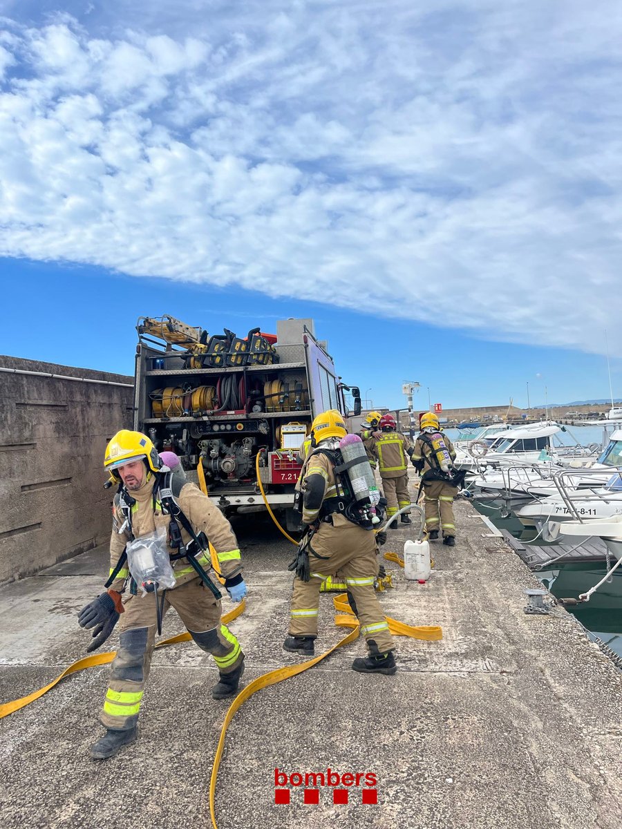
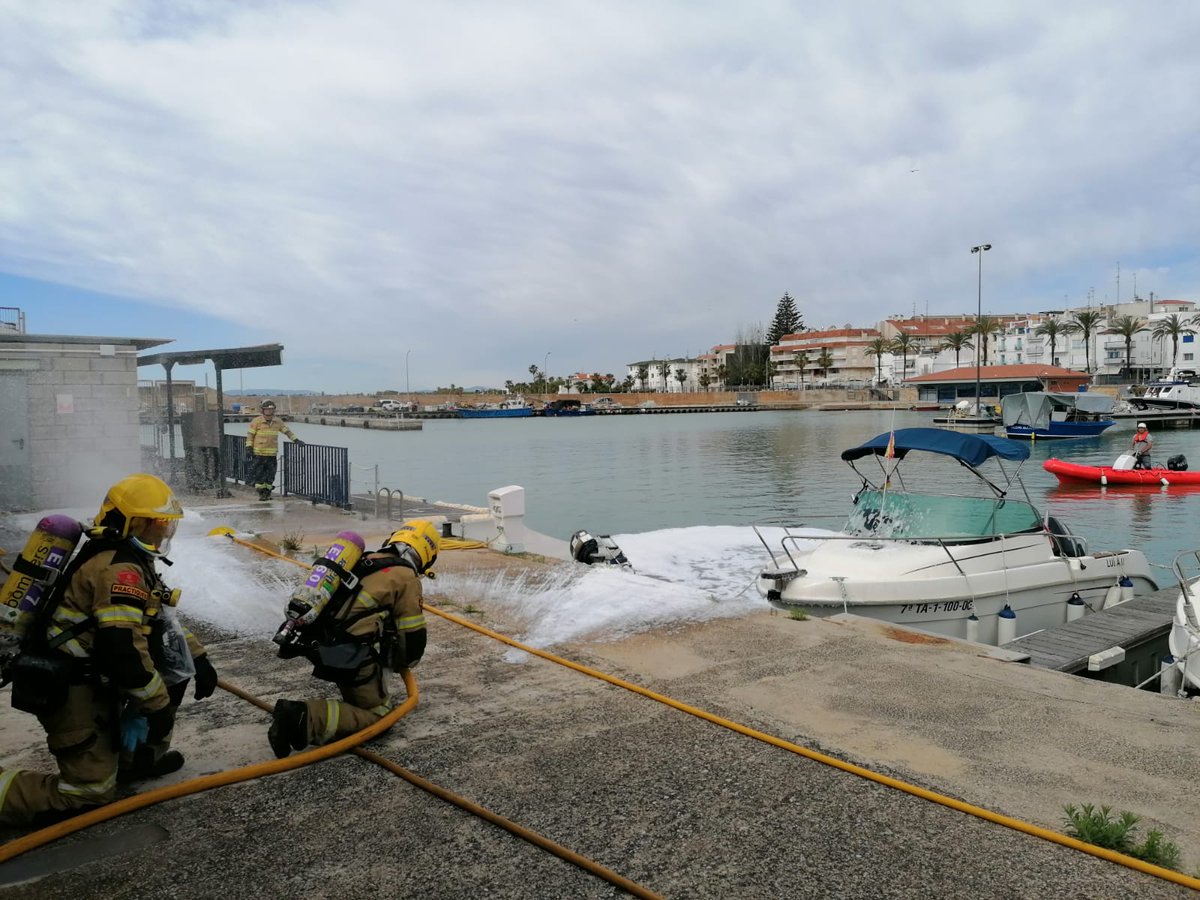
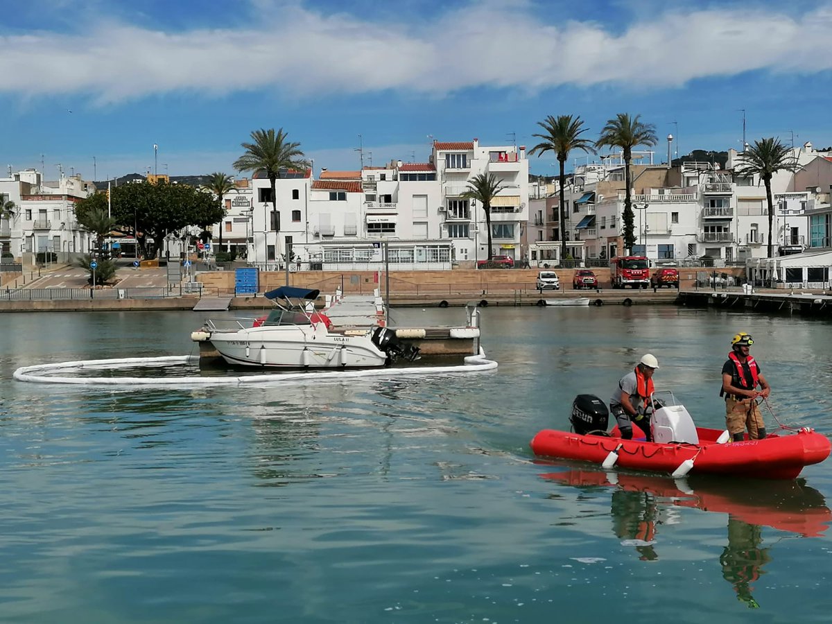
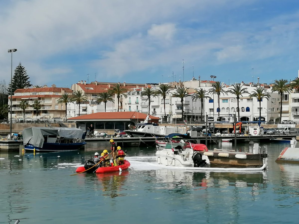
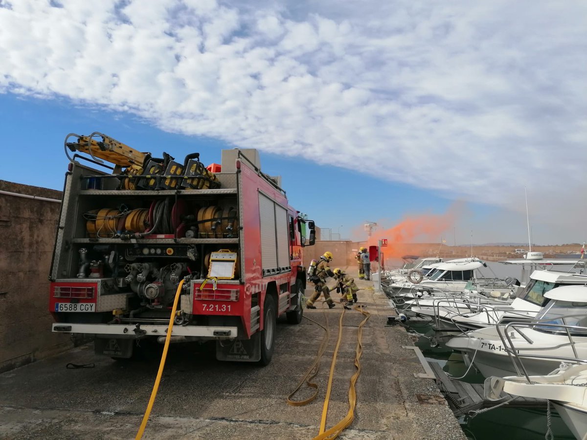
Avui, #bomberscat #RETerresEbre, conjuntament amb el Port de les Cases d’Alcanar —que ha activat el PAU— hem dut a terme un exercici d’extinció d’incendi en una embarcació d’oci amb vessament.
Hem practicat maniobres de protecció, extinció i desplegament de barreres absorbents.
2
29
2,785

Avui els @bomberscat dels parcs d'#Ulldecona i #Amposta han fet un exercici per activar el PAU del port de #lescasesdalcanar, per practicar maniobres de protecció, extinció i col·locació de barreres absorbents.
El supòsit ha estat l'incendi d'un vaixell amb vessament.
1
3
342

Apr 27
🩹♻️ El tèxtil sanitari, a la resta!
⚫️Amb el canvi de contenidors, ara el tèxtil sanitari ha d'anar al contenidor de la fracció resta 👇
Bolquers, compreses i tampons, bastonets per netejar les orelles, corones absorbents de lactància, tovalloles humides, fil dental o petits residus de cures domèstiques.
Tota la informació sobre les fraccions:
web.girona.cat/netejairesidu…

4
4
4
1,242