
Symmetry Breaking in the U-Net: Hybrid Deep-Learning Multi-Class Segmentation of HeLa Cells in Reflected Light Microscopy Images
✏️ Ali Ghaznavi et al.
🔗 brnw.ch/21x1hJY
Viewed: 3438; Cited: 5
#mdpisymmetry #semanticsegmentation #cellsegmentation
@JihoceskaUni

1
2
44

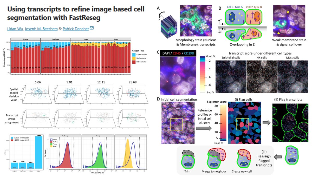
#SpatialTranscriptomics #CellSegmentation
FastReseg
github.com/Nanostring-Biosta…
Using transcript scoring "spatial doublet" test to rectify segmentation errors🥸
vs transcript-dominant approaches: Baysor Proseg
@brukerspatial @SciReports 2025
nature.com/articles/s41598-0…
12
56
6,252

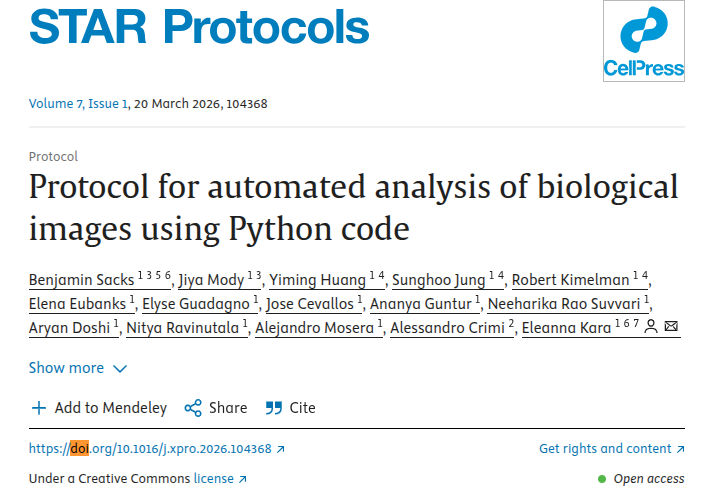
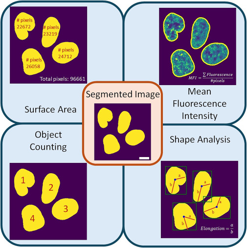
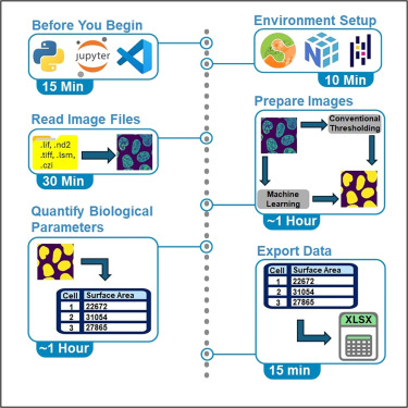
Check our yet another "Protocol for automated analysis of biological images using #Python code" sciencedirect.com/science/ar…
-Steps for the analysis of confocal images through Python
-Guidance on the usage of machine learning for #cellsegmentation
-Troubleshooting of common issues
5
15
1,487

20 Aug 2025
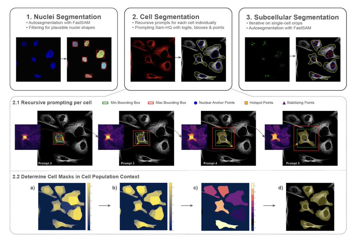
subCellSAM: Zero-Shot (Sub-)Cellular Segmentation for Hit Validation in Drug Discovery
1. Introducing subCellSAM, a novel method for zero-shot segmentation of cellular and subcellular structures in high-content screening (HCS) for drug discovery. This novel approach leverages a pre-trained segmentation foundation model without any fine-tuning, guided by an in-context learning strategy that incorporates morphological and topological priors of cell images.
2. The core innovation of subCellSAM lies in its three-step process for nuclei, cell, and subcellular segmentation. It employs a self-prompting mechanism that uses growing masks and strategically placed foreground/background points to guide the segmentation process, ensuring biologically plausible and accurate results without dataset-specific tuning.
3. subCellSAM demonstrates superior performance in cell segmentation across diverse datasets, outperforming specialized methods such as CellPose 3, DeepCell, and CellSAM. It achieves a mean Dice Score (DSC) of 0.901 and Intersection over Union (IoU) of 0.832 on the BBBC008 dataset, showcasing its robustness and generalizability.
4. In the context of hit validation for drug discovery, subCellSAM effectively segments subcellular structures without any parameter tuning, resulting in high-quality downstream results. It achieves Z'-factor values comparable to baseline methods and accurately calculates EC50 values for compound potency, highlighting its potential for automated HCS analysis pipelines.
5. The method's modular design allows for flexible integration of different models, and its reliance on pre-defined morphological and topological priors makes it a viable strategy for reducing manual configuration in HCS analysis. This approach paves the way for more efficient and accurate drug discovery processes.
📜Paper: arxiv.org/abs/2508.13701
#ComputationalBiology #DrugDiscovery #CellSegmentation #ZeroShotLearning #InContextLearning
2
5
895
13 Apr 2025
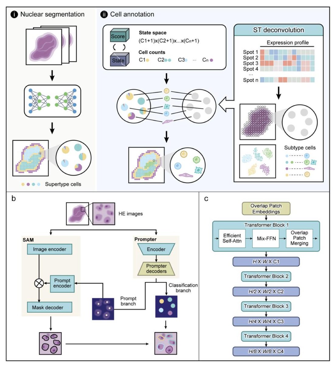
SpatioCell: A Deep Learning Algorithm for High-resolution Single-cell Mapping through Deep Integration of Histology Image and Sequencing Data
1. SpatioCell is a novel deep learning framework that achieves single-cell resolution from multicellular-resolution spatial transcriptomics (ST) by integrating histological images and sequencing data, redefining spatial cell mapping in complex tissues.
2. It combines fine-tuned Segment Anything Model (SAM) with an automatic prompt generator based on SegFormer, enabling accurate nuclear segmentation and morphological classification even in dense, overlapping, or weakly stained H&E images.
3. SpatioCell integrates image-derived morphological profiles with transcriptome-derived cell type compositions via dynamic programming, modeled as a knapsack problem, to assign high-confidence cell types at single-cell resolution within each spatial spot.
4. In benchmarks on six public datasets (e.g., PanNuke, CellbinDB), SpatioCell outperformed state-of-the-art tools like Hover-Net, Cellpose, StarDist, and Mask R-CNN in segmentation and classification metrics, including AJI, PQ, and detection quality.
5. The framework is highly adaptable, demonstrating robust performance on both H&E and DAPI-stained images, including FFPE and fresh-frozen samples, across normal and cancerous tissues from multiple species.
6. On simulated ST datasets (from single-cell resolution data), SpatioCell significantly outperformed image-only or deconvolution-only approaches in cell-type annotation accuracy across four cancers, with gains over 10% in both supertype and subtype levels.
7. Crucially, SpatioCell imputes cell types in unsequenced regions between ST spots by leveraging morphological similarity, enabling continuous whole-slide annotation and recovery of tissue structures like tumor-stroma boundaries and lymphocyte niches.
8. Applied to real tumor ST data (e.g., breast and ovarian cancers), SpatioCell reconstructed microanatomical features like vasculature, tumor margins, and immune infiltration zones with high fidelity, outperforming traditional deconvolution methods.
9. To correct errors from sequencing-based deconvolution, SpatioCell introduces the Competitive Balance Index (CBI), a dynamic correction mechanism that adjusts cell type assignments using morphological predictions, improving annotation consistency.
10. Overall, SpatioCell sets a new standard for spatial transcriptomic analysis by integrating segmentation, morphology, transcriptomics, and dynamic modeling to achieve accurate and biologically informative single-cell mapping across entire tissue sections.
📜Paper: biorxiv.org/content/10.1101/…
#SpatialTranscriptomics #SingleCell #DeepLearning #ComputationalPathology #Histology #CellSegmentation #TissueMicroenvironment #CancerBiology #Bioinformatics #SpatioCell

3
24
1,866
13 Apr 2025
SpatioCell: A Deep Learning Algorithm for High-resolution Single-cell Mapping through Deep Integration of Histology Image and Sequencing Data
1. SpatioCell is a novel deep learning framework that achieves single-cell resolution from multicellular-resolution spatial transcriptomics (ST) by integrating histological images and sequencing data, redefining spatial cell mapping in complex tissues.
2. It combines fine-tuned Segment Anything Model (SAM) with an automatic prompt generator based on SegFormer, enabling accurate nuclear segmentation and morphological classification even in dense, overlapping, or weakly stained H&E images.
3. SpatioCell integrates image-derived morphological profiles with transcriptome-derived cell type compositions via dynamic programming, modeled as a knapsack problem, to assign high-confidence cell types at single-cell resolution within each spatial spot.
4. In benchmarks on six public datasets (e.g., PanNuke, CellbinDB), SpatioCell outperformed state-of-the-art tools like Hover-Net, Cellpose, StarDist, and Mask R-CNN in segmentation and classification metrics, including AJI, PQ, and detection quality.
5. The framework is highly adaptable, demonstrating robust performance on both H&E and DAPI-stained images, including FFPE and fresh-frozen samples, across normal and cancerous tissues from multiple species.
6. On simulated ST datasets (from single-cell resolution data), SpatioCell significantly outperformed image-only or deconvolution-only approaches in cell-type annotation accuracy across four cancers, with gains over 10% in both supertype and subtype levels.
7. Crucially, SpatioCell imputes cell types in unsequenced regions between ST spots by leveraging morphological similarity, enabling continuous whole-slide annotation and recovery of tissue structures like tumor-stroma boundaries and lymphocyte niches.
8. Applied to real tumor ST data (e.g., breast and ovarian cancers), SpatioCell reconstructed microanatomical features like vasculature, tumor margins, and immune infiltration zones with high fidelity, outperforming traditional deconvolution methods.
9. To correct errors from sequencing-based deconvolution, SpatioCell introduces the Competitive Balance Index (CBI), a dynamic correction mechanism that adjusts cell type assignments using morphological predictions, improving annotation consistency.
10. Overall, SpatioCell sets a new standard for spatial transcriptomic analysis by integrating segmentation, morphology, transcriptomics, and dynamic modeling to achieve accurate and biologically informative single-cell mapping across entire tissue sections.
📜Paper: biorxiv.org/content/10.1101/…
#SpatialTranscriptomics #SingleCell #DeepLearning #ComputationalPathology #Histology #CellSegmentation #TissueMicroenvironment #CancerBiology #Bioinformatics #SpatioCell
1
10
1,329

24 Feb 2025
Day 1 of #AGBT25, ft. exciting @10xGenomics announcements!
Later this year, #Flex coming out with 96 barcodes then eventually scaling up to 384, #Xenium #protein capabilities, and #VisiumHD #cellsegmentation.... #DukeMGC can't decide which we're most excited about! 🧬🧠💻


1
4
9
2,514

6 Sep 2024
🎯 Precise #CellSegmentation is challenging. @vizgen_inc uses cell boundary stains and robust algorithms for accuracy.
See these #DataImages of FFPE mouse breast tumor by researchers at @NeuroAlc, showcasing cell boundary staining and segmentation revealing natural cell shapes.
4
10
1,674
7 Aug 2024
🎯 Precise #CellSegmentation is challenging. @vizgen_inc uses cell boundary stains and robust algorithms for accuracy.
See these #DataImages of FFPE mouse breast tumor by researchers at @NeuroAlc, showcasing cell boundary staining and segmentation revealing natural cell shapes.
1
5
7
889
15 May 2024
💻Missed the #Primetech & @vizgen_inc Live Webinar? Watch It On-Demand! 🇯🇵
Explore the #MERSCOPE 1000-plex fully custom gene panel & advanced #CellSegmentation methods using #Cellpose2.
🔗 hubs.ly/Q02wYfP60
#MERSCOPE #CellSegmentation #MachineLearning #JapaneseWebinar
1
3
567
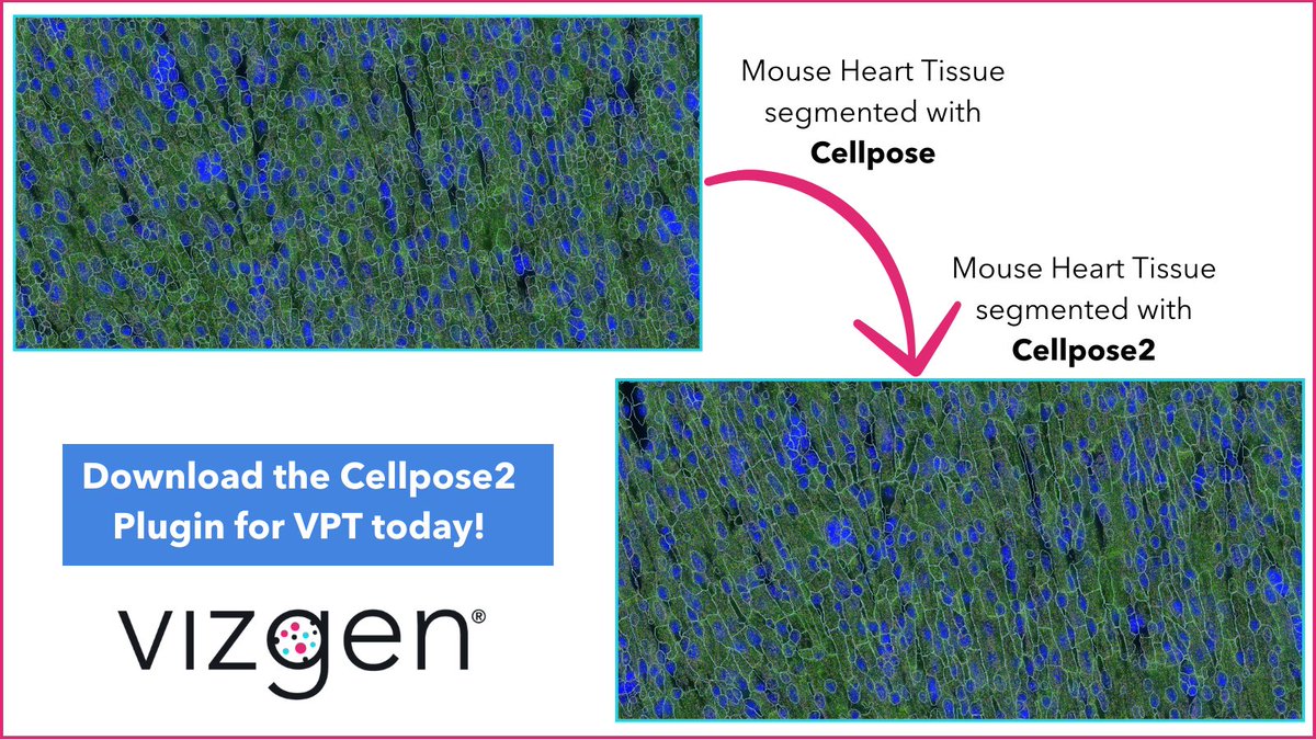
26 Apr 2024
Struggling with cell segmentation in complex tissues like multinucleated muscle or abnormally shaped cells? Enhance your #CellSegmentation with @vizgen_inc's #Cellpose2 Plugin! Learn more about VPT and the plugin on our website- hubs.ly/Q02v04-l0
#MERSCOPE #CellSegmentation
4
9
1,108
22 Apr 2024
💻Webinar Alert for #SpatialTranscriptomics Customers in Japan!
Developments in Spatial Genomics Analysis and Advanced Cell Segmentation
📅 Wednesday, April 24, 2024
⏰15:00 - 15:45 JST
🎙️Wenbin Gu, Primetech Co.
🔗 hubs.ly/Q02tBwP_0
#CellSegmentation #MERSCOPE
1
5
794
15 Apr 2024
💻Webinar Alert for #SpatialTranscriptomics Customers in Japan!
Developments in Spatial Genomics Analysis and Advanced Cell Segmentation
📅 Wednesday, April 24, 2024
⏰15:00 - 15:45 JST
🎙️Wenbin Gu, Primetech Co.
🔗 hubs.ly/Q02sQSrr0
#CellSegmentation #MERSCOPE

1
1
9
925
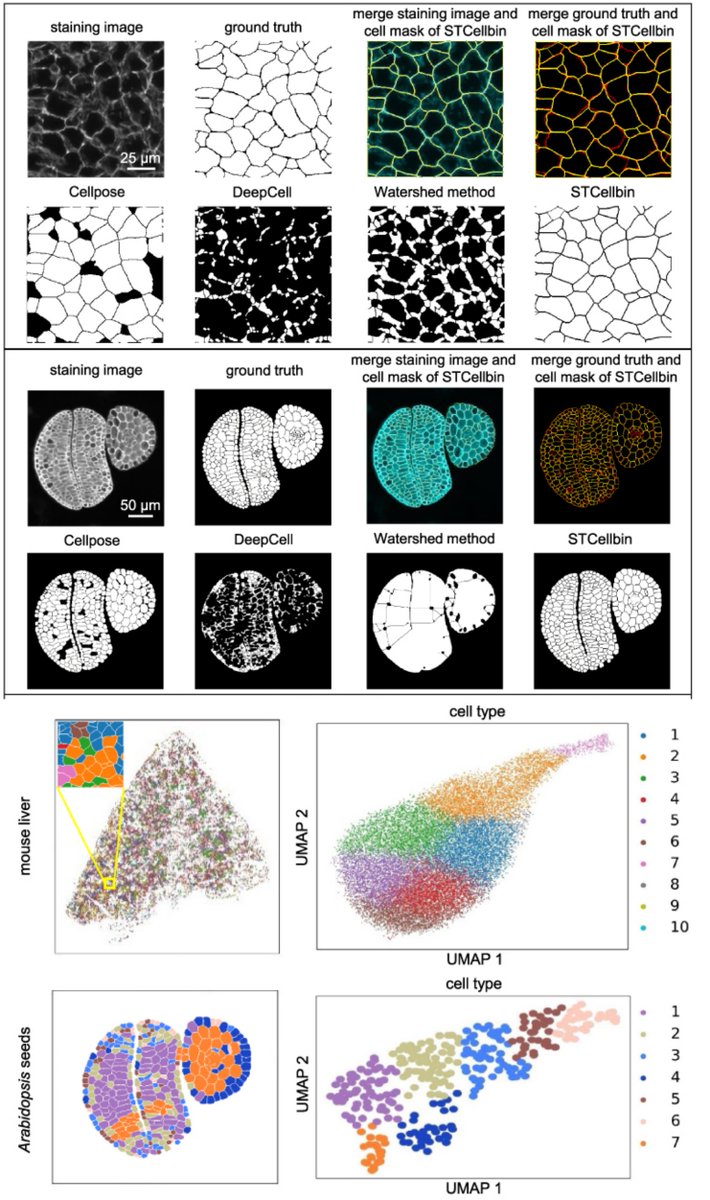
STCellbin
Use Nuclei Cell Membrane/Wall stains to improve #CellSegmentation & single-cell-resolved #SpatialTranscriptomics
Tailored for #StereoSeq, but likely implementable for other ST methods
Susanne Brix & Xun Xu labs @GigaByteJournal 2024
gigabytejournal.com/articles…
2
18
87
8,370

10 Mar 2024
And the O.S.C.A.R. for the Best Cell Makeup and Styling role goes to.....
@10xGenomics Xenium Cell Segmentation Add-On Kit for "12 Angry Reviewers"!
Congratulations!
#xenium #spatialbiology #oscars2024 #cellsegmentation
2
3
351
4 Mar 2024
Customize your #CellSegmentation with the new #Cellpose2 Plugin for the #Vizgen Post-Processing Tool!
Visit our website to learn how you can improve segmentation results for challenging tissues with the plugin for #VPT:
hubs.ly/Q02mN7ZV0
#MERSCOPE #MERFISH #SpatialOmics
2
8
857
26 Feb 2024
Customize your #CellSegmentation with the new #Cellpose2 Plugin for the Vizgen Post-Processing Tool!
Visit our website to learn more about #VPT and the Cellpose2 cell segmentation plugin: hubs.ly/Q02m2Xf80
#Vizgen #MERSCOPE #MERFISH #Spatialomics
1
3
10
1,002
14 Feb 2024
A spatial haiku from us to you this #ValentinesDay💖
Genes whisper their tales,
Segmentation unveils truth,
Cells' spatial ballet.
Share your #MERSCOPE #SpatialTranscriptomics ❤️ poems with us today!
#AcademicTwitter #Vizgen #MERFISH #CellSegmentation #Cellpose2 #VPT
2
2
7
820
9 Feb 2024
Customize your #CellSegmentation with the new #Cellpose2 Plugin for the #Vizgen Post-Processing Tool!
Visit our website to learn how you can improve segmentation results for challenging tissues with the plugin for #VPT:
hubs.ly/Q02k2S000
#MERSCOPE #MERFISH #Spatialomics
6
17
1,525

22 Jan 2024
Why is #CellSegmentation so challenging? It can be a daunting task due to several factors:
1. Heterogeneous Shapes
2. Variation in Size and Shape
3. Weak Boundary Gradients
4. Makeshift Nature of Segmentation Approaches.
More at bit.ly/3vUFole
#SpatialBiology
3
6
696