
Jun 13
BI実践ラボ|アメリエフ技術ブログに新規記事を投稿しました。
「空間的遺伝子発現解析入門③」
staffblog.amelieff.jp/entry/…
空間的遺伝子発現解析のうち、イメージングベース (Imaging-based) の代表的なプラットフォーム (Xenium, CosMx) を解説します。
#enrichplot #GO解析 #可視化 #新規記事 #技術ブログ

1
8
704

We're hiring at Xenium HR! Open roles in Tualatin, OR (hybrid) and remote:
🔹 Employee Relations Partner II
🔹 HR Business Partner II
🔹 Implementation Specialist
🔹 Employee Experience Rep
🔹 Benefits Specialist II
🔹 HR Account Rep
Apply today! ➡️na2.hubs.ly/H065-hg0

13

In '24 we brought "Cepelia" and managed to snatch 1st on Xenium.
Last weekend we released "The Lab" - and...
#c64 #demoscene #Elysium
youtu.be/9g91vnJ6Bz4?is=8NQo…

1
6
273
Dr.ジャスティス☺︎🦖 retweeted

Jun 9
空間トランスクリプトームやりたいって言ったのは確かだけど。タイムラインギチギチの共同研究のXenium立ち上げ責任者みたいな立ち位置に祭り上げられそうで、日々胃が痛い。
1
9
1,343

Xenium 空間オミックス解析(spatial omics)主に高解像度spatial transcriptomics/RNA解析と付随タンパク解析

レイニンXLS+LTSピペットとLTS低吸着フィルター滅菌チップが推奨されています。ビデオの中でも登場します。
216

3/
NephroBase - the first kidney foundation AI model:
🔅 40 million cells, 1 billion parameters across human, pig, rat & mouse
🔅 Integrates snRNA, snATAC, XENIUM, OSMX spatial data

1
2
78

Jun 4
【昨日の注目リリースまとめ】
📷 資金調達
■ フォースタートアップス株式会社
prtimes.jp/main/html/rd/p/00…
フォースタートアップスが2026年5月のスタートアップ資金調達金額ランキングを発表。1位は240億円を調達したOishii Farm(次世代植物工場)、2位は48億8000万円を調達したmovus technologies(自動車サブスクリプションサービス)、3位は45億円を調達したIVRy(電話自動応答サービス)。
■ Direava株式会社
prtimes.jp/main/html/rd/p/00…
Direavaは未来創生3号ファンド、ニッセイ・キャピタル、SBIインベストメントから第三者割当増資を実施。外科領域のフィジカルAI開発と海外展開を加速する。
📷 業務提携・協業
■ 浜松ホトニクス株式会社
prtimes.jp/main/html/rd/p/00…
浜松ホトニクス、NKT Photonics、Yaqumoの3社が量子コンピュータ産業化に向けた先端光学システムの共同開発とグローバルサプライチェーン構築の覚書を締結。
■ 株式会社第一興商
prtimes.jp/main/html/rd/p/00…
第一興商は、インディペンデントレコード会社ULTRA-VYBEと提携し、2026年6月4日から原盤カラオケ「ANTENNA!」への楽曲提供を開始。アーティストとの接点を拡大し、コンテンツを強化する。
■ NTT株式会社
prtimes.jp/main/html/rd/p/00…
NTTはアイルランドのMBRYONICSと宇宙向け光通信分野のパートナーシップを締結。IOWN技術の宇宙適用を進める。
■ コクヨ株式会社
prtimes.jp/main/html/rd/p/00…
コクヨはピコシステムと提携し、電子帳票配信システム「@Tovas」と販売管理システム「はんなり匠」「Spicα」の連携を開始。帳票データの自動配信を実現し、事務工数とコストの削減を図る。
■ 株式会社博報堂プロダクツ
prtimes.jp/main/html/rd/p/00…
博報堂プロダクツと大和が業務提携し、AEMを活用した高品質なデジタルマーケティング基盤の協働提案を開始。
■ 株式会社商船三井
prtimes.jp/main/html/rd/p/00…
商船三井は米国初の世界最大級FLNGプロジェクトに約3億米ドルを出資。Delfin Midstream、Global Infrastructure Partners、Vitolらと提携し、2030年頃の生産開始を目指す。
■ DHLサプライチェーンジャパン株式会社
prtimes.jp/main/html/rd/p/00…
DHLサプライチェーンと東邦ホールディングスが、日本におけるヘルスロジスティクスの強化を目的とした基本合意を締結。両社のネットワークと専門性を活用し、医薬品流通のワンストップサービスなどを展開する。
■ 株式会社Wiz
prtimes.jp/main/html/rd/p/00…
WizとMS&Consultingが業務提携。リサーチ&コンサルティングとコスト削減BPO、AI/DX支援を組み合わせ、企業の事業成長を支援。Wizの山﨑CEOがMS&Consultingの取締役会長に就任。
■ 三井住友トラストクラブ株式会社
prtimes.jp/main/html/rd/p/00…
三井住友トラストクラブは2026年6月から、京都観光向け人力車サービス「京都 忍者屋 人力車優待」を開始。ダイナースクラブ会員を対象に、割引料金で特別な観光体験を提供する。
■ 株式会社PKSHA Technology
prtimes.jp/main/html/rd/p/00…
東大松尾研、PKSHA、Anthropicが協力し、AIが日本社会に与える影響を可視化する「Japan AI Index」の構築を開始。雇用・産業・教育データとLLM利用統計を統合して分析を進める。
■ 日本PCサービス株式会社
prtimes.jp/main/html/rd/p/00…
日本PCサービスの子会社・スマホスピタルが「ドコモ提携修理取次店舗」に認定され、ドコモショップで受付した修理を全国9店舗で即時対応する。
■ ボンディッシュ株式会社
prtimes.jp/main/html/rd/p/00…
ボンディッシュとサンニンが提携し、「食」と「空間」を融合させた次世代ワークプレイスの共同提案を開始。出社したくなるオフィスづくりを支援。
■ 株式会社Relic
prtimes.jp/main/html/rd/p/00…
RelicとRUFUが連携し、海外トレンドと実売データを活用した新商品企画支援サービス「IDEATION Cloud Goods」を提供開始。
■ 日本空輸株式会社
prtimes.jp/main/html/rd/p/00…
日本空輸が「日和ホテル松山」と提携、2026年4月開業。松山への航空券と宿泊のセット予約を開始。
■ WHILL株式会社
prtimes.jp/main/html/rd/p/00…
WHILLはヒースロー空港第3ターミナルで自動運転サービスの実証を開始。英国のABM社と提携し、航空業界の移動サービスをDX化し、旅客の利便性向上とスタッフの負担軽減を目指す。
■ ソースネクスト株式会社
prtimes.jp/main/html/rd/p/00…
ソースネクストは、Genspark Inc.との提携に基づき、次世代AIエージェント「Genspark Team」の国内販売を開始。法人・チーム向けに最適化し、日本独自の商習慣に対応した請求書払いなど、導入の利便性を高めた。
■ ローデ・シュワルツ・ジャパン株式会社
prtimes.jp/main/html/rd/p/00…
ローデ・シュワルツとQuantum Systems社は、次世代無人システムにEW・C-UAS機能を統合するための業務提携を締結。防衛能力の向上を目指す。
■ 株式会社バイウィル
prtimes.jp/main/html/rd/p/00…
福岡市とバイウィルがJ-クレジット活用事業で連携。脱炭素化推進を目的に環境価値の創出・流通を進める。
■ ファンズ株式会社
prtimes.jp/main/html/rd/p/00…
ファンズは台湾の販売金融・BNPL企業AMFCを連結子会社化。Funds AMFCとして再編し、金融包摂を推進する「成長循環」モデルを強化する。
■ FastLabel株式会社
prtimes.jp/main/html/rd/p/00…
FastLabelとMWは「住宅・家事AIロボット開発」に向け、大規模で信頼性の高いデータセット構築を目的に基本合意書(MoU)を締結し協業を開始しました。
■ 株式会社リーピー
prtimes.jp/main/html/rd/p/00…
リーピーはKatawaraと業務提携し、働き方の多様化に対応した採用支援体制を強化する。
■ 株式会社WHERE
prtimes.jp/main/html/rd/p/00…
大東建託がWHEREの不動産AIツールを導入し、衛星データとAIを活用した用地開拓を推進。Deal Techとして一気通貫で支援。
■ RE100電力株式会社
prtimes.jp/main/html/rd/p/00…
RE100電力は、ウィルと系統用蓄電池のアグリゲーション業務代行契約を締結。電力市場での最適運用を通じて再生可能エネルギーの活用と電力系統の安定化を図る。
■ 株式会社エンミッシュ
prtimes.jp/main/html/rd/p/00…
エンミッシュ、AI電話サービス「nocall.ai」と販売パートナー契約。Salesforce連携でコール業務の自動化を支援。
■ 株式会社Yaqumo
prtimes.jp/main/html/rd/p/00…
Yaqumo、NKT Photonics、浜松ホトニクスが量子コンピュータ産業化に向け、先端光学システムの共同開発とサプライチェーン構築を目的とした覚書を締結。
■ 株式会社Nature Innovation Group
prtimes.jp/main/html/rd/p/00…
京阪グループと傘シェアリングサービス「アイカサ」が業務提携し、関西で初となる駅と商業施設など計60スポットでの展開を開始。SDGs達成と環境負荷削減を目的に、使い捨て傘の削減と雨天時の移動インフラを強化する。
■ データセクション株式会社
prtimes.jp/main/html/rd/p/00…
データセクション、タイのAIデータセンター構築に向けASRock Rackと連携。NVIDIA B200搭載サーバー587台を導入し、処理能力を強化。
■ Kudan株式会社
prtimes.jp/main/html/rd/p/00…
KudanとMRDVSは協業し、KudanのVisual SLAM技術をMRDVSのスマートセンサーモジュールに統合。ロボットやフリート向けに、自律的な空間認識を実現する。
■ 株式会社WIZE
prtimes.jp/main/html/rd/p/00…
WIZEはSBI VCトレードと提携し、ソラナ(SOL)の取得・運用体制を強化。国内屈指のソラナ・トレジャリー企業を目指す。
■ 株式会社biomy
prtimes.jp/main/html/rd/p/00…
biomyは10x Genomics社とパートナーシップを締結し、Xeniumプラットフォームのデータを活用したAI駆動型Virtual Cellモデルの開発を進める。
■ 少数株ドットコム株式会社
prtimes.jp/main/html/rd/p/00…
少数株ドットコムが支援するオムライス専門店「オムライスランド」が、東北展開の一歩として仙台駅前に新拠点を開設。ブランドの浸透と地域経済への貢献を目的とする。
■ 株式会社ウィズモー
prtimes.jp/main/html/rd/p/00…
ウィズモーが店舗設計のふなとみ氏と協業し、SNSを活用した受注支援サービスを開始。フォロワー数ではなく本業の売上に直結する運用を提供。
■ 株式会社パロンゴ
prtimes.jp/main/html/rd/p/00…
パロンゴは米Horizon3.aiとパートナーシップを締結し、AI駆動型セキュリティ検証プラットフォーム「NodeZero」の取り扱いを開始。日本企業のサイバーレジリエンス強化を支援する。
■ 一般社団法人ドローンサービス推進協議会
prtimes.jp/main/html/rd/p/00…
ドローンサービス品質認証(DSQ認証)を運営するDSPAと、ドローンプラットフォーム「XROSS」が連携。認証情報がプラットフォーム上で可視化され、発注者が事業者を客観的に選定できる環境を整える。
■ プレミアムウォーター株式会社
prtimes.jp/main/html/rd/p/00…
プレミアムウォーターは、こどもっちパークを運営するF.K.Solutionsと業務提携し、子育て世帯を応援する取り組みを推進。2026年6月からこどもっちパークの利用料金をより使いやすくする。
1
9
10,906

Jun 3
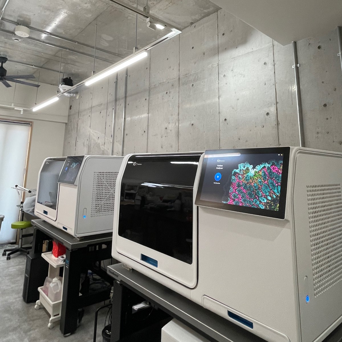
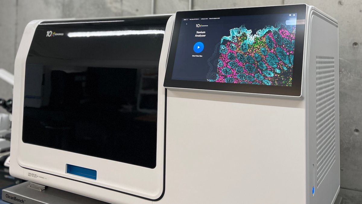
【CyberomiX 機器図鑑 #01 Xenium In Situ】
CyberomiXでは、空間解析プラットフォーム
「Xenium In Situ(ゼニウム イン サイチュー)」
という最新の機械を導入しています🧬
現在は2台体制で稼働中🤖🤖
研究者のみなさまの多様なニーズに対応できる環境を整えています。
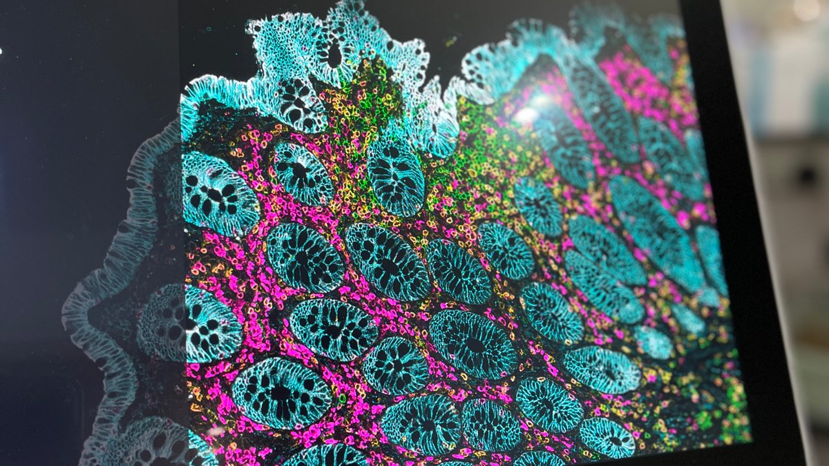
Xenium In Situとは、
『どんな細胞がどこで何をしているか?』を組織の中で可視化できる技術です。
例えば腫瘍の中でも、
・増殖が活発な場所
・免疫細胞が集まる場所
・薬剤が効きにくい場所
などが大きく異なります。
Xeniumを用いることで、
こうした "細胞の局所環境"を高解像度で解析でき
💊新薬開発
👀バイオマーカー探索
👤個別化医療
などへの応用が期待されています。
CyberomiXでは、最新の解析技術を通じて研究者のみなさまの「もっと知りたい」に応えられる環境づくりを進めています。
#遺伝子解析 #ゲノム解析 #CyberomiX #機器図鑑
2
18
1,471

Jun 1
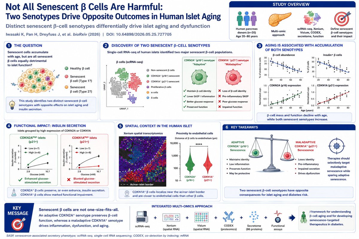
Not All Senescent β Cells Are Harmful: A Human Pancreas Atlas Reveals Adaptive and Maladaptive Senotypes
Cellular senescence is increasingly recognized as a driver of aging and age-related disease. Yet senescent cells are not a uniform population. A new human pancreas atlas now demonstrates that senescent β cells exist in at least two fundamentally different states—one adaptive and one maladaptive.
Using integrated single-cell RNA-seq, Xenium spatial transcriptomics, Visium spatial transcriptomics, CODEX spatial proteomics, secretome profiling, and functional insulin secretion assays, investigators analyzed pancreatic tissue from donors spanning ages 20–80 years.
The study identified two major senescent β-cell populations:
🟢 CDKN2A⁺ (p16⁺) senotype
🔴 CDKN1A⁺ (p21⁺) senotype
The distinction proved biologically important.
The CDKN2A⁺ senotype retained β-cell identity, preserved expression of key transcription factors such as MAFA and PDX1, maintained glucose responsiveness, and displayed a relatively low-inflammatory secretory phenotype. Functional studies showed that islets enriched for CDKN2A expression exhibited superior glucose-stimulated insulin secretion.
In contrast, the CDKN1A⁺ senotype exhibited many hallmarks of pathological aging.
These cells showed:
▪ Loss of insulin gene expression
▪ Reduced MAFA, PDX1, GK and ABCC8 expression
▪ Impaired glucose-stimulated insulin secretion
▪ Pro-inflammatory SASP programs
▪ Increased immune-cell infiltration within islets
Perhaps the most striking result came from direct functional testing.
Human islets with high CDKN1A expression failed to appropriately increase insulin secretion during both static and dynamic glucose stimulation assays. By contrast, CDKN2A-high islets remained functionally competent and even demonstrated enhanced secretory responses.
The spatial analyses revealed additional insights.
CDKN1A⁺ β cells preferentially localized near the acinar–islet border and were significantly closer to endothelial cells than non-senescent β cells. This raises the possibility of bidirectional signaling between vascular aging and β-cell senescence.
Aging was associated with progressive accumulation of both senotypes, but the consequences differed dramatically. As shown in the lifespan analyses, β-cell abundance, insulin content, and C-peptide levels declined with age, while CDKN1A and CDKN2A expression increased. However, only the CDKN1A program was consistently linked to loss of endocrine identity and dysfunction.
The implications extend beyond diabetes biology.
Current senolytic approaches often treat senescent cells as uniformly harmful. These findings suggest that indiscriminate elimination of all senescent β cells may be biologically misguided. Instead, future therapies may need to selectively target the maladaptive CDKN1A⁺ senotype while preserving or even supporting the adaptive CDKN2A⁺ population.
More broadly, the study provides compelling evidence that aging tissues are composed of distinct senescence programs with divergent physiological consequences—a concept likely relevant far beyond the endocrine pancreas.
Reference
Iwasaki K, Pan H, Dreyfuss J, et al. Distinct senescent β-cell senotypes differentially drive islet aging and dysfunction. bioRxiv (2026). DOI: 10.64898/2026.05.25.727705.
1
6
13
484

May 25
あくまで一例かつ簡易的ですが以下のような感じ。昔研究室でXenium(sp-rna-seq)の機械初めて見たときすごい感動した、ちなみに費用はめっちゃかかる、一応10x-genomicsがleading company
ゲノムの発現状況を位置情報込みで取れるってのが学生だった自分的に革命的でした

1
3
579

May 22
🎓 Position: Doctoral Fellow – Molecular Muscle Biology | 🇧🇪 PhD Opportunity at Ghent University
🏫 University: Ghent University
📍 Location: Ghent, Belgium 🇧🇪
🏢 Department: Movement and Sports Sciences
👨🏫 Supervisor: Prof. Wim Derave
⏰ Deadline: 30 June 2026
📅 Duration: Up to 4 years
💰 Salary: 100% net salary (AAP equivalent, tax-free fellowship)
Interested in understanding how muscles adapt to exercise at the molecular level?
This PhD project explores how skeletal muscle responds to exercise by studying transcriptional activity within and between muscle fibers. Using cutting-edge techniques like spatial transcriptomics (RNAscope & Xenium), you’ll investigate how different muscle fibers adapt—even when not directly activated.
The research combines animal and human studies, aiming to uncover fundamental mechanisms behind muscle plasticity, with implications for health, performance, and chronic disease prevention. You’ll collaborate with international experts in bioinformatics, molecular biology, and neurobiology across leading research groups.
👤 Ideal Candidate:
• Master’s in biomedical sciences, biology, bioinformatics, medicine, or related fields
• Strong interest in muscle physiology and molecular biology
• Experience with lab techniques (e.g., transcriptomics, immunohistochemistry)
• Basic programming/data analysis skills (R/Python)
• Independent, detail-oriented, and collaborative mindset
• Proficient in English
🌟 Why Apply:
• Work on high-impact research in exercise biology and health
• Access state-of-the-art facilities (genomics, imaging, bioinformatics)
• Join an internationally recognized research lab (Ghent Muscle Lab)
• Strong collaboration across disciplines and countries
• Structured PhD training and career development support
• Excellent work-life balance with 36 days leave benefits
🌍 About Ghent:
A vibrant student city in Belgium, Ghent offers a rich cultural scene, historic charm, and a strong international research environment—making it an ideal place to live and study.
🔗 More Info:
phdscanner.com/opportunities…
#PhDOpportunity #BiomedicalResearch #MuscleBiology #PhDPositions #GhentUniversity #Belgium #LifeSciences #Bioinformatics #ResearchCareers
2
354


A recent independent study published in Nature evaluated imaging-based #spatialtranscriptomics platforms in inflammatory bowel disease (IBD) tissues, CosMx SMI demonstrated higher detection efficiency than Xenium across commercially available panels. 🔗 nature.com/articles/s41467-0…

1
3
136
May 17
🧫 PDAC Has Entered the Era of Integrated Tumor Microenvironment Atlases
Pancreatic cancer spatial biology is rapidly evolving from descriptive single-cell profiling into multi-modal ecosystem mapping capable of defining actionable therapeutic niches.
A new generation of integrated PDAC atlases is now combining:
• scRNA-seq
• Visium spatial transcriptomics
• Xenium high-resolution imaging
• bulk RNA deconvolution
• paired primary/metastatic spatial datasets
into unified tumor microenvironment frameworks. (PMC)
The scale is becoming enormous.
Recent PDAC atlas efforts collectively span:
🔹 >180,000–700,000 single cells
🔹 multi-cohort spatial transcriptomics
🔹 metastatic paired sampling
🔹 and increasingly subcellular-resolution imaging platforms.
But the most important shift is conceptual.
The field is moving away from:
“Which cell types exist?”
toward:
“Which spatially organized cellular programs drive progression, immune collapse, fibrosis, and therapy resistance?”
One emerging axis appears repeatedly across datasets:
🧱 POSTN fibroblasts
×
🧫 SPP1 macrophage programs
These stromal–myeloid ecosystems are consistently associated with:
• ECM remodeling
• EMT activation
• invasive phenotypes
• immune suppression
• and poor prognosis.
POSTN signaling in particular is becoming one of the dominant stromal drivers in PDAC biology. Integrated single-cell and spatial analyses show that POSTN-enriched fibroblasts interact with tumor cells through integrin signaling pathways including ITGAV/ITGB5, activating PI3K/AKT/β-catenin programs linked to aggressive disease. (IJBS)
At the opposite end of the spectrum, immune-active niches enriched for:
• CCL4 T cells
• plasma-cell programs
• antigen presentation signatures
appear associated with more immunologically permissive microenvironments.
This suggests that future PDAC therapy may require simultaneous remodeling of both stromal and immune ecotypes.
Not simply “kill tumor cells.”
But spatially re-engineer the ecosystem.
The implications are enormous.
With Xenium and MERFISH-scale imaging now approaching subcellular and junction-level resolution, PDAC atlases are evolving into functional maps for:
• theranostic imaging
• spatial biomarker selection
• resistance prediction
• and ecotype-guided trial design.
PDAC research is no longer entering the atlas era.
It is entering the ecosystem engineering era.
#PDAC #SpatialTranscriptomics #SingleCell #TumorMicroenvironment #Xenium #MERFISH #CancerAtlas

5
12
40
2,502
May 16
The era of static tumor atlases is ending.
We are entering the 11th dimension of oncology: Non-Invasive cfDNA-based Spatial Ecotypes. 🩸🔬
For years, decoding the tumor microenvironment (TME) required invasive spatial profiling platforms like Xenium, CosMx, Visium, and Stereo-seq. These technologies transformed cancer biology, but they remained expensive, tissue-dependent, and difficult to repeat longitudinally.
Now, two landmark studies have fundamentally shifted the field.
A massive Stanford/Harvard consortium study published in Nature (2026; DOI: 10.1038/s41586-026-10452-4) integrated over 10 million spatial transcriptomic spots and single cells across carcinomas and melanomas. Using machine learning, the team identified 9 conserved Spatial Ecotypes (SEs)—multicellular “neighborhoods” defined by distinct cellular compositions, signaling programs, and spatial topology.
These SEs directly correlated with:
• overall survival (OS)
• progression-free survival (PFS)
• immune checkpoint inhibitor (ICI) response
• invasive tumor fronts and immune niches
But the real breakthrough came next.
Each spatial ecotype carried a unique DNA methylation signature. Using deep learning on plasma cfDNA, investigators reconstructed the tumor’s spatial ecosystem directly from blood. In melanoma patients, a simple liquid biopsy predicted immunotherapy response by recovering SE composition non-invasively.
At nearly the same time, the Wang Lab’s Nature Cancer 2025 “TabulaTIME Pan-Cancer” atlas (DOI: 10.1038/s43018-025-01039-5) established the pan-cancer structural backbone:
• 4.48 million integrated cells
• 36 cancer types
• 103 harmonized studies
• conserved immunosuppressive barriers including $CTHRC1^ $ CAFs and $SLPI^ $ macrophages
Together, these studies redefine spatial oncology:
Tissue atlas → Pan-cancer ecotypes → cfDNA spatial recovery
We are no longer limited to watching tumors through a single biopsy snapshot.
The tumor microenvironment can now be serially monitored through blood.
The future of spatial biology is not only on glass slides.
It is circulating through our veins. 🩸✨
#SpatialBiology #LiquidBiopsy #SingleCell #CancerResearch #Oncology #Bioinformatics #MachineLearning

3
12
783

Excited to present the first pre-print from our group, an investigation the human bone marrow microenvironments in patients with myelodysplastic syndromes (MDS) and normal age-matched subjects using Xenium genotype-informed spatial transcriptomics:
biorxiv.org/content/10.64898…
6
37
141
12,595

May 12
Evidence of off-target probe binding affecting 10x Genomics Xenium gene panels compromise accuracy of spatial transcriptomic profiling
elifesciences.org/articles/1…
4
13
3,923
May 11
Phoenix may be a major inflection point for spatial biology
A new bioRxiv preprint introduces Phoenix, a generative AI system that predicts single-cell spatial transcriptomics directly from routine H&E pathology slides — potentially transforming how we study cancer ecosystems at population scale.
The scale is striking.
Phoenix was trained on:
22.2 million cell-image/expression pairs
16 organ systems
79 Xenium slides 924 tissue microarray cores
10,000 GPU hours on a pre-exascale supercomputer
using high-resolution Xenium spatial datasets rather than lower-resolution Visium data.
The key advance is not simply “predicting genes from images.”
Many previous methods failed to generalize across:
institutions,
tissues,
stain variability,
unseen cohorts,
and batch effects.
Phoenix instead demonstrates:
zero-shot prediction,
transfer across unseen organs,
pan-cancer spatial inference,
and clinically relevant biomarker discovery.
Importantly, this is not just benchmark engineering.
The authors used Phoenix to recover meaningful biology across multiple systems.
Examples include:
breast cancer subtype ecosystems,
colorectal tumor budding evolution,
pancreatic PanIN progression,
immune-fibroblast spatial interactions,
chemotherapy/radiotherapy response,
sarcoma immune remodeling,
and KRAS-driven spatial states in mouse PDAC.
One of the most interesting concepts is the emergence of pan-cancer spatial ecotypes.
Across 9,544 TCGA patients, Phoenix identified recurrent multicellular ecosystems:
EC1 → dysfunctional inflammatory state
EC2 → immune competent state
EC3 → fibrotic/remodeling state
with distinct prognostic implications.
The EC3 “stromal remodeling” ecosystem is especially notable because it echoes growing evidence across:
fibrosis,
aging,
CAF biology,
and chronic wound repair
that maladaptive stromal niches may actively sustain disease progression.
Another major implication:
Phoenix enables virtual spatial transcriptomics on archived pathology collections.
Instead of running expensive Xenium/Visium workflows on thousands of samples, existing H&E archives could potentially become:
spatial atlases,
biomarker discovery engines,
therapy-response maps,
and longitudinal ecosystem datasets.
The paper is also a reminder that the future of pathology may not be “AI replaces pathology,” but rather:
AI converts morphology into systems biology.
There are still important limitations:
dependence on Xenium-quality training data,
restricted gene panels,
uncertainty across platforms,
and the need for prospective clinical validation.
But conceptually, this feels important.
Spatial omics may be transitioning from a boutique assay into a scalable computational layer on top of routine histology.
bioRxiv DOI: 10.64898/2026.04.25.720812

1
1
2
98

May 9
Resolving sensitivity, specificity and signal contamination in Xenium spatial transcriptomics | Nature Methods nature.com/articles/s41592-0…
3
12
963