
4 Nov 2025
Via #OPG_Optica: Optical attention mechanism for high-resolution computational imaging bit.ly/4nzPz3W #HighresolutionImaging #OpticalAttention @HIT_1920
1
7
312

24 Sep 2025
🔜 #UnveilingSoon
From complexity to clarity — where decisions begin with clear information.
Designed to support smarter, faster clinical decisions.
A new era of innovation is on the horizon. Stay tuned.
#SamsungHealthcare #AIHealthcare #SmartDiagnostics #HighResolutionImaging
2
62

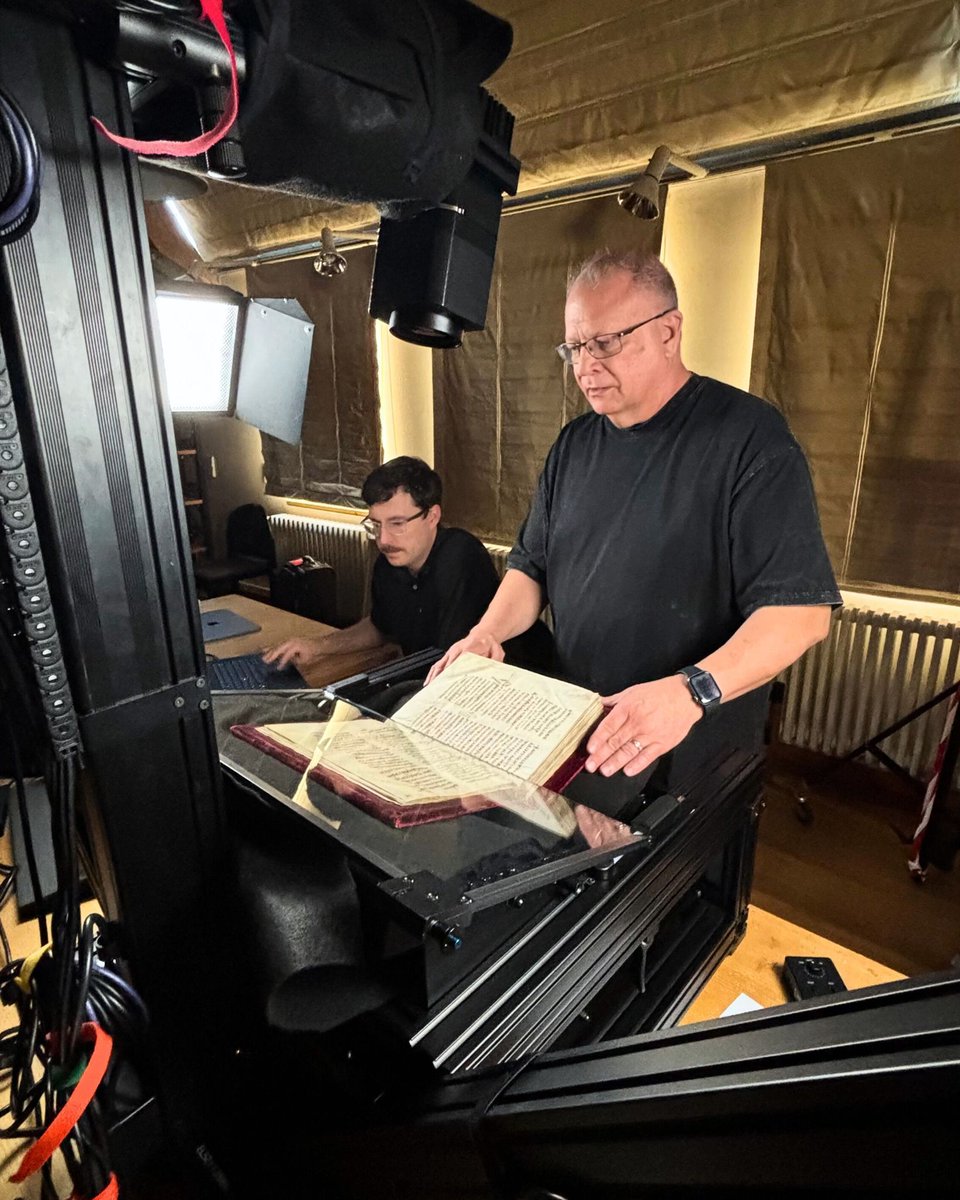
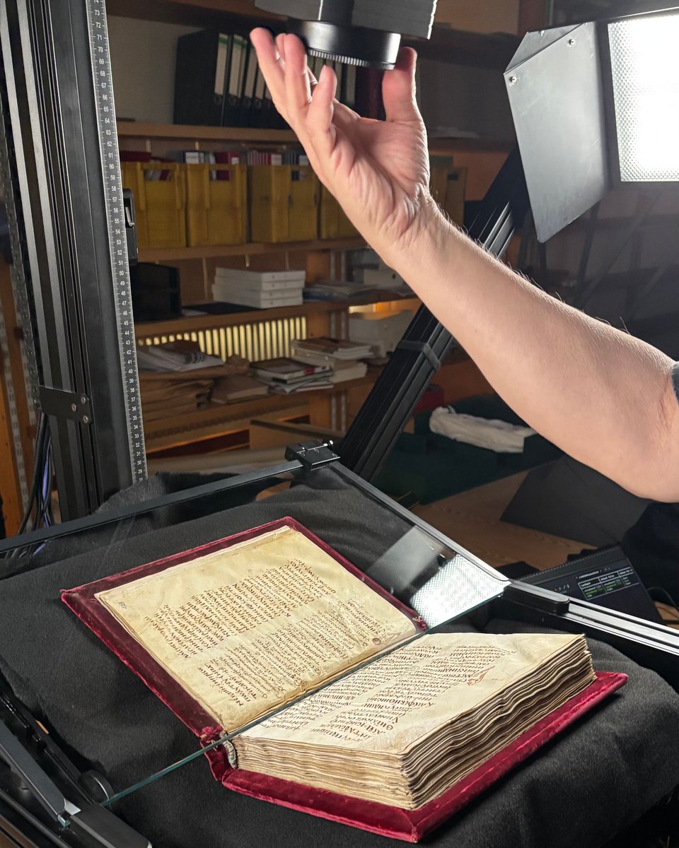
Before traveling to the St Nicholas Hospital in Bernkastel, our CSNTM team collaborated with the Museum am Dom Trier (@museumamdom) to digitize a 9th-century Greek lectionary (GA L179) using our state-of-the-art imaging technology. The manuscript originally belonged to the treasury of St. Simeon's Abbey and was later donated to the cathedral treasury by Canon Peter Josef von Hontheim.
#CSNTM #digitization #greekmanuscript #triergermany #testament #stsimeon #trierdom #cathedraltreasury #HighResolutionImaging #bistumtrier #9thcentury #museumtrier #griechischesmanuskript #lectionary #NewTestament #cathedralmuseum

2
12
443

28 Nov 2024
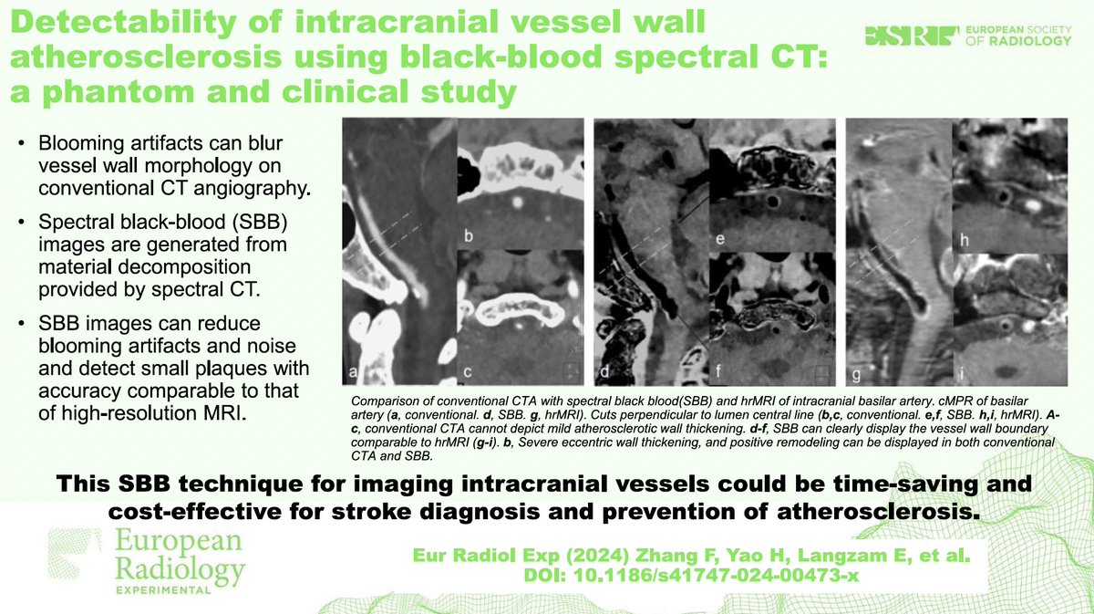
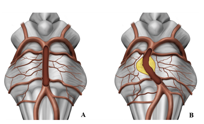
SPECTRAL CTA FOR BRAIN IMAGING
Detectability of intracranial vessel wall atherosclerosis using black-blood spectral CT: a phantom and clinical study. Fan Zhang et al. Eur Radiol Exp. 2024 Jul 3;8(1):78. doi: 10.1186/s41747-024-00473-x.
Objective
The study aimed to evaluate the effectiveness of spectral black-blood (SBB) imaging, generated through material decomposition from dual-layer spectral computed tomography (CT), in detecting intracranial vessel wall atherosclerosis. It compared SBB with conventional CT angiography (CTA) and high-resolution magnetic resonance imaging (hrMRI), which served as the gold standard.
Methods
1. Phantom Study:
• A custom-designed phantom mimicked intracranial artery conditions, with hollow tubes of varying diameters and wall thickness.
• SBB and conventional CTA images were compared for wall detectability, contrast-to-noise ratio (CNR), and measurement accuracy.
2. Clinical Study:
• Enrolled 34 patients with ischemic stroke or transient ischemic attack.
• Patients underwent both SBB imaging and hrMRI, with measurements of diagnostic certainty, vessel conspicuity, and plaque detectability.
3. Quantitative and Qualitative Analyses:
• Diagnostic certainty and plaque conspicuity were assessed using Likert scales.
• Plaque burden, remodeling index, and eccentricity were quantitatively compared between SBB and hrMRI.
Results
Phantom Study:
• Wall Detectability:
• SBB identified all walls (100%) compared to 75% detectability in conventional CTA (p < 0.001).
• For walls thinner than 1 mm, conventional CTA failed to detect them, while SBB achieved full detectability.
• Accuracy:
• SBB demonstrated higher measurement accuracy for wall thickness (mean absolute error [MAE] 3% vs. 8% for conventional CTA).
• Inner tube diameter measurements were also more accurate with SBB (MAE 2% vs. 19%).
Clinical Study:
• Diagnostic Certainty:
• SBB outperformed conventional CTA in vessel wall detection (median diagnostic certainty score: 3 vs. 0; p < 0.001).
• Plaque Detectability:
• Sensitivity, specificity, and accuracy of SBB were 94%, 98%, and 96% for plaques >1 mm, compared to 24%, 74%, and 50% with conventional CTA.
• Inexperienced readers significantly improved their performance with training, approaching the accuracy of experienced radiologists when using SBB.
• CNR:
• SBB provided significantly higher CNR for wall/lumen compared to hrMRI (p < 0.001), although CNR for wall/periarterial CSF was comparable between SBB and hrMRI.
• Morphological Features:
• Measurements of remodeling index, plaque burden, and eccentricity from SBB closely matched hrMRI (intraclass correlation coefficients 0.85–0.94).
Conclusions
• SBB imaging improves the detection and characterization of intracranial atherosclerotic plaques, particularly for thin walls and small plaques.
• It offers diagnostic accuracy comparable to hrMRI but with the advantages of faster acquisition times and reduced motion artifacts.
• SBB may become a valuable, cost-effective tool for stroke prevention and management, especially in patients unsuitable for hrMRI.
Clinical Implications
• SBB imaging could enhance stroke prevention by identifying culprit plaques missed by conventional CTA.
• The technique has potential applications in other vascular conditions, such as coronary and carotid artery assessments.
Limitations
1. Small sample size, requiring validation in larger cohorts.
2. Focused only on posterior circulation, necessitating studies on anterior circulation.
3. Excluded calcified and mixed plaques due to challenges with calcium suppression.
4. Limited generalizability due to use of a specific CT platform.
This study demonstrates the promise of SBB imaging for early detection and management of intracranial atherosclerosis, combining the benefits of CT’s accessibility with advanced diagnostic capabilities.
Open access at: eurradiolexp.springeropen.co…
#SpectralBlackBlood #CTImaging #IntracranialAtherosclerosis #StrokePrevention #AtherosclerosisDetection #HighResolutionImaging #BlackBloodCT #MedicalImaging #PlaqueDetection #StrokeManagement #CTInnovation #VascularHealth #IntracranialPlaque #RadiologyResearch #AdvancedCTTechniques
1
1
334

12 Jun 2024
Jan-Philipp Burchert et al.: X-ray phase-contrast tomography of cells manipulated with an optical stretcher #OpticalStretcher #XRayPhaseContrastTomography #HighResolutionImaging @uniGoettingen @TU_Muenchen... #IUCr journals.iucr.org/paper?S160…
3
141

9 Mar 2023
#NewNProt: Preparation of whole #human organs for #highresolutionimaging using hierarchical phase-contrast #Xraytomography go.nature.com/3kSBUL3
2
1,754

16 Feb 2023
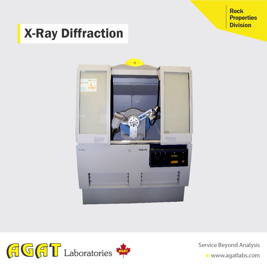
Did you know, x-ray diffraction, or XRD, can be used to identify and quantify 1000’s of different materials? Combining XRD and high resolution imaging, there is virtually nothing we can’t identify. #xrd #highresolutionimaging #laboratoryservices #agatlabs
1
39

#Melanoma cells (green) in cleared mouse #lung tissue. #Nuclei in magenta. Scale bar 40 microns. #HighResolutionImaging #Microscopy #Metastasis
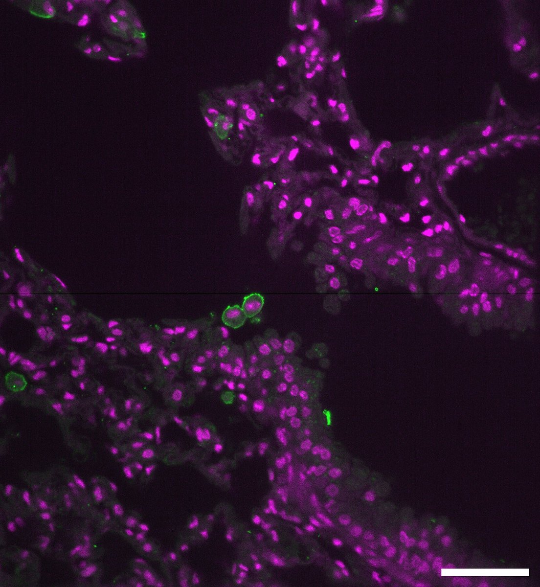
ALT Melanoma cells (green) in cleared mouse lung tissue. Magenta represents all cell nuclei.
1
2
8
8 Feb 2022
#FeaturedProtocol this week is for Vertical Cell Imaging by Nanostructured Immobilisation (VerCINI), which enables #highresolutionimaging of #bacterialcell features, such as #proteindynamics, by trapping the cells vertically in a #nanofabricated mould go.nature.com/3L49GFE
3

9 Sep 2021
Have you heard of #TSV technology?
ASI's R&D manager, Erik Hogenbirk explains what it is and how ASI uses it to enable higher resolution imaging and diffraction.
#electrondiffraction #microED #HighResolutionImaging #HighSensitiveArea #CheeTahM3Mega
lnkd.in/gY-bsQxA
1
2

5 Jul 2021
From the @MRSBulletin: The initial work on #HighResolutionImaging and #ChemicalAnalysis of #LiquidCellSamples. #imaging #NanoscaleMaterials #MaterialsScience @BerkeleyLab qoo.ly/3cz97n
2

19 Jun 2021
Utilizing #highresolutionimaging, direct observation of density peaks, which signal solidity, were possible and led researchers to discover an unexpected ‘crystal’-type state that occurs prior to the #superfluid state.
bit.ly/3vnNUTZ
@iqoqivienna #supersolids
2
11 May 2021
Via #OSA_Optica: Limiting the incident NA for efficient wavefront shaping through thin anisotropic scattering media ow.ly/Tk8U50EEzc5 #HighResolutionImaging
3
8

24 Feb 2021
This paper describes a straightforward practical approach for high-resolution imaging of PTFE/Teflon sample by using #AFM bit.ly/37Ija7g #polymerscience #polymers #spm #highresolutionimaging
1

5 Dec 2020
Interesting findings on #highresolutionimaging May help us defining better treatment options for these challenging cases #dolichoectasia @BAFOUND @TAAF @PennNSG

Latest Content - Neuroimaging
Dobrocky et al: Absence of pontine perforators in vertebrobasilar dolichoectasia on ultra-high resolution cone-beam computed tomography. ow.ly/1bRb50Cuzrt
1
3

22 Sep 2020
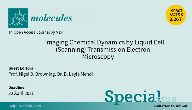
📢New Special Issue Open for Submissions: Imaging Chemical Dynamics by Liquid Cell (Scanning) Transmission Electron Microscopy
✏️Guest edited by Prof. Nigel D. Browning and Dr. B. Layla Mehdi
🔗mdpi.com/journal/molecules/s…
📌#LiquidCellTEM #HighResolutionImaging #QuantitativeChemistry
1
3

7 Mar 2020
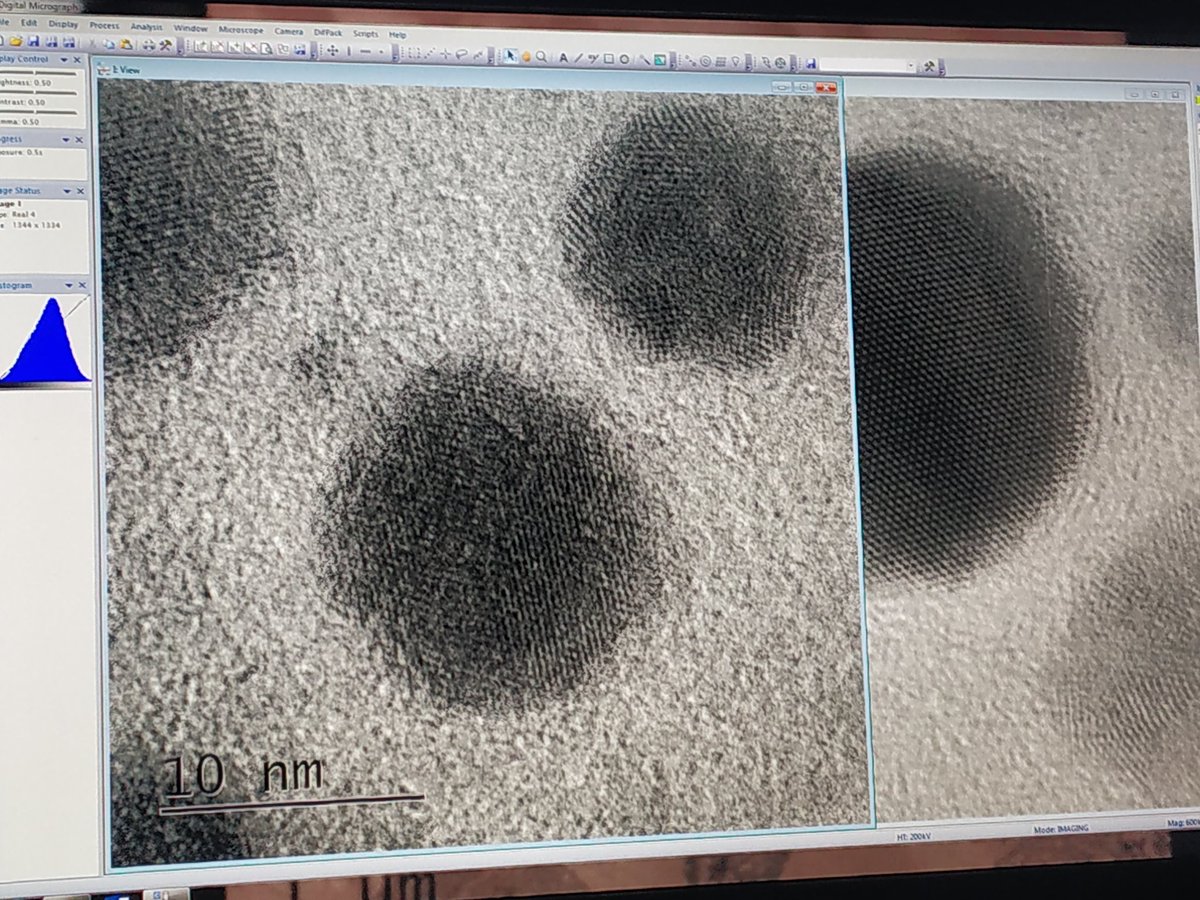
HR imaging Gold nanoparticles
Trying to get rid of astigmatism
#electronmicroscope #transmissionelectronmicroscope #highresolutionimaging #jeol #jeolindia #materialscharacterisation
2
2

New graphene-based metasurface capable of independent amplitude and phase control of light
sciencedaily.com/releases/20…
@versarien @2DTECH #graphene #2dmaterials #lightmodulation #holography #highresolutionimaging #opticalcommunicationsystems
2
9

21 Feb 2020
New graphene-based metasurface capable of independent amplitude and phase control of light
sciencedaily.com/releases/20…
@versarien @2DTECH #graphene #2dmaterials #lightmodulation #holography #highresolutionimaging #opticalcommunicationsystems
1
2
13

18 Apr 2013
Discovery paves the way for ultra fast #highresolutionimaging in real time phy.so/285500609
7
1