


Apr 24
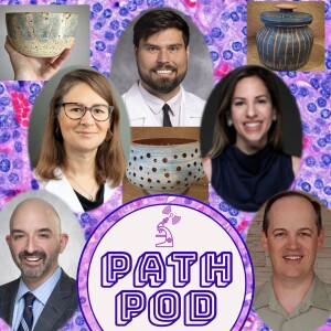
⭐ NEW #PathPod #IHCTalk!
@MArnold_PedPath
@IHC_guy
@sanamloghavi
speak w/ Dr. Grayson Cole and Dr. Nicole Cipriani! Hear about their paths to Oral and Head and Neck Pathology, as well as their 2025 article in Head and Neck Pathology!
🔊pathpod.podbean.com/e/ihc-ta…
9
24
2,381

Can pathology be learned through audio?
A new Academic Pathology paper says yes.
PathPod reached 95K downloads across 70
countries, and 30 pathology podcasts are part
of this shift. Full paper in comments.
#DigitalPathology
2
26
Mar 24
⭐ NEW #PathPod!
Host @MArnold_PedPath speaks with medical students @taranjit_kaur_ and Harnoor Galani about the @grosslyspeaking podcast!
We listen to their episode about autopsy, public health and forensics featuring their guest @NicoleJacksonMD
🔊pathpod.podbean.com/e/pathpo…

4
3
432
Feb 10
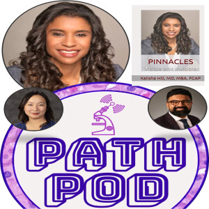
⭐ NEW #PathPod BeyondTheScope!
Hosts @KMirza and @Sara_Jiang speak w/
Dr. @KalishaHillMD
Hear their conversation about Dr. Hill's book PINNACLES: Visible and Invisible and her journey as a leader in Pathology and President Elect for @Pathologists
🔊pathpod.podbean.com/e/beyond…
9
11
962
Jan 30
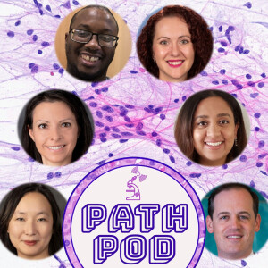
⭐ NEW #PathPod AroundTheScope!
Hosts @MArnold_PedPath & @Sara_Jiang speak w/
Dr. Michael Williams
Dr. @NedaWickEdPath
Dr. Maria Gubbiotti
Dr. Simmi Patel
Hear what is unique about neuropathology training and practice!
🔊pathpod.podbean.com/e/around…
6
10
684

What a pleasant surprise soundtrack to this morning’s workout. Always a delight hearing from @MeredithKHerman and learning more about her colleagues on workforce development. Thanks, @PathPod!!
#PathTwitter #PathSIG
19 Dec 2025

⭐ NEW #PathPod AroundTheScope!
Hosts @MArnold_PedPath and @Sara_Jiang speak with Drs. @MeredithKHerman, Paul Kowalski, @Aberry_Med, and Peace Preston about how DO trainees gain exposure to the field of Pathology.
🔊pathpod.podbean.com/e/around…
👉Article: researchgate.net/publication…

1
4
3
765

19 Dec 2025
Pathology needs talent—not labels.
We’re talking DOs in pathology, breaking barriers, and what it really takes to get here. 🎙️
Thank you @PathPod for discussing this important topic!
Unfiltered. Honest. Data-driven.
#DOsInMedicine #PathTwitter #MedEd
19 Dec 2025
⭐ NEW #PathPod AroundTheScope!
Hosts @MArnold_PedPath and @Sara_Jiang speak with Drs. @MeredithKHerman, Paul Kowalski, @Aberry_Med, and Peace Preston about how DO trainees gain exposure to the field of Pathology.
🔊pathpod.podbean.com/e/around…
👉Article: researchgate.net/publication…
1
8
27
3,488
19 Dec 2025
⭐ NEW #PathPod AroundTheScope!
Hosts @MArnold_PedPath and @Sara_Jiang speak with Drs. @MeredithKHerman, Paul Kowalski, @Aberry_Med, and Peace Preston about how DO trainees gain exposure to the field of Pathology.
🔊pathpod.podbean.com/e/around…
👉Article: researchgate.net/publication…
4
15
5,309
21 Nov 2025
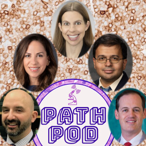
⭐ NEW #PathPod #IHCTalk!
@MArnold_PedPath, @IHC_guy, and @sanamloghavi speak with Dr. Pranav Patwardhan and Dr. Erika Moore about the Utility of TRBC1 Immunohistochemistry in the Evaluation of T-Cell Lymphomas!
🔊pathpod.podbean.com/e/ihc-ta…
1
8
21
3,925

14 Nov 2025
What did you enjoy most about The Case of Leo's Missing Energy? Was there a particular moment in the story that stood out to you?
37

12 Nov 2025
Today on #InternationalPathologyDay we honour the silent heroes behind every diagnosis — the scientists, clinicians and lab teams who turn data into answers. 🧬🔬
Dive into the world of pathology with these curated resources from FeedSpot:
Podcasts: podcast.feedspot.com/patholo…
YouTube Channels: videos.feedspot.com/patholog…
Blogs: bloggers.feedspot.com/pathol…
Top picks: @ASCP_Chicago , @olkazuraw , @peopleofpath , @ARUPLabs , @dpatweet , @PathPod , @deadmendo , @DiversifyInPath , @mayocliniclabs , @DrTravisBrown , @TheUSCAP , @OsmoseIt , @RCPath , @Pathologists , @Dr_Rabiul_Haque , @Medscape , @medpagetoday , @tissuepathology , @VijayPatho , @Voicebrook , @ParasiteGal ...many more
#InternationalPathologyDay #Pathology #LabMedicine #Diagnostics
1
1
242

12 Nov 2025
On #InternationalPathologyDay we celebrate the dedicated professionals behind every diagnosis—the scientists, clinicians, and laboratory teams guiding patient care.🧬🔬
Dive into these insightful discussions: podcast.feedspot.com/patholo…
Top picks: @ASCP_Chicago , @olkazuraw , @peopleofpath , @ARUPLabs , @dpatweet , @PathPod , @deadmendo , @DiversifyInPath , @mayocliniclabs , @DrTravisBrown , @TheUSCAP ...many more
#InternationalPathologyDay #Pathology #LabMedicine #Diagnostics
58

7 Nov 2025
cc: @sanamloghavi @Sara_Jiang @MArnold_PedPath @ALBoothMD @cullen_lilley @AadilAhmedMD @MeredithKHerman @Papa_Heme @acweyand @ShikhaJainMD @UMichPath @umichmedicine @UMichMedSchool @ledje @Path_SIG @PathElective @PathPod @RodneyRohde @IHC_guy @loyolapathology @UChicagoPath @UMichRISE @AliBrownMD @MottChildren @BloodCancerUtd @MyPatientStory @HemOncFellows @JMGardnerMD
1
2
432

5 Nov 2025
This is sooo amazing!!! Congratulations! Can't wait to get this for my little sister :')
1
57


4 Nov 2025
Get better every day and solve this!
Category: Nephrology: Kidney Disease Diagnosis and Management > Glomerulonephritis: Causes, Diagnosis, and Management > Complement-Mediated Glomerulonephritis
A 12-year-old boy is brought to the clinic because of facial puffiness and dark, “cola-colored” urine that began 3 days ago. Ten days ago he completed a course of amoxicillin for culture-confirmed group A streptococcal pharyngitis.
Vital signs: temperature 37 °C, pulse 80 beats/min, respiratory rate 16 breaths/min, blood pressure 135/85 mm Hg (consistent with stage 1 hypertension, ≥95th percentile for age, sex, and height), oxygen saturation 98 % on room air.
Physical examination shows periorbital edema and mild bilateral pedal pitting edema; the remainder is unremarkable.
Urinalysis: 3 blood, 2 protein, negative leukocyte esterase; microscopy reveals numerous dysmorphic erythrocytes and many red blood cell casts.
Serum creatinine: 1.1 mg/dL (reference 0.3–0.7 mg/dL).
Serum albumin: 3.2 g/dL (reference 3.5–5.0 g/dL).
Estimated GFR: 75 mL/min/1.73 m² (reference > 90 mL/min/1.73 m²).
Complement levels: C3 = 55 mg/dL (low); C4 normal.
Anti-streptolysin O titer: elevated.
Antinuclear antibody and ANCA: negative.
Renal biopsy findings
• Light microscopy – enlarged, hypercellular glomeruli with numerous neutrophils and obliteration of capillary lumina.
• Electron microscopy – large, irregular subepithelial electron-dense “hump-like” deposits.
Which immunofluorescence pattern on the renal biopsy is most consistent with the glomerulonephritis in this patient?
**Absence of immune deposits with diffuse podocyte foot-process effacement**
**Linear deposition of IgG along the entire glomerular basement membrane**
**Granular deposition of C3 and immunoglobulins (predominantly IgG with lesser IgM) along glomerular capillary walls and mesangium ("starry-sky" pattern)**
**Predominant mesangial deposition of IgA with C3 co-deposition**
The answer and explanation will be shared as a reply to this post once it closes. Visit endlessmedical.academy/auth?… to discover the correct answer and detailed explanation right now.
#RenalPath #Step2CK #FOAMedKidney #MedStudentLife #MicroscopyMonday #CaseFiles #StrepThroatComplications #USMLE
@PathProfs @AcademicIntern @FOAMedForum @KidneyBoy @MedEdSociety @PathPod
*Generated by AI. May contain errors. Use at own risk. Full disclaimer: endlessmedical.academy/auth?…
🔗 endlessmedical.academy/auth?…
29