
Jun 10
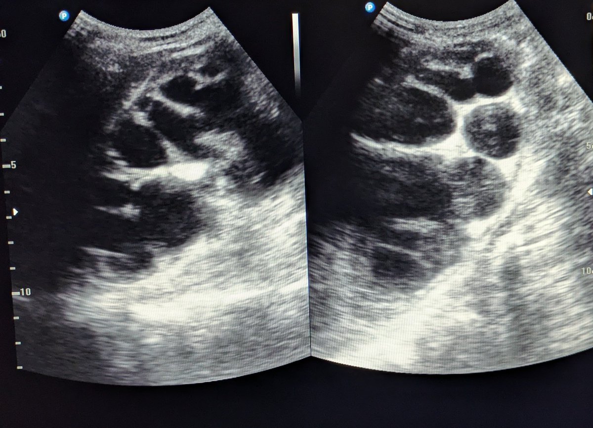
What is marked in red is severe hydronephrosis with probable pyonephrosis or dysplastic multicystic kidney.
192

May 30
الاحتباس مش دايما بيتقسطر بسهولة وبتحتاج ساعات تركب قسطرة فوق العانة
Intractable hematuria
Torsion
Priapism
Urethral stone
Pyonephrosis ممكن يحتاج تركب قساطر كلوية
AKI due to postrenal cause بيحتاج تركب دعامات او تركب قساطر كلوية
بس هتفضل كام سنة في حياتك تشتغل طوارئ يعني
1
2
114

May 25
youtu.be/sK-zbEOKMss?si=Gmt1…
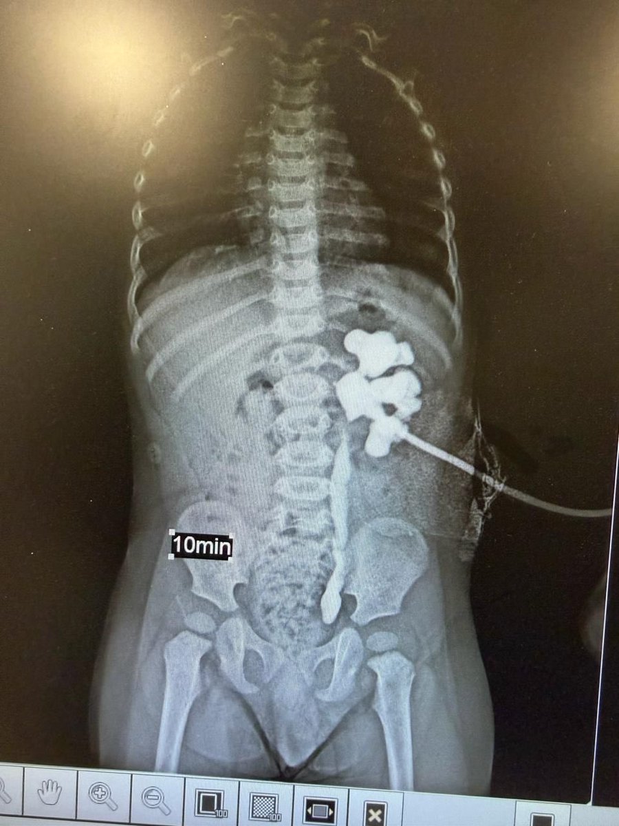
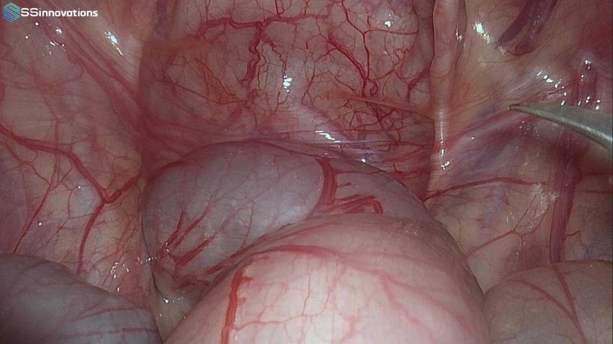
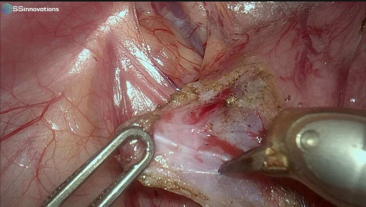
A challenging and important learning point in pediatric urology.
When PUJO and VUJO coexist, missing the distal obstruction can lead to serious postoperative complications. In this infant case, only PUJO was identified initially and an open pyeloplasty was performed elsewhere based on ultrasound findings. However, the associated UVJO was missed.
After stent removal, the child presented with pyonephrosis. Emergency PCN was performed, and subsequent nephrostogram demonstrated complete cut-off at the UVJ. We proceeded with robotic ureteric reimplantation, and the child recovered well.
This case highlights the importance of evaluating the entire urinary drainage pathway before intervention. In combined obstruction cases, distal obstruction should generally be addressed first.
Video: Infant Robotic Reimplantation Case
youtu.be/sK-zbEOKMss?si=Gmt1…
#RoboticSurgery #PediatricUrology #Urology #RoboticUrology #PUJO #UVJO #Pyeloplasty #UretericReimplantation #InfantSurgery #MinimallyInvasiveSurgery #Hydronephrosis #Pyonephrosis #PreetiUrology #DrChandraMohanVaddi #AssociationOfSouthernUrologists

2
3
36
4,592

May 13
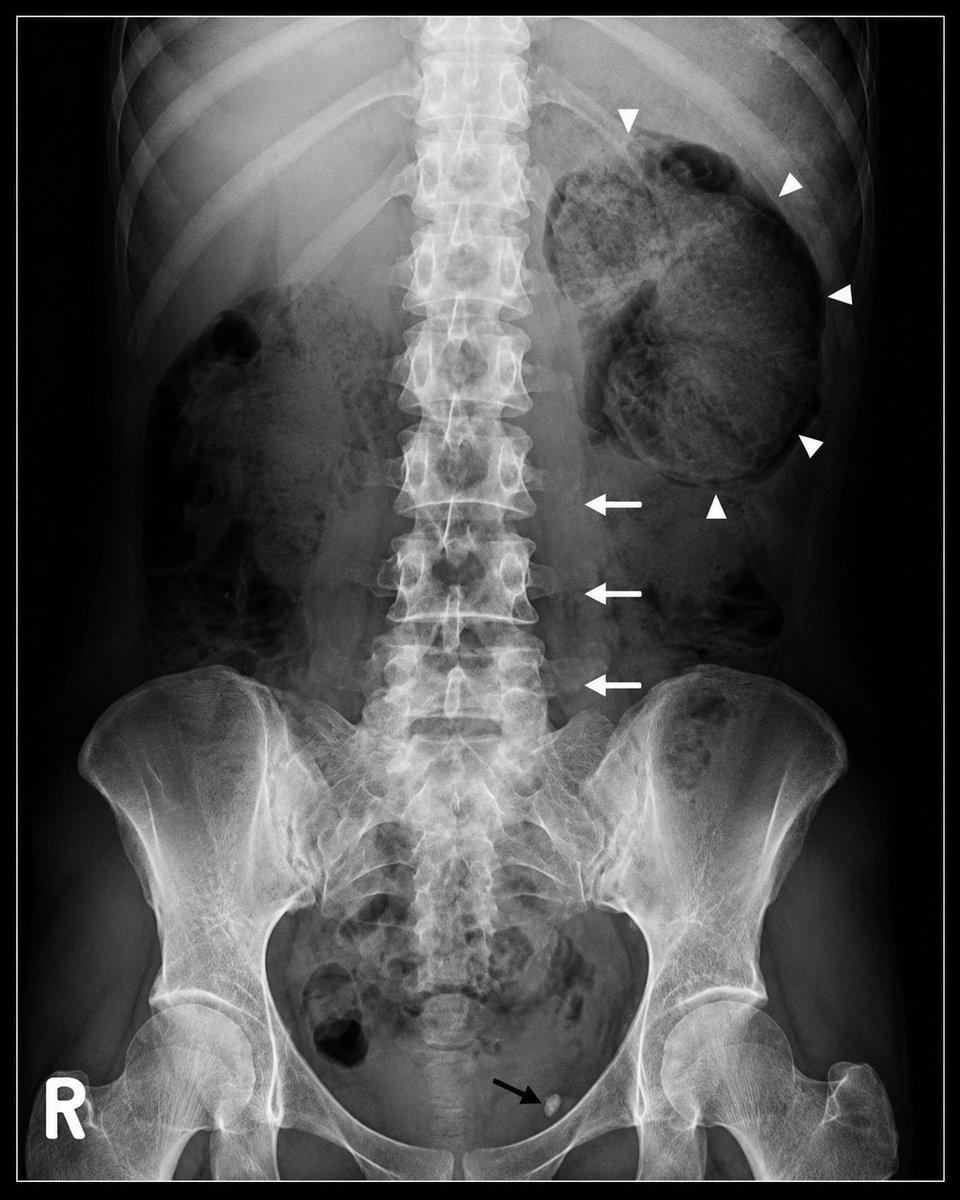
XGP & Pyonephrosis
➡ Primary Diagnosis: The most likely cause of sepsis is Xanthogranulomatous Pyelonephritis (XGP) or pyonephrosis.
➡ Etiology: Sepsis is secondary to an obstructive renal calculus leading to severe, chronic infection.
➡ Radiographic Clues:
Renal Mass: Significant enlargement of the left renal shadow.
Psoas Sign: Loss of the left psoas margin, indicating retroperitoneal inflammation.
Infection Evidence: Presence of gas-containing areas suggesting gas-forming organisms.
Calculi: Visible urinary tract calcifications supporting obstructive uropathy.
➡ Pathophysiology: Chronic obstruction causes urinary stasis, leading to destruction of renal parenchyma and suppurative inflammation.
➡ Microbiology: Typically associated with Proteus or E. coli.
➡ High-Yield Pearl: A CT scan would classically reveal the "Bear Paw Sign," which is pathognomonic for XGP.
2
523

Most likely cause of sepsis: Xanthogranulomatous pyelonephritis (XGP) / pyonephrosis secondary to obstructive renal calculus 🪨🦠
📌 Key radiographic clues:
• Enlarged left renal shadow/mass
• Loss of the left psoas margin → retroperitoneal inflammation
• Gas-containing appearance suggesting severe infection with gas-forming organisms
• Associated urinary tract calcification supporting obstructive uropathy
🧠 Pathophysiology:
Chronic urinary obstruction causes:
→ urinary stasis
→ recurrent infection
→ destruction of renal parenchyma
→ suppurative inflammation and sepsis
⚠️ Common organisms:
• Proteus
• E. coli
📚 High-yield pearl:
CT classically demonstrates the “Bear Paw Sign” in XGP.
✅ Most likely diagnosis:
Sepsis secondary to infected obstructed kidney (XGP/pyonephrosis).

2
2
502
Most likely cause of sepsis: Xanthogranulomatous pyelonephritis (XGP) / pyonephrosis secondary to obstructive renal calculus 🪨🦠
📌 Key radiographic clues:
• Enlarged left renal shadow/mass
• Loss of the left psoas margin → retroperitoneal inflammation
• Gas-containing appearance suggesting severe infection with gas-forming organisms
• Associated urinary tract calcification supporting obstructive uropathy
🧠 Pathophysiology:
Chronic urinary obstruction causes:
→ urinary stasis
→ recurrent infection
→ destruction of renal parenchyma
→ suppurative inflammation and sepsis
⚠️ Common organisms:
• Proteus
• E. coli
📚 High-yield pearl:
CT classically demonstrates the “Bear Paw Sign” in XGP.
✅ Most likely diagnosis:
Sepsis secondary to infected obstructed kidney (XGP/pyonephrosis).
1
2
645


Mar 19
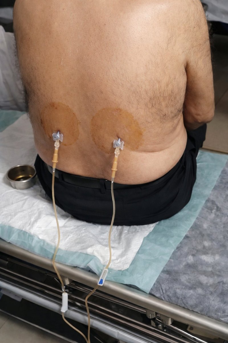
Here is the Arabic text you can overlay on your infographic (clean, exam-focused, ready for your #MedEd posts 👇):
🟠 فغر الكلية عبر الجلد ثنائي الجانب
(Bilateral Percutaneous Nephrostomy)
🧠 ما هو الإجراء؟
إجراء تدخلي يتم فيه إدخال قساطر عبر الجلد إلى حوض الكلية لتصريف البول في حالات انسداد الجهاز البولي.
📌 متى يُستخدم؟
✔ انسداد الحالب (حصوات، أورام، تضيّق)
✔ فشل كلوي انسدادي (Obstructive uropathy)
✔ موه الكلية الشديد (Hydronephrosis)
✔ إنتان بولي مع انسداد (Pyonephrosis – حالة طارئة)
⚙️ كيف يتم؟
✔ تحت توجيه الأشعة (Ultrasound / Fluoroscopy)
✔ إدخال القسطرة عبر الخاصرة إلى حوض الكلية
✔ يتم التصريف إلى كيس خارجي
✔ غالبًا تحت تخدير موضعي
🔑 معلومات مهمة
✔ ثنائي (Bilateral) = كلا الكليتين
✔ عبر الجلد (Percutaneous) = من خلال الجلد
✔ Nephrostomy = تصريف مباشر من الكلية
⚠️ المضاعفات المحتملة
⚠ نزيف
⚠ عدوى
⚠ انسداد أو انزلاق القسطرة
⚠ تسرب البول حول القسطرة
💡 Pearl (معلومة ذهبية)
👉 إجراء منقذ للحياة في حالات الانسداد مع الإنتان
👉 يجب عدم تأخير التصريف في pyonephrosis
4
1,031

Mar 7
A 72-year-old diabetic patient presents with fever, hypotension, and confusion.
Labs:
Lactate: 5 mmol/L
WBC: 19,000
CT abdomen shows obstructed infected kidney (pyonephrosis).
Despite fluids and antibiotics, the patient remains hypotensive.
What is the most important next step?
A. Add vancomycin
B. Start vasopressors
C. Urgent urinary drainage
D. Repeat CT scan
E. Dialysis

1
2
80


23 Dec 2025
A. Malecot's catheter.
• It is a self‑retaining drainage tube with a characteristic mushroom/flower‑shaped winged tip used mainly for suprapubic cystostomy and nephrostomy.
∆ Main uses-:
• Suprapubic cystostomy for bladder outlet obstruction, urethral injury, urethral stricture, neurogenic bladder.
• Percutaneous nephrostomy for upper urinary tract obstruction, post‑PCNL drainage, pyonephrosis..
∆ Advantages-:
• Wide lumen and winged tip allow efficient drainage of thick viscous fluids (pus, clots) compared with Foley.
• Good retention with low risk of accidental dislodgement when correctly positioned.
1
2
5
1,059

20 Dec 2025
Acute urinary retention
Hematuria
Ureteric stone
Obstructing stone with infection
Pyonephrosis
Hydronephrosis
Acute pyelonephritis
Urosepsis
Acute bacterial prostatitis
Epididymo-orchitis
Fournier’s gangrene
Testicular torsion
Scrotal abscess
فيه اشياء كثير بس هذي ال most common
16 Dec 2025

السلام عليكم
عندي فكره رهيبه راح تفيد الكل ان شاءالله ، ايش رايكم كل واحد رزدنت او انتيرن في روتيشن معين يقولنا most common case اللي يشوفونها خلال الروتيشن ايش يصير نقرا عنها قبل بدايه الروتيشن ونعرف الاكزامنيشن والمانجمنت etc، مثلا Gs appendicitis وهكذا! ويكون البوست مرجع لنا كلنا
16
2,016

29 Nov 2025
Moderate is fair
But the fluid content is echo rich , mostly likely infected and now pyonephrosis

1
5
87

10 Sep 2025
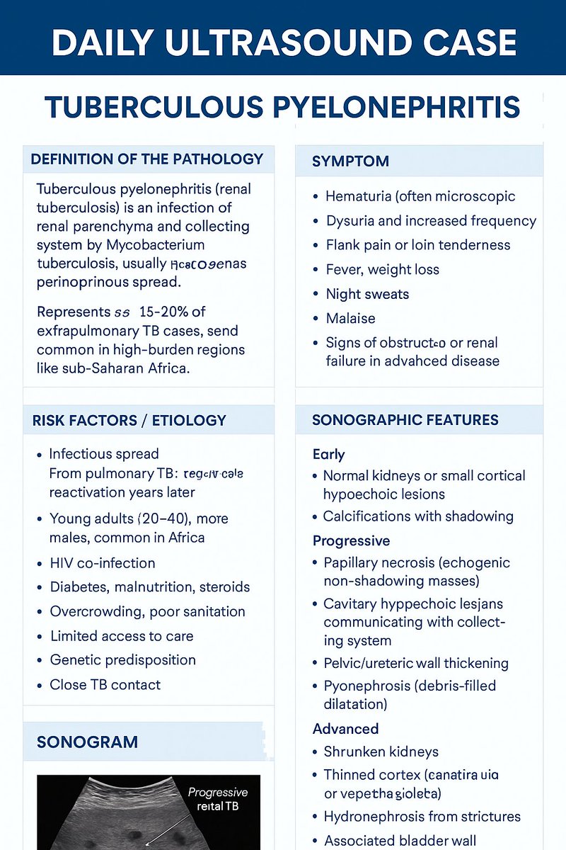
#DailyUltrasoundCase
☢️ Tuberculous Pyelonephritis
⏩️ Definition
Tuberculous pyelonephritis (renal tuberculosis) is infection of the renal parenchyma and collecting system by Mycobacterium tuberculosis, usually via hematogenous spread. It causes granulomas, necrosis, cavitation, fibrosis, and calcification. It represents 15–20% of extrapulmonary TB cases and is common in high-burden regions like sub-Saharan Africa.
⏩️ Symptoms
Hematuria (often microscopic)
Dysuria and increased frequency
Flank pain or loin tenderness
Fever, weight loss, night sweats, malaise
Signs of obstruction or renal failure in advanced disease
⏩️ Risk Factors / Etiology
Infectious spread: Hematogenous from pulmonary TB, reactivation years later
Demographics: Young adults (20–40), more in males, common in Africa
Immunocompromise: HIV co-infection, diabetes, malnutrition, steroids
Socioeconomic: Overcrowding, poor sanitation, limited access to care
Other: Genetic predisposition, close TB contact
☢️ Sonographic Features
Early: Normal kidneys or small cortical hypoechoic lesions; calcifications with shadowing
Progressive:
Papillary necrosis (echogenic non-shadowing masses)
Cavitary hypoechoic lesions communicating with collecting system
Pelvic/ureteric wall thickening
Pyonephrosis (debris-filled dilatation)
Advanced:
Shrunken kidneys with thinned cortex
Dense calcifications (“putty kidney”)
Hydronephrosis from strictures
Associated bladder wall thickening
Color Doppler: Reduced flow in necrotic areas, hyperemia in active inflammation🔚
8 Sep 2025

#DailyUltrasoundCase
☢️ HIV-Associated Nephropathy (HIVAN)
⏩️ Definition
HIV-associated nephropathy (HIVAN) is a collapsing form of focal segmental glomerulosclerosis in patients with HIV. It leads to rapid progression to ESRD due to direct infection of renal epithelial cells. Pathology shows glomerular collapse, microcystic tubular dilatation, and interstitial inflammation. It is common in sub-Saharan Africa where HIV prevalence is high.
⏩️ Symptoms
Heavy proteinuria with rapid renal function decline
Microscopic or gross hematuria
Hypertension and peripheral edema
Often in advanced HIV, but may appear earlier
Systemic HIV-related symptoms: fatigue, weight loss
Severe disease: uremic symptoms (nausea, confusion, pruritus)
⏩️ Risk Factors / Etiology
Cause: HIV infection with viral gene expression in renal cells
Demographics: Strong link to Black African ancestry (APOL1 variants); higher burden in sub-Saharan Africa
Disease Stage: Advanced or untreated HIV (low CD4); may occur in acute infection
Other: Co-infections (hepatitis), malnutrition, delayed ART
☢️ Sonographic Features
Bilaterally enlarged kidneys (>12–13 cm)
Diffusely hyperechoic parenchyma, loss of corticomedullary differentiation
Decreased/absent sinus fat echogenicity (renal edema)
Late stage: small, shrunken kidneys with cortical thinning (ESRD)
Doppler: Normal or reduced renal blood flow

3
6
246

18 Jun 2025
Gross pyonephrosis is secondary to obstructing calculus at uretero pelvic junction.
2
16
88
2,456

🛑 A nephrostomy is a procedure where a thin tube (called a nephrostomy tube) is inserted through the skin into the kidney to drain urine directly from the kidney.
♦️Indications :
🔶️It's done when urine flow is blocked and can't pass from the kidney to the bladder, such as in:
🔷️Kidney stones (obstructing the ureter)
🔷️Tumors (bladder, cervix, or colon)
🔷️Infections with pus buildup (pyonephrosis)
🔷️Ureteric injury or stricture
🔷️Congenital anomalies (in children).
1
4
19
1,483


25 Feb 2025
Both kidneys : Normal site, enlarged in size up to 14x8 cm , irregular shape, lost
parenchymal thickness and increased echogenecity-,no masses . There is
marked back pressure changes with turbidity and internal echo suggesting
pyonephrosis
#ultrasound
#sonologist

15
77
3,934