Hartmann’s Top Guy retweeted

10h
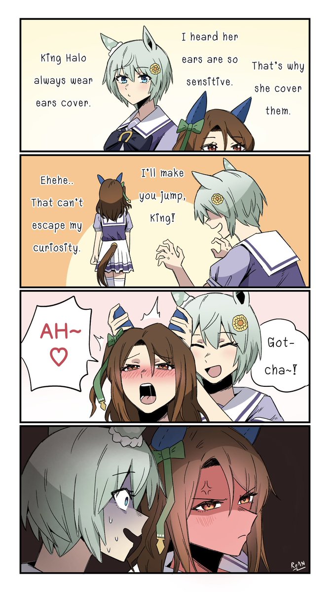
The Sensitive Ears
#umamusume #umamusumeprettyderby
24
627
10,462
79,190
Judy retweeted

May 6
Do you think small boobs are more sensitive? I think yes
46
1,840
3,320
187,002


Tau the Explorer 🛩 retweeted

this is a sensitive topic for me.

45
1,102
8,077
330,174


22s
Sunday ECG Academics
U-turn on ECG and Surawicz Criteria for Hypokalemia
By DR. D.P. KHAITAN,MD (MEDICINE) FCGP (IND) FIAMS (MEDICINE) FICP FICCMD
FIACM
@ecgandrhythmRoe @ECG_BUDDY_ARPI
@EcgsOnly @ecgrhythms @ECGWeekly
@medflutter_ @ekg
One often hesitates to make a U-turn while driving a car. Yet experience teaches that a
timely U-turn may avert danger and lead to a safer destination.
Similarly, recognition of a prominent U wave on the ECG may redirect the clinician's
attention toward hypokalemia and help prevent its potentially serious consequences. Thus, the ECG "U-turn" may become a clinical "life-saving turn."
In hypokalemia U wave is a prominent deflection that follows the T wave on the
ECG and is thought to reflect delayed repolarization of Purkinje fibers and/or
mid-myocardial (M) cells.
A triphasic ST–T–U complex, characterized by ST-segment depression, followed
by T-wave flattening or inversion, and culminating in a prominent U wave denotes
a valuable ECG pattern suggestive of hypokalemia.
The Surawicz criteria represent an important milestone in the electrocardiographic diagnosis of hypokalemia, highlighting the significance of this triphasic "ST–T–U" pattern as a manifestation of altered cardiac repolarization. Although modern laboratory testing remains the diagnostic standard, the ECG continues to serve as a valuable bedside tool for recognizing the cardiac effects of hypokalemia.
See this ECG
A 58 years female , with no history of any previous significant medical illness presented with the complaints of weakness in limbs that made her unable to get up from the bed after she woke up in the morning (Low serum potassium 1.90 mEq/L with no subnormal
Findings :
Triphasic repolarization abnormalities : ST segment depression – attenuated T wave –
prominent U wave , most marked over V4,V5 and V6,best at V4.
Incomplete RBBB ,please see V1 which is followed by negative T and U waves in the
same negative direction (In hypokalemia U waves remain concordant with the T wave).
Q-U interval is prolonged.
Discussion :
Three elements here are important to look at : the clinical history of vomiting , ECG findings as discussed and low serum potassium level.
The Triphasic abnormalities such as ST segment depression – the attenuated T-wave –prominent U-wave : this is very characteristic of hypokalemia.
Severe hypokalemia (Serum K < 2.5 mEq/dL is considered to be severe) causes Right
Bundle Branch Block (RBBB) by altering the electrical gradients of cardiomyocytes,
leading to prolonged repolarization and conduction delay. The right bundle branch is
particularly sensitive to these ionic shifts, which delay the electrical signal from
moving simultaneously down the left and right.
💢Take-Home Message
▪️The electrocardiographic manifestations of hypokalemia result primarily from delayed and heterogeneous ventricular repolarization arising from reduced outward potassium currents.
▪️The resulting consecutive changes—ST depression, T-wave flattening, and prominent U waves reflect progressive stages of repolarization disturbances rather than isolated electrocardiographic abnormalities.
▪️Recognizing the historical progression of hypokalemic ECG patterns—which shifted from viewing ST-segment, T-wave, and U-wave abnormalities as independent features to understanding them as a single, unified repolarization disorder—is a critical component of electrocardiographic interpretation.
▪️Surawicz and colleagues systematically described the ECG pattern of hypokalemia and thus , helped in establishing the relationship between potassium depletion and ST-T-U abnormalities.
▪️In nutshell to say , the ECG changes in hypokalemia present a continuum of repolarizationabnormalities: from ST-segment depression and T-wave flattening to prominent U waves,
which may reflect delayed Purkinje and / or mid-myocardium recovery.

2
J❤️ retweeted

12h
sensitive young man eating his goyslop when suddenly attacked by evil foid:

22
763
13,293
532,979
Azekhaime🤴🏿 retweeted

6h
charlamagne folding under pressure and telling on kenny, the most sensitive rapper alive that can’t handle ONE mf criticizing him ✌🏼😂
11
72
559
33,900
Ksa retweeted

Jun 12
I don’t care how sensitive you are your going to take it and be a good boy for mommy, so adorable.
#overstimulation #femdom #cnc #blowjob #handjob #overstim #horny #mommy
 MACROMASTIGOON
4
840
15,230
134,450
ܛܔܔܔܛܔܛܔܛ retweeted

9h
NPM Malware >
- Obfuscated postinstall hook (.prepare.cjs) uses char-code arrays to hide sensitive variable names
- Credential harvesting targeting AWS_ACCESS_KEY_ID, AWS_SECRET_ACCESS_KEY, GITHUB_TOKEN, NPM_TOKEN
- Exfiltration endpoint at open[.larksuite[.com (Feishu bot webhook) acts as C2
- Explicit sandbox evasion logic checking for SANDYCLAW, OPENCLAW, PERMISO, CHAINRADAR env
https[://www[.npmjs[.com/package/ash-claw/v/1.7.13
https[://sandyclaw[.permiso[.io/shared/5PVyO_QI-Y0ENhwni99JtULn_f8oVSMpqWXiYktq5E4
@TekDefense 😂
7
18
714
JIPONG GB retweeted

Apr 14
Mau bgt punya cowok clingy yang suka mainin pentil sensitive ceweknya sendiri sampe ngacung… when ya… 🥺
 ゆりか
ゆりか
2
20
182
17,314
