
12 Jan 2025
Basics of Reading Slides for Leukemias in Hematopathology 🧵
🔬 A guide with mnemonics, and practical tips for diagnosing leukemias on slides. Whether you’re reviewing bone marrow biopsies or peripheral smears, here’s what to look for:
1️⃣ Start with the Big Picture (Low Power) 🔍
•Use 10x magnification to assess:
•Cellularity: Hypercellularity suggests leukemia 📈.
•Look for abnormal clusters or infiltrates 🧱.
•Identify necrosis or fibrosis if present.
📝 Mnemonic: “Locate the Leukemia!”
💡 Tip: Compare the sample with normal marrow to catch subtle abnormalities.
2️⃣ Go Higher Power (40x/100x) 👀
•Examine morphology:
•Blasts: Large size, high N:C ratio, fine chromatin, nucleoli.
•Auer rods: Pathognomonic for AML, especially APL 🚨.
•Granules: Common in myeloid blasts.
•Cytoplasmic vacuoles: Seen in ALL or Burkitt lymphoma.
📝 Mnemonic: “Big Blasts Have Auer Rods.”
💡 Tip: Document cell sizes, inclusions, and other features systematically.
3️⃣ Identify Key Features by Leukemia Type 🩸
•Acute Myeloid Leukemia (AML):
•20% blasts in bone marrow or peripheral blood.
•Presence of Auer rods and granules 🧫.
•Subtypes:
•APL (M3): Multiple Auer rods, bundles (“faggot cells”), t(15;17).
•Myelomonocytic (M4): Mix of myeloid and monocytic blasts.
•Monocytic (M5): Prominent monoblasts with vacuoles.
•Acute Lymphoblastic Leukemia (ALL):
•Small, round blasts with scant cytoplasm.
•B-ALL: Common in kids 👶, CD10 , CD19 .
•T-ALL: Often presents with a mediastinal mass 🚩, CD3 , TdT .
•Chronic Myeloid Leukemia (CML):
•Hypercellular marrow with granulocytic hyperplasia 📊.
•Increased neutrophils, myelocytes, basophils.
•Look for blasts in advanced or blast phase.
•Chronic Lymphocytic Leukemia (CLL):
•Small lymphocytes with clumped chromatin.
•Smudge cells: A hallmark on peripheral smear 💔.
•Often co-expresses CD19/CD5.
📝 Mnemonic:
•“AML = Auer Rods”
•“ALL = A Little Lymphoblast”
•“CML = Crowded Marrow”
•“CLL = Clumped Chromatin”
4️⃣ Special Stains & Markers 🎨
•Cytochemical Stains:
•MPO: Positive in myeloid blasts ✅.
•Sudan Black B: Stains lipids in myeloid cells 🖤.
•PAS: Positive in lymphoblasts 🟣.
•Immunohistochemistry (IHC):
•Blasts: CD34, CD117.
•Lymphoblasts: TdT 🧬.
•B-cell Markers: CD19, CD20.
•T-cell Markers: CD3, CD7.
•Flow Cytometry:
•AML: CD13, CD33, CD34 .
•B-ALL: CD19, CD10, TdT .
•T-ALL: CD3, CD7, TdT .
•CLL: CD19, CD5 co-expression.
📝 Mnemonic: “MPO = Myeloid, TdT = Tiny Lymphoblasts.”
5️⃣ Correlate with Clinical Data 📋
•Peripheral Smear: Check for circulating blasts, neutrophils, lymphocytes, or dysplastic cells.
•CBC Trends:
•WBC 📈 in CML or leukemic phases.
•Pancytopenia in advanced marrow infiltration 🔻.
•Cytogenetics:
•AML: t(8;21), t(15;17) (APL), inv(16).
•ALL: t(12;21) (good prognosis), t(9;22) (poor).
•CML: t(9;22) (BCR-ABL).
💡 Tip: Clinical age helps:
•ALL = Pediatric 👶.
•AML = Elderly 👵.
6️⃣ Exam Pearls 📚
•AML Auer rods DIC: Think APL, start ATRA 🚑.
•ALL mediastinal mass: T-ALL 🚩.
•CML splenomegaly: Confirm with BCR-ABL 🧬.
•CLL smudge cells: Classic feature 💔.
•High WBC Basophilia: Often CML 📈.
Key Summary for Slide Reading:
FeatureAML 🧫ALL 🟣CML 📊CLL 💔
BlastsLarge, Auer rodsSmall, roundFew in chronic phaseAbsent
MarkersMPO, CD33TdT, CD10, CD19BCR-ABLCD19/CD5
HallmarkAuer rodsMediastinal mass (T)↑ BasophilsSmudge cells
For more pathology guidance, bookmark this thread! 🔬🩺
#Hematopathology #Leukemia #SlideReview
4
22
1,016

8 Mar 2023
Going to USCAP?
Visit @Roche at booth #228.
Sign up to attend slide review or educational sessions.
go.roche.com/USCAP2023-Twitt…
#roche #USCAP2023 #slidereview #educational #conference #pathology #PathTwitter #SESSION

2
695
13 Feb 2023
Going to USCAP?
Visit @Roche at booth #228.
Sign up to attend slide review or educational sessions.
go.roche.com/USCAP2023-Twitt…
#roche #uscap #slidereview #educational #conference #pathology #PathTwitter #session

2
990

30 Sep 2020
What did you think of the latest episode of #SlideReview? Hoping to do something similar this week as I am currently offsite, which can make a live webinar more challenging. Let me know your thoughts!
#GUPath
Slides: bit.ly/SlideReviewGU04b
Video: bit.ly/SlideReviewGU04
1
2
30 Sep 2020
Thanks everyone for your patience - a new #SlideReview is now LIVE! The final chapter of our first #GUpath series can be found at the link below!
#ChallengeCases coming out later this week!
Slides; bit.ly/SlideReviewGU04b
Video: bit.ly/SlideReviewGU04
2
3
5
30 Sep 2020
#SlideReview
@KMirza @ALBoothMD @mydermpath @JMGardnerMD @Williamson_SR @SaraEWobker @priyaraomd @LiangChengMD @AkgulMd @slusagar @pembeoltulu @rovingatuscap @Teclis82 @RoncinMD @Dr_Brian_Cox @anueru432 @arshna101 @RabailRai @mingfei_yan @OsmeAbdullah
bit.ly/SlideReviewGU04

3
4
16 Sep 2020
Missing your dose of #GUpath #SlideReview? Check out the #ShortReview on a bladder biopsy with a component of Small Cell Carcinoma at the link below! #VirtualPath
Video: bit.ly/ShortReviewSmCCBladde…
We'll be back next week with new content - thanks everyone for your patience 🙏
1
5
9
15 Sep 2020
Anyone else missing their weekly dose of #SlideReview?
On my last week of #coagulation, which has been great but definitely taking far more time than I’m used to!
Hang on for one more week so that we can finish up the #GUpath series and dive into #GYNpath! #VirtualPath
2
6
2 Sep 2020
Some days you just have to learn when to slow down.
Today is one of those days - loving my #Coagulation rotation but it’s definitely keeping me on my toes!
Because of this, there will be a special edition of #SlideReview this week...
1
2
4
2 Sep 2020
Tonight on #SlideReview we'll be completing our #GUpath series with prostate and testicular lesions.
Want to join the live Zoom (5:30pm EST)?
bit.ly/SlideReviewZoom
Joining Manually?
Meeting # 734 1168 1509
PASSWORD CHANGES WEEKLY. PLEASE DM PRIOR TO 5:15PM EST TODAY
1
5
2 Sep 2020
New #SlideReview is LIVE! Check out our bladder lecture (links below) #ChallengeCases out Friday! #GUpath #VirtualPath @KMirza @ALBoothMD @mydermpath @JMGardnerMD @Williamson_SR @LiangChengMD @SaraEWobker @RoncinMD
Slides: bit.ly/SlideReviewGU03b
Lecture: bit.ly/SlideReviewGU03
1
5
10
1 Sep 2020
Tomorrow on #SlideReview we'll be completing our #GUpath series with prostate and testicular lesions.
Want to join the session tomorrow (5:30pm EST)?
bit.ly/SlideReviewZoom
Joining Manually?
Meeting # 734 1168 1509
PASSWORD CHANGES WEEKLY. PLEASE DM PRIOR TO 5:15PM EST WEDS
2
3
26 Aug 2020
Tonight on #SlideReview we'll be continuing our #GUpath series with lesions involving the bladder.
Want to join the live Zoom (5:30pm EST)?
bit.ly/SlideReviewZoom
Joining Manually?
Meeting # 734 1168 1509
PASSWORD WILL BE CHANGING WEEKLY. PLEASE DM PRIOR TO 5:15PM EST
1
11
12
26 Aug 2020
New #SlideReview is LIVE! Please see the links👇🏻for part 2 of our kidney lesions #GUpath #VirtualPath
#ChallengeCases coming your way on Friday!
Slides: bit.ly/SlideReviewGU02b
Video: bit.ly/SlideReviewGU02
1
3
5
19 Aug 2020
Almost ready to start #SlideReview!
Part 2 of the #GUpath series (and part 2 of renal lesions)
Want to join the live Zoom (5:30pm EST)?
bit.ly/SlideReviewZoom
Joining Manually?
Meeting # 734 1168 1509
Password: 4VxhpN
1
1
19 Aug 2020
Tonight on #SlideReview we'll be going back to our #GUpath series for a second helping of renal lesions!
Want to join the live Zoom (5:30pm EST)?
bit.ly/SlideReviewZoom
Joining Manually?
Meeting # 734 1168 1509
Password: 4VxhpN
2
4
17 Aug 2020
New #ChallengeCases for #GUpath 01 is LIVE! Please give a watch/like/share (and sub) #VirtualPath #SlideReview
@KMirza @ALBoothMD @mydermpath @Teclis82 @AkgulMd @slusagar @Williamson_SR
@LiangChengMD @RoncinMD
@mingfei_yan @ArshnaQ @RabailRai @anueru432
bit.ly/SlideReviewGU01c
10
11
14 Aug 2020
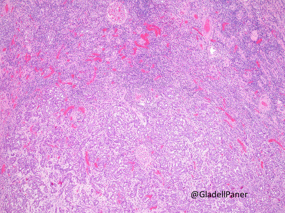
Great example of a rare case (anyone recognize the features from this week’s #SlideReview?) #GUpath
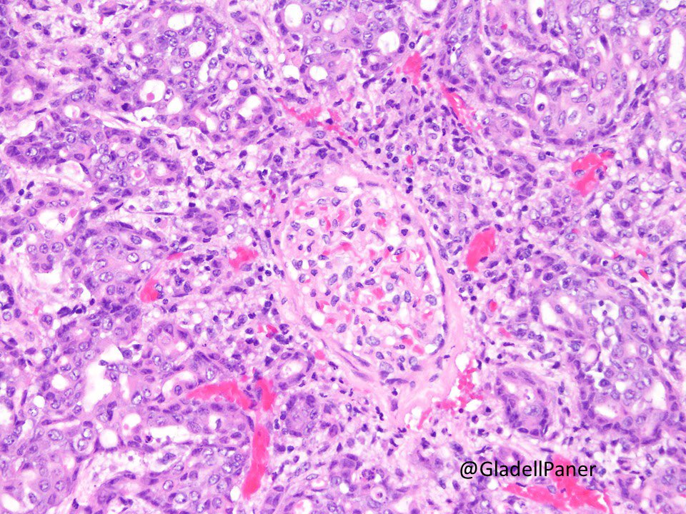
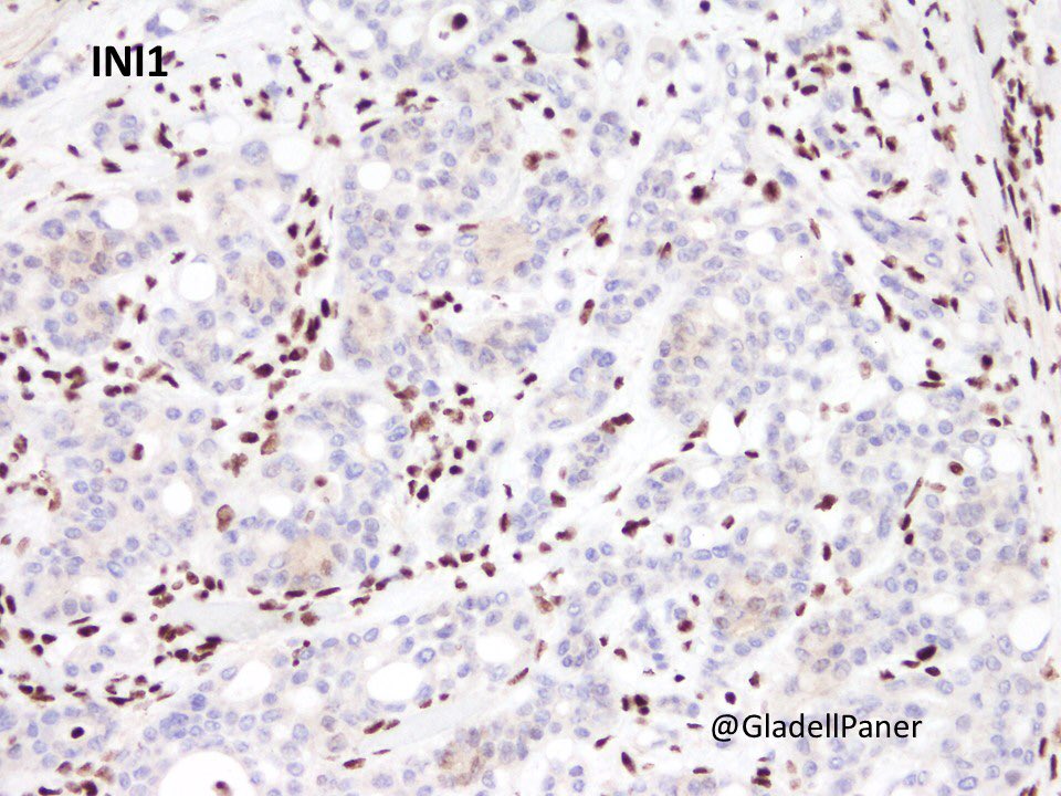
14 Aug 2020

Medullary carcinoma of #kidney in a young patient with sickle cell trait. Infiltrative tumor with reticular or cribriform as most common pattern. Loss of INI1 nuclear expression important for diagnosis. #GUPath

2
4
14 Aug 2020
New #SlideReview is LIVE! Part 1 of #GUpath - Kidney lesions. Please give a watch/like/share (and sub) #VirtualPath
@KMirza @ALBoothMD @mydermpath @Teclis82 @AkgulMd @slusagar @Williamson_SR @LiangChengMD
@RoncinMD
Slides: bit.ly/SlideReviewGU01b
Video: bit.ly/SlideReviewGU01
4
4
13 Aug 2020
New #SlideReview is LIVE! The last video for the #GIpath series covers malignant colon/appendix lesions #VirtualPath
@KMirza @ALBoothMD @mydermpath @CArnold_GI @JMGardnerMD @Teclis82 @ArshnaQ @RabailRai @anueru432
Slides: bit.ly/SlideReviewGI04b
Video: bit.ly/SlideReviewGI04
10
13