
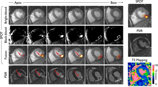
📄 SPOT-MAPPING: combining oedema and scar assessment in a single CMR sequence
🔗 DOI: doi.org/10.1093/ehjimp/qyag0…
🫀 CMR is powerful—but often fragmented:
👉 T2 mapping for oedema
👉 LGE for scar
➡️ Multiple sequences, multiple breath-holds, longer scans
This study introduces a potential game changer: SPOT-MAPPING.
✨ What is SPOT-MAPPING?
👉 A novel CMR sequence combining:
✔ T2 mapping (oedema)
✔ Black-blood LGE (scar clarity)
✔ Bright-blood imaging (anatomy)
➡️ All in a single, co-registered acquisition
📊 As illustrated in the graphical abstract (page 2):
👉 simultaneous visualization of oedema scar anatomy
✨ Key findings:
⏱️ Acquisition time ↓ by ~50%
➡️ ~6 min vs ~12 min with conventional sequences
📈 Excellent agreement with reference techniques:
✔ LV mass (ICC ≥ 0.93)
✔ Scar quantification (minimal bias)
✔ T2 values (no significant difference in oedematous myocardium)
🧠 Why this matters:
👉 Traditional workflows:
❗ multiple breath-holds
❗ misaligned datasets
❗ time-consuming analysis
👉 SPOT-MAPPING:
✔ perfectly co-registered data
✔ faster workflow
✔ improved patient comfort
📊 Added clinical value:
👉 Enables simultaneous assessment of:
oedema vs irreversible injury
scar transmurality
microvascular obstruction / IMH
➡️ Particularly relevant in acute MI and complex cardiomyopathies
⚠️ Important nuance:
👉 Slight overestimation of T2 values vs reference
👉 Requires validation in larger cohorts
💡 Clinical take-home message:
👉 SPOT-MAPPING simplifies CMR without compromising accuracy
➡️ From multiple sequences → one integrated acquisition
👉 A step toward:
✔ faster scans
✔ standardized workflows
✔ more scalable CMR
🚨 Bottom line:
One scan, multiple insights—SPOT-MAPPING may redefine how we acquire and interpret CMR.
#Cardiology #CMR #CardiacImaging #T2Mapping #LGE #MyocardialInfarction #AIinMedicine #MedTech #PrecisionMedicine 🫀📊

1
12
26
2,247
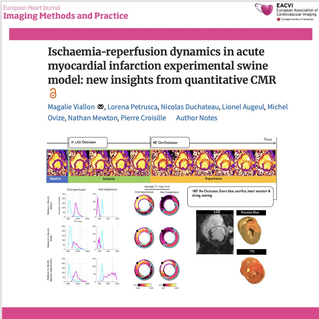
📄 Ischaemia–reperfusion injury: new insights from quantitative CMR
🔗 DOI: doi.org/10.1093/ehjimp/qyaf1…
🫀 Reperfusion saves myocardium—but it can also cause additional injury.
This experimental CMR study provides high-resolution, time-resolved insights into what really happens in the myocardium during ischaemia and early reperfusion.
✨ Study design:
🔹 Swine model of acute MI (LAD occlusion → reperfusion)
🔹 Continuous CMR acquisition at:
baseline
during ischaemia
immediately after reperfusion
up to 2–3 hours
🔹 Quantitative T1 & T2 mapping pixel-wise analysis
✨ Key findings:
📈 During ischaemia:
➡️ Moderate increase in T1 and T2 ( ~11%)
👉 reflecting early oedema
📈 After reperfusion:
➡️ Marked increase in T1 and T2 ( ~23%)
👉 indicating rapid and significant tissue water accumulation
📊 Critical insight:
👉 The biggest myocardial change happens AFTER reperfusion
➡️ Not during ischaemia
➡️ But immediately after restoring flow
🧠 Three distinct reperfusion patterns identified:
1️⃣ Reperfusion MVO (microvascular obstruction)
👉 severe injury, largest signal increase
2️⃣ Reperfusion without MVO
👉 less severe but still significant oedema
3️⃣ No effective reperfusion
👉 blunted signal changes
📌 As shown in Figures 2–4 (page 5), each pattern has distinct T1/T2 evolution profiles
⚠️ Key paradigm shift:
👉 Post-reperfusion oedema:
❗ does NOT reflect pre-ischaemic tissue status
❗ does NOT predict final tissue outcome
➡️ Early imaging interpretation may be misleading
🤖 Methodological innovation:
✔ Pixel-wise standardised analysis
✔ Eliminates ROI bias
✔ Enables true spatial and temporal mapping of injury
💡 Clinical implications:
👉 Reperfusion injury is:
dynamic, heterogeneous, and not fully captured by traditional imaging timing
👉 Timing of CMR matters:
➡️ early post-reperfusion scans may reflect transient inflammation, not final infarct size
🚨 Bottom line:
In acute MI, the most dramatic myocardial changes occur after reperfusion—not during ischaemia—and early oedema does not define final injury.
#Cardiology #CMR #MyocardialInfarction #ReperfusionInjury #T1Mapping #T2Mapping #CardiacImaging #PrecisionMedicine #TranslationalResearch 🫀📊
17
36
2,072

Feb 18
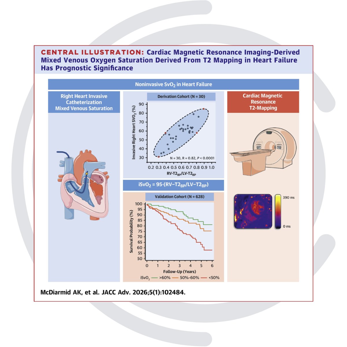
🫀📊 New in JACC: Advances: Routine CMR T2 mapping can noninvasively estimate mixed venous O₂ saturation and independently predict mortality & HF hospitalization. A promising step toward practical, noninvasive hemodynamic assessment in HF.
🔗loom.ly/ajvcfLA
#HeartFailure #CardiacMRI #T2Mapping #Hemodynamics
1
5
642

24 Oct 2025
#EHJIMP
What's your possible diagnosis in a patient with #chestpain, raised #troponin and normal #coronary_angiography?
Can you see the difference between #SPOT and #PSIR and #T2mapping sequences?
doi.org/10.1093/ehjimp/qyaf1…
@JGrapsa @alessia_gimelli @EHJIMPEiC @pabloplopez_
1
7
16
1,617

10 Sep 2025
📢 #SpecialIssue
Cardiac Imaging and Heart Diseases: Recent Progress
📅 30 May 2026
👨🔬 Guest Editor:
Dr. Francesco Secchi from University of Milan, Italy
🔗 mdpi.com/journal/applsci/spe…
#cardiacimaging #cardiovascularMRI #cardiacCT #4DFlowMRI #T1mapping #T2mapping #lateenhancementCT #fractionalflowreserve #FFRCT #artificialintelligenceincardiacimaging #machinelearningincardiology #radiomics #3Dimaging #hybridimaging #perfusionimaging #motionanalysis #automatedsegmentation #deeplearningforcardiacdiagnosis #functionalassessment #prognosticevaluation #bigdataincardiovascularimaging #automatedimageprocessing #clinicalimpactofimagingtechnologies #cardiovasculardiseasediagnosis

1
2
63

28 Jan 2024
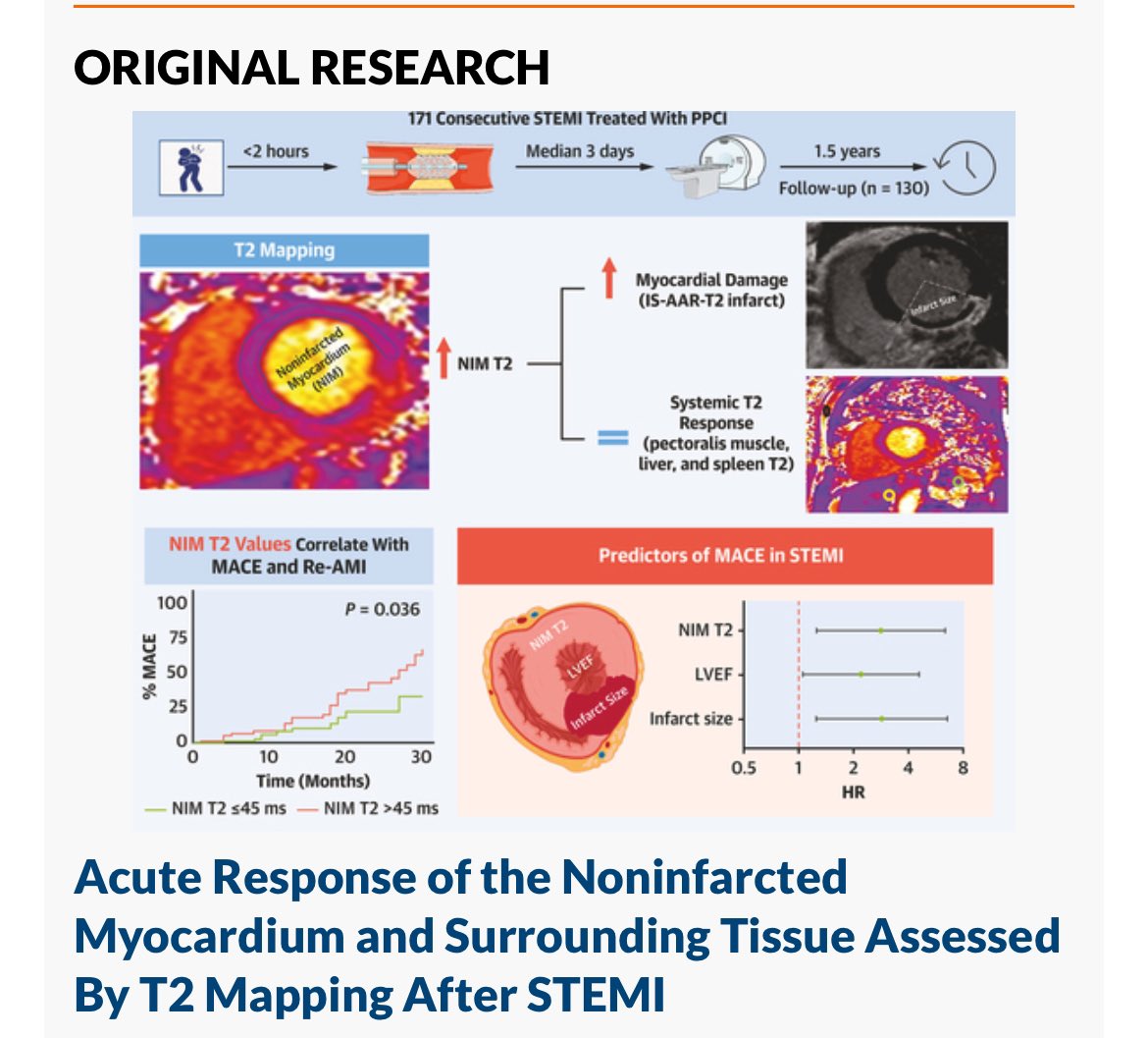
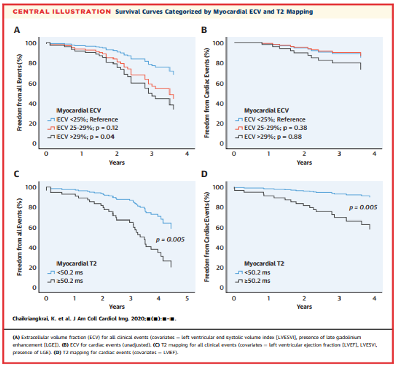
心筋梗塞の時の非梗塞心筋におけるT2mappingの意義。同様のことはT1もECVも報告あります。加えて心筋梗塞後の何日目に撮るかは重要です。
28 Jan 2024

Higher T2 values 🧲 after STEMI in Noninfarcted Myocardium (NIM) & Surrounding Tissue are independently associated with worse CV outcomes, mainly because of higher risk of MI. #whycmr @SCMRorg @JACCJournals @AnnaGiuliaPavon
4
549


15 Nov 2023
今回の話にはありませんでしたが,これらに加えて
「冠動脈CTみたいなMRCA」
「負荷心筋シンチみたいな負荷perfusion」
「T1/T2mapping」
など,さまざまなシークエンスの情報を追加できます
しかも非侵襲的に
心エコー+Gaシンチ+病理
って考えただけでもワクワクしませんか?
1
1
5
1,637

26 May 2023
Wie wichtig ist die Früherkennung von Knieknorpelveränderungen im Nachwuchsleistungssport?🏐
Die Ergebnisse der prospektiven #T2Mapping-studie wurden kürzlich veröffentlicht!🤓🎉 @UKL_Leipzig @ThiemeGruppe
DOI: 10.1055/a-2081-3245
#Röfo #cartilage #pediatric #volleyball #knee

3
158

11 Mar 2023
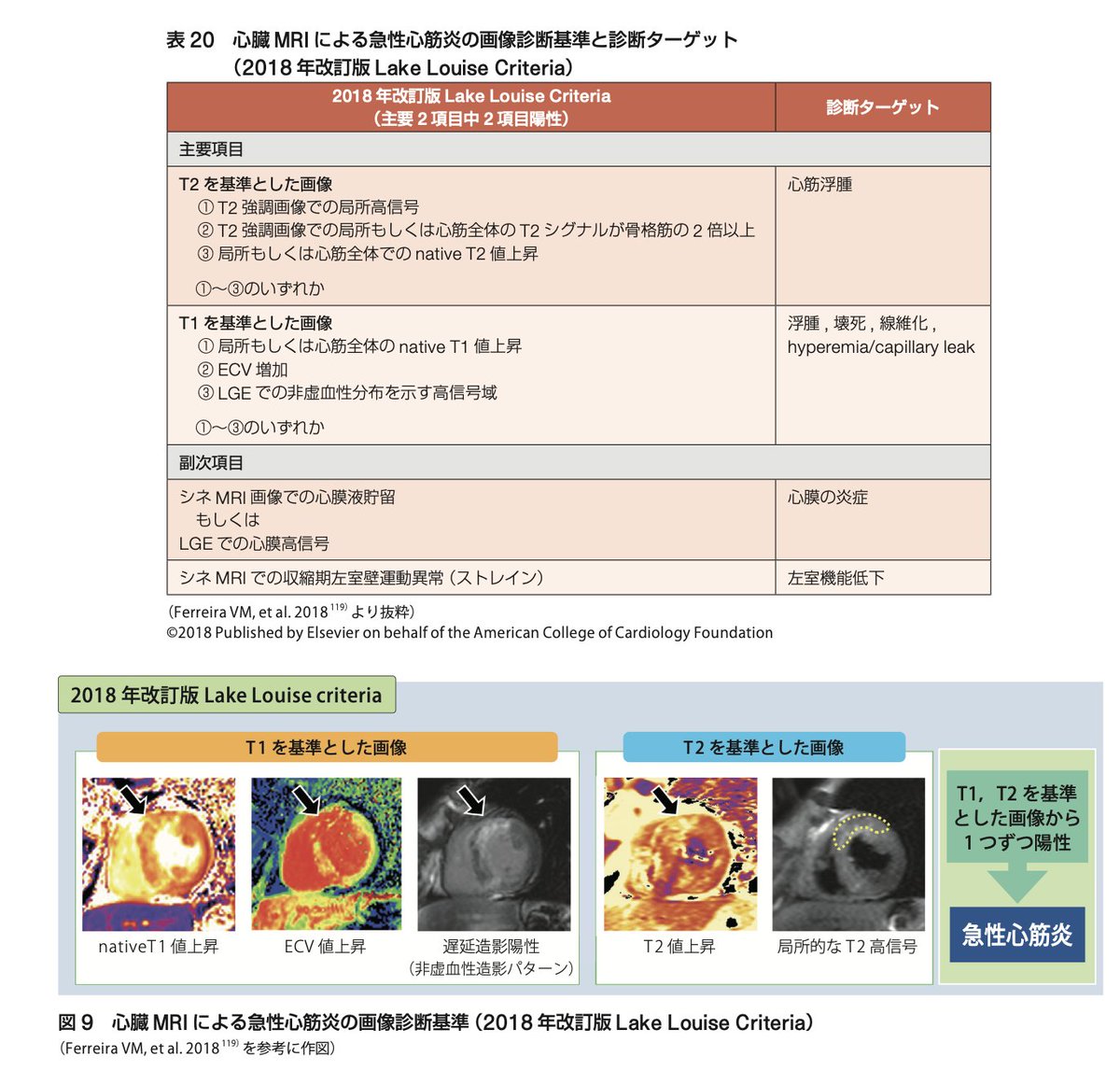
心臓MRIに関して
✔︎T1(LGE/T1mapping)での非侵襲的心筋障害
✔︎T2(T2強調画像/T2mapping)での心筋炎症
を組み合わせた改訂版Lake Louise Criteriaが診断に用いられる
発症後2-3週以内の検査を推奨
5/n
1
3
21
1,989

14 Aug 2020
Valor pronóstico del #VEC y #T2mapping en Trasplante Cardiaco
/
ECV & T2 mapping in Heart Transplant
@JACCJournals bit.ly/3hCVhjZ
@SIAC_cardio @onco_cardiology @purviparwani @LopezOpitz @ecocardio_cl @urmeneta87 @DiegoChango1 @chemaimagencv @HugoMartinezCMR @iamritu
3
5
6 Aug 2020
Valor pronóstico del #VEC y #T2mapping en Trasplante Cardiaco
/
ECV fraction & T2 mapping in Heart Transplant
@JACCJournals bit.ly/3hCVhjZ
@iamritu @chemaimagencv @onco_cardiology @mariovar55 @SIAC_cardio @purviparwani @LopezOpitz @DrSergioBarros @JournalASEcho

3
3

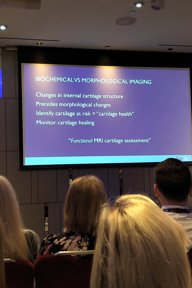
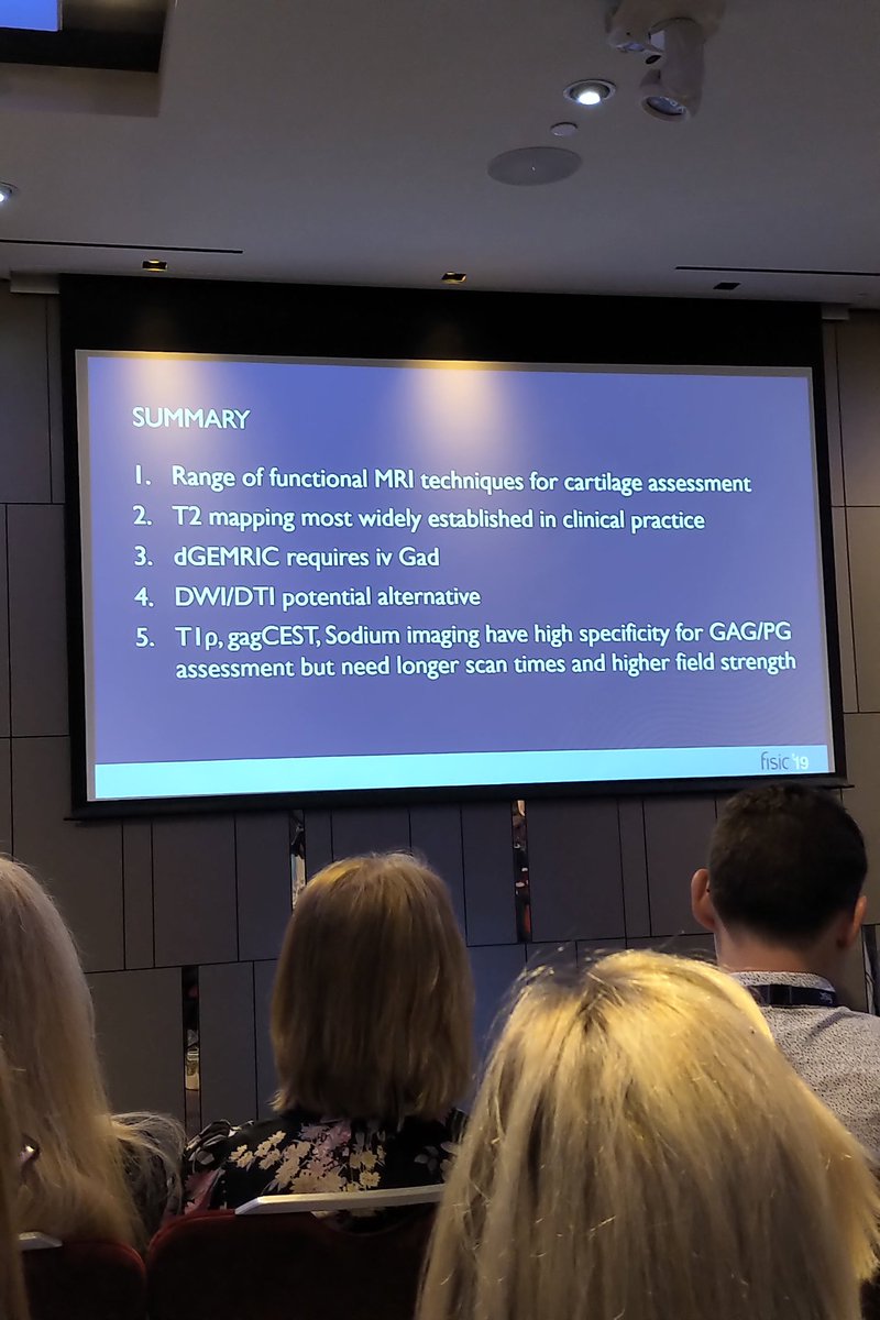
6 Nov 2019
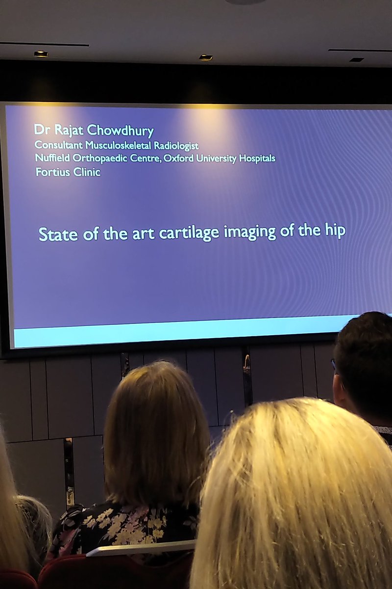
State of the art imaging of the hip, dr. Rajat Chowdhury. The goal is to prevent damage before it's done... So we want to do "functional MRI cartilage assessment". T2mapping, T1ro, DWT/DWI, dGEMRIC, etc. #sportsmedicine #sportsradiology @FortiusClinicUK @FortiusFISIC @AxiomClinic
2

10 Jul 2019
Memo(for me):
@JACCJournals
#CMR
#T2mapping
#IntracardiomyocyteEdema
#EarliestMarker
#Anthracycline
#Cardiotoxicity
#CardioOnc
Serial Magnetic Resonance Imaging to Identify Early Stages of Anthracy... sciencedirect.com/science/ar…
#CardioJapan
3
4

17 Jun 2019
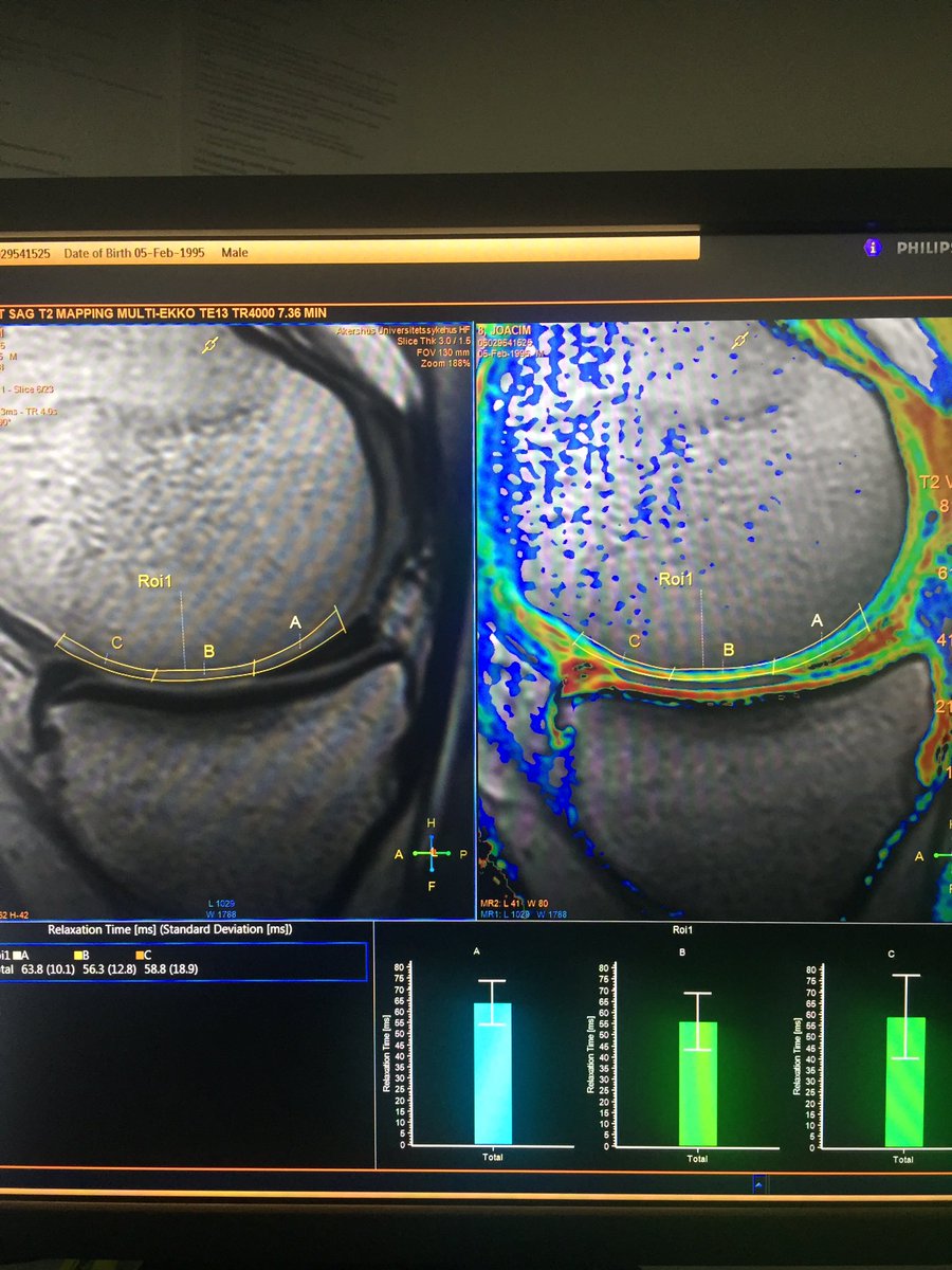
T2 relaxation time mapping can be a potential biomarker for cartilage deterioration. Changes in these times can be the result of changes in hydration or disruptions in the network of collagen. T2 maps are acquired by performing a spin echo based multi-echo acquisition #T2mapping
5
14

27 May 2019
How reliable is T2 mapping? We’re looking into the reliability and clinical consequences of T2 mapping results in #cartilage surgery patients. To be continued... @CartilageRepair #t2mapping
1
7

18 Feb 2019
Reduction of Global Longitudinal Strain (GLS) in echo is an early sign of chemotherapy toxicity, before EF goes down. Garan-Ariola et al now show in 20 pigs that myocardial edema seen by CMR T2mapping may be the earliest sign. onlinejacc.org/content/73/7/…. #cardiotwitter @escardio
1
63
142

Really interesting paper on #CMR in #CardioOncology - does #T2mapping have a key role to play in detecting #anthracycline #cardiotoxicity? Well done to @cgalanarriola and colleagues!
onlinejacc.org/content/73/7/…
1
2
12
2 Oct 2018
T2 relaxation time mapping has shown to be a potential biomarker for cartilage deterioration. T2 maps are acquired by performing a spin echo based multi-echo acquisition.
#imagingU, #T2mapping, #CartilageMapping
4

19 Jul 2018
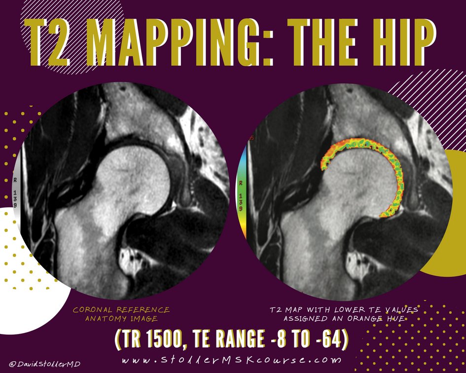
T2 Mapping (TR 1500, TE range -8 to -64) Articular cartilage of acetabular roof thicker at lateral acetabular rim. Fibrocartilaginous labrum included in thresholding of TE values StollerMSKcourse.com
#StollerMSKcourse #TheHip #T2Mapping #MRI #orthopaedics #sportsmedicine
4
7