
Jun 9
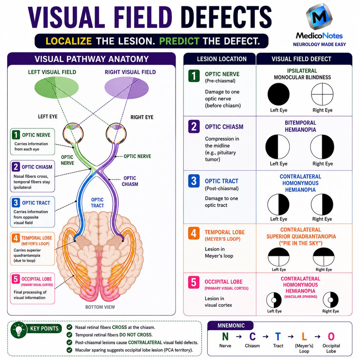
🧠 VISUAL FIELD DEFECTS
Localize the Lesion. Predict the Defect.
⸻
1️⃣ Optic Nerve
➊ Pre-chiasmal lesion
➋ Ipsilateral monocular blindness
➌ Vision loss in one eye only
💡 Think: “One Nerve = One Eye”
⸻
2️⃣ Optic Chiasm
➊ Compression of crossing nasal fibers
➋ Classically caused by pituitary adenoma
➌ Bitemporal hemianopia
💡 Think: “Can’t see the sides”
⸻
3️⃣ Optic Tract
➊ Post-chiasmal lesion
➋ Contralateral homonymous hemianopia
➌ Same visual field lost in both eyes
💡 Think: “Tract = Opposite Side Lost”
⸻
4️⃣ Temporal Lobe (Meyer’s Loop)
➊ Contralateral superior quadrantanopia
➋ Loss of upper visual field quadrant
➌ Temporal lobe lesion
💡 Think: “Pie in the Sky”
⸻
5️⃣ Occipital Lobe
➊ Contralateral homonymous hemianopia
➋ Macular sparing often present
➌ PCA territory infarction is a classic cause
💡 Think: “Occipital = Opposite Field Lost”
⸻
🎯 High-Yield Rules
✅ Nasal retinal fibers cross at the chiasm
✅ Temporal retinal fibers do NOT cross
✅ Post-chiasmal lesions cause contralateral defects
✅ Macular sparing suggests occipital lobe involvement
⸻
🧠 Easy Mnemonic
N → C → T → L → O
👁️ Nerve → Monocular Blindness
✖️ Chiasm → Bitemporal Hemianopia
🔵 Tract → Homonymous Hemianopia
🥧 Loop (Meyer’s) → Pie in the Sky
🎯 Occipital → Homonymous Hemianopia Macular Sparing
⸻
📚 Master Neurology the high-yield way with the MedicoNotes Neurology Book.
🌐 mediconotes.com
#Neurology #VisualFieldDefects #Neuroanatomy #MedicalEducation #MedicoNotes
54
107
2,802

24 May 2024
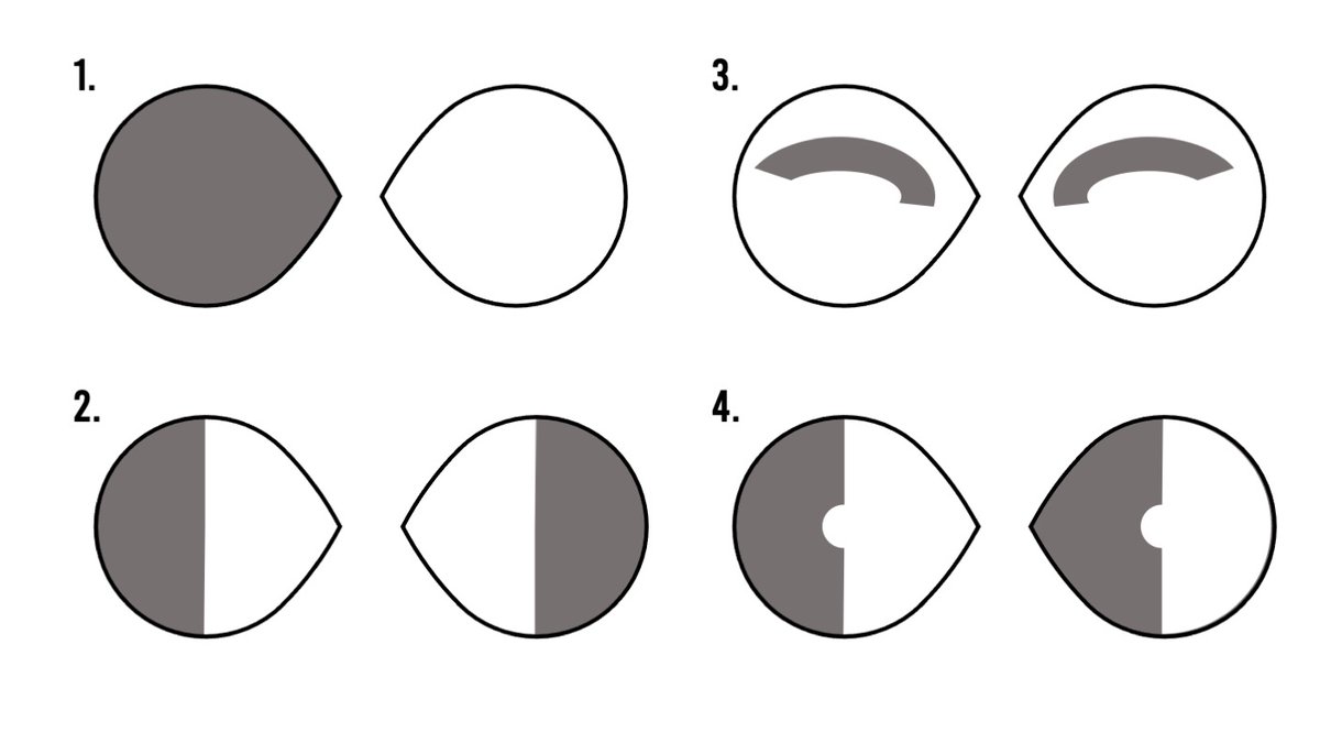
Learn about visual field defects and associated lesions with this TrueLearn infographic! Save to review later!
#vision #visualfielddefects #lesions #resident #healthcare #healthcarestudents #healthcareworker #truelearn #infographic

1
163

25 May 2023
I’m not saying I have poor spatial awareness these days, but when I walk down a busy street I channel
strong Richard-Ashcroft-in-the-Bittersweet-Symphony-video vibes.
#AZOOR #visualfielddefects
3
478

What is a likely cause of these #VisualFieldDefects?
Potential answers...
A. #PituitaryTumor 🍒
B. #CRAO ☄️
C. #MCAStroke 🧠
D. #Glaucoma ♎️
@EyeRounds @Eyes4Ears
✅ lensophthalmology.com/questi…
1
3
7

22 Aug 2020
The student team that built this great model was made up of:
🧠 Dhritica Bora 👁️🛠️
🧠 Sam Boxwell 👁️🛠️
🧠 Meng Jiang 👁️🛠️
🧠 Demi Oke 👁️🛠️
🧠 Conor Ring 👁️🛠️
#VisualFieldDefects #Neuro #Vision
(with names spelled correctly this time 🙈)
1
6