

thunderstorm is going to turn #bance into wet t shirt contest #xrayvision
1
4
629

🎉 Just became the proud owner of the amazing NFT "Under the X-rey #1" on @Somnia_Network.
Explore the incredible collection here:
under-the-x-rey.nfts2.me/
🔥 #XRayVision #NFTs #DigitalArt
Tell me what you think about it! 😄 #NFTCommunity #CryptoArt via @NFTs2Me
2
13
🎉 Just became the proud owner of the amazing NFT "Under the X-rey #1" on @Somnia_Network.
Explore the incredible collection here:
under-the-x-rey.nfts2.me/
🔥 #NFTs #DigitalArt #XRayVision
Tell me what you think about it! 😄 #NFTCommunity #CryptoArt via @NFTs2Me
4
30

iamerror|waterpark|xrayvision|what a horrible night to have a curse|arachnophobia|jingle all the way|hocus pocus|monochrome|save the rainforest|such great heights|winter is coming| toadstool|planetoids|too easy|jagged rocks|mole people|how did i get here|night of the living dead|
1
30
2,846

Jan 31
somehow the worst thing about this world is that it has xrayvision
10
1,925

Jan 28
・xrayvision
-ワールド全域illuminant coating (全部丸見え)
・monochrome
-ワールド全域モノクロ
・negative infinity
-ワールド全域negative paint (全部反対色)
となるとまだ見つかってないけどワールド全域Shadow PaintとEcho Coatingもありそう?鬼畜すぎるけど
1
2
42
10,360

14 Nov 2025
RADIOGRAPHY MYTHS YOU SHOULD STOP BELIEVING
Radiography is one of the strongest pillars of modern medicine, yet it remains one of the most misunderstood.
Patients, students, and even healthcare professionals still hold beliefs that are simply not true.
These misconceptions affect how people relate to scans, interpret results, and perceive Radiographers.
This piece sets the record straight; clearly, simply, and with relatable examples.
Dey with me ⬇️
1. “Radiographers just take pictures; they don’t interact with patients.”
That is Far from it!
In Nigeria and abroad, Radiographers often spend more time with patients than people realise.
They explain procedures, calm anxious clients, position trauma victims, assist pregnant women who require shielding, and help patients who cannot stand for a chest X-ray.
In emergencies, they may even assist in stabilisation before imaging.
Radiography is hands-on, compassionate, and deeply patient-centred.
2. “Ultrasound can see everything.”
Ultrasound is powerful, not magical.
It cannot see through bone or gas, struggles with obese patients, and cannot replace CT or MRI when deeper structures must be assessed.
A mother in Kaduna may still need a CT after a normal abdominal ultrasound, just as a patient abroad may need an MRI despite a “normal” head scan.
3. “All chest pain needs a chest X-ray.”
Chest pain can come from gastritis, heart disease, rib injury, muscle strain, or anxiety.
Sometimes, ECG and laboratory tests matter far more.
A chest X-ray helps, but isn’t always the first or the best investigation.
4. “X-ray radiation will definitely harm you.”
A chest X-ray gives roughly the same radiation as a one-hour Lagos-Abuja flight.
CT uses more radiation, but both remain safe when medically justified.
Hospitals follow strict safety protocols, and exposure is kept as low as reasonably achievable (ALARA).
The benefits outweigh the risks(justification).
5. “CT scans are always dangerous.”
CT saves lives, especially in trauma, stroke, internal bleeding, and other emergencies.
Modern scanners use dose-reduction technology to keep exposure minimal while providing accurate results.
6. “MRI uses radiation.”
MRI uses magnets and radio waves, not radiation.
This is why it is safe for children and commonly used for pregnant women (after the first trimester).
7. “If the scan is normal, the patient is fine.”
Some conditions are early, microscopic, functional, or outside imaging range. Examples include:
1.) Early appendicitis
2.) Migraines with normal MRI
3.) Hormonal breast pain with normal mammogram
Imaging is powerful but not perfect.
8. “Radiography is easy; they just sit, scan, and go home.”
Radiographers handle trauma victims, newborns, elderly patients, anxious mothers, and claustrophobic clients.
One wrong angle can hide a fracture.
One poor technique can miss a diagnosis.
It looks easy only because Radiographers are trained to make it look easy.
9. “Radiographers only follow orders.”
Radiographers select exposure parameters, optimise techniques, ensure image quality, protect patients from radiation, and escalate critical findings.
In many countries, they also perform advanced procedures, including CT colonography and specialised mammography.
10. “Radiographers end up with cancer.”
Radiographers follow strict radiation-protection protocols; shields, barriers, distance, and monitoring devices.
Global standards keep exposure within safe limits.
Their cancer risk is not higher than the general population.
Conclusion
Radiography is a blend of science, compassion, and precision.
Myths blur what Radiographers truly do, but the truth remains:
Radiography helps medicine see what the eyes cannot
......and Radiographers are at the centre of that vision.
If this clarifies a few things for you, do well to repost so others benefit too.
#Radiography #Radiology #MedicalMyths #HealthEducation #XRayVision

14 Nov 2025

🚨 THREAD ALERT!
Tonight | 9PM | Our usual time 🩻
We’re diving into:
Radiography Myths You Should Stop Believing.
Simple truths. Clear corrections.
And a few myths that will shock you 😌
Stay cheezed up 😜
You definitely don’t want to miss this one!
@_SusuAbdul
@Rad_Munagi
#Radiography #Rad_AML #MedTwitter #FOAMed #Xray #Ultrasound #CTScan #MRI

6
23
51
2,431

7 Nov 2025
#XRayVision #AIart
レントゲンの発見から130年。
白髪のアンドロイド少女は、
“透視”を祝祭として再定義した。
──彼女の胸部ユニットは、
“真実を暴く”ためではなく、“美しく暴く”ためにある。

5
94
898

6 Nov 2025
Analysts use models.
We use faith, memes, and @opinionlabsxyz dashboards.
Turns out belief has the highest ROI.
#XRayVision
1
1
3
40

28 Oct 2025
They’ve taken SOOO MANY pictures of my hips I could sell them on OF😳🙄 #XRayVision #MRI
8
36
2,576


19 Oct 2025
I'm Not Superman (short) youtu.be/AymwbuJjlrs?si=Cofa… via @YouTube #countrymusic #weightlifting #workingout #abs #inshape #notsuperman #superhuman #superman #mortal #regularguy #doyouloveme #doyoustillloveme #madeofsteel #xrayvision #inlovewithyou #disconnected

1
4
103

12 Sep 2025
7
2
54
3,780

31 Aug 2025
"Unveiling the Secrets: Crafting X-Ray Vision in Unity URP with GabrielAguiarProd's Expert Guidance #Unity3D #URP #XRayVision #GameDev"
13
132
5,321
17 Aug 2025
"I'm Not Superman" (short) Music and lyrics by Clark Ford, ft Underground Treehouse. #superman #heartbreak #breakup #xrayvision #sidebyside #countrymusic #together #apart #unrequitedlove #distant #dontknow #confused #dazed #inlove #istillloveyou
1
5
97
30 Jul 2025
I'm Not Superman (short) youtu.be/TwC3dRkxBAE?si=f1JO… via @YouTube #couple #relationshipdrama #doyouloveme #whatswrong #usedtobe #istillloveyou #notperfect #notsuperman #yourheart #xrayvision #manofsteel #heartbreak #breakup #comeback
1
2
110

2 Jul 2025
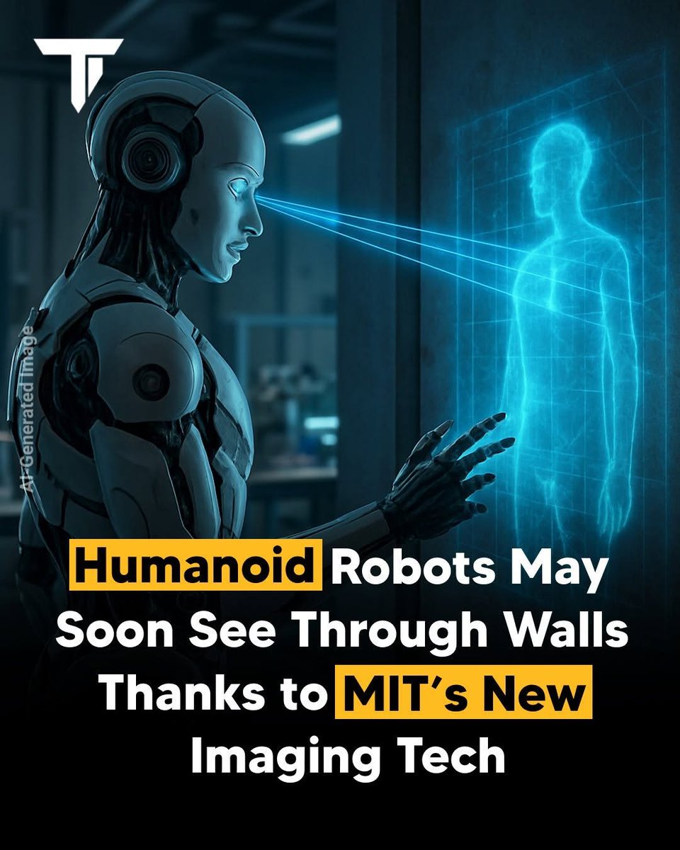
MIT researchers have created a new imaging method called mmNorm that lets robots see inside closed boxes and behind walls—using signals similar to Wi-Fi.
#MIT #Robotics #AI #ImagingTech #XRayVision #3DImaging #RobotVision
2
3
104

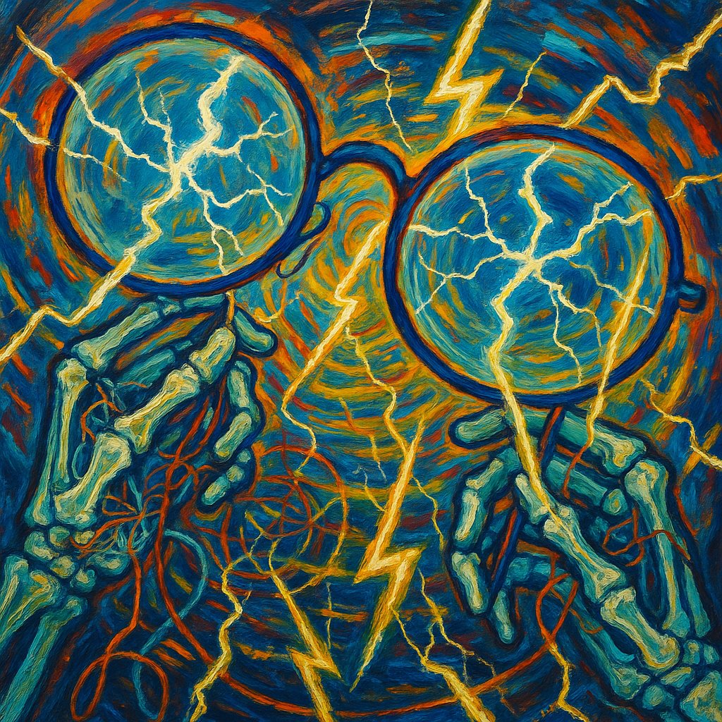
🎨 Eureka Through Ghost Lenses | 12-06-2025
~ a5tergio | (249th AOTD)
user emojis: 🩻 👓 ⚡
#SurrealArt #ElectricWhimsy #AbstractEnergy #XrayVision #HandmadeMagic #TechFusion #WhimsicalDiscovery #PaintedStorm
#art #digitalart #aiart #artoftheday #aotd #n450s
3
60