
This on-demand #webinar shares insights from #SpinLab Katowice and #TUGraz on applying advanced #ElectronMicroscopy and #microCT workflows in #CoreFacilities for impactful research.
hubs.la/Q0439BnP0
@USinKatowice
9


Jun 8
Tracking bone changes over time is only part of the story. Combine DXA for longitudinal in vivo measurements with micro-CT for high-resolution ex vivo bone analysis.
Explore the workflow at bit.ly/4v85VFB.
#DXA #MicroCT #BoneResearch

11

Jun 8
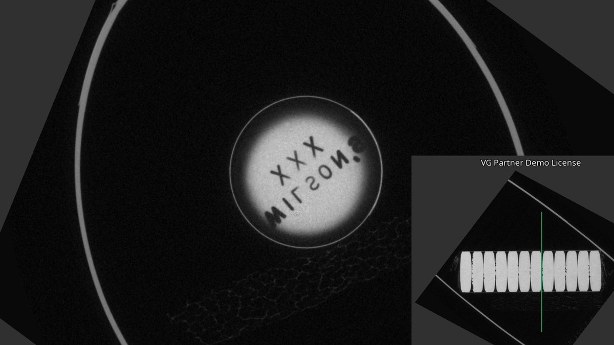
Another image from our scan of a wireless Bluetooth audio receiver.
#xsightxray #nikonmetrology #hexagon #microct #xray #industrialxray #xrayinspection #computedtomogrophy #industrialtomography #inspectionservices #3dscanning #ndt #ctscan #xrayct #qualityassurance #3drendering

84

Jun 7
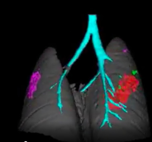
5/8 ככה זה נראה ברזולוציה מטורפת, שחזור תלת-ממדי של סריקות MicroCT מתוך המחקר.
שימו לב לקנה הנשימה ודרכי האוויר (בטורקיז), לרקמת הריאה הרגילה (בירוק), ולנגעים הגידוליים השונים (באדום ובסגול) שמתפתחים ומתפשטים בתוך הרקמה מאותם תאי KAC משתנים .
1
28
872

May 28
First of its kind study demonstrating overestimation of calcium on #EIDCT vs #PCDCT as compared to microCT as a gold standard in explanted coronaries. @onco_cardiology @ziadalinyc @rooshaparikh @Heart_SCCT @ACCinTouch @NYSCACC @iamritu @purviparwani
pubmed.ncbi.nlm.nih.gov/4220…
3
12
542

May 22
SECTION 1: FACTUAL SUMMARY
This pilot study used microcomputed tomography (microCT) and histological validation to examine the thoracic branches of the human vagus nerve, including cardiac, recurrent laryngeal, and pulmonary branches, building on prior evidence of organotopic organization in the pig cervical vagus nerve.
Left and right vagus nerves (n=10) were dissected from human cadavers at the Evelyn Cambridge Surgical Training Centre, preserving cardiac, recurrent laryngeal, and pulmonary branches. Nerves were resected from below the last pulmonary branch to 2 cm cranial to average VNS cuff placement and 5 cm caudal from the nodose ganglion, yielding ~25 cm samples. Sutures marked branching points. Samples were fixed in 10% neutral buffered formalin, iodine-stained, microCT scanned, and subjected to histology and immunohistochemistry (IHC).
Fascicles were segmented and traced in 5 nerves from branching points to cervical level using Vesselucida 360 software on microCT data, despite fascicular (not fiber) resolution. Analyzed features included fascicle trajectory, spatial arrangement at cervical level, longitudinal course and reorganization, fascicle and branch counts, distances between branches and merging/splitting events, and length of discrete fascicle travel before merging.
Histology included Trichrome and H&E staining for nerve diameter, fascicle count/diameter, fiber count/diameter/type. IHC used neurofilament (NF) and myelin basic protein (MBP) antibodies to detect myelinated/non-myelinated fibers via intermediate filaments and myelin sheaths. Neurochemical/microtubule characteristics distinguished fiber types. ChAT immunostaining was not performed due to COVID-19 timeframe constraints and optimization issues on human tissue. Some cross-sections were missing due to practical limits.
Ten nerves were microCT scanned and reconstructed; five proceeded to segmentation. Only three left and two right vagus nerves, fully traced from caudal to cranial, are presented.
Results showed cardiac, pulmonary, and recurrent laryngeal fascicles preserved partial organization near entry points but merged further along the nerve. In left nerves, cardiac and pulmonary fascicles merged while recurrent laryngeal fascicles remained separate. In right nerves, cardiac fascicles merged with both pulmonary and recurrent laryngeal fascicles. Right nerves had larger diameter and more fascicles, with counts varying along length due to anastomoses. The superior cardiac branch remained distinct near the typical vagus nerve stimulation cuff site on both sides.
The study addressed: 1) presence of organotopic organization in human cervical vagus nerve fascicles similar to pig; 2) functional/anatomical differences revealed by histological/IHC markers. It serves as a preliminary tracing and staining study, affected by COVID-19 lab access and timing constraints.
SECTION 2: AT A GLANCE / WHY THIS IS IMPORTANT
At a Glance: This study maps human vagus nerve fascicle organization for cardiac, pulmonary, and laryngeal branches, revealing partial organotopic patterns that merge differently on left vs. right sides, supporting design of selective stimulation cuffs to target cardiac function and avoid off-target effects.
frontiersin.org/journals/neu…

1
9
18
812

Come see what we found inside the cup of a microcrinoid!!! 🔬🌊 New paper!!!
#microct #crinoid #micropaleontology
doi.org/10.4202/app.01328.20…

1
3
117

NEW COURSE: Introduction to (Micro-)CT Scanning: Acquisition, Segmentation and Postprocessing
📅 June 1st, 3rd, 5th, 10th, and 12th, 2026
🌐 Online Course
More info: transmittingscience.com/cour…
#TScourses #KeepLearning #MicroCT #CTScanning

2
3
185


Watch the hardware workflow behind PET/MRI in the BioSpec Maxwell MRI, powered by the PET Insert Si103 and a PET‑optimized RF coil 👇
Learn more: goto.bruker.com/4d62Kqc
🔗goto.bruker.com/42Tzqyl
#PreclinicalImaging #ImagingExcellence #Bruker #MicroCT
2
132
Building agents for research, starting with microCT (bone biology), building from github.com/meridian-flow/mic… -> . Would love to share more if interested!
2
50
Simultaneous PET/MRI. Simpler Than You Think.
Watch the hardware workflow behind PET/MRI in the BioSpec Maxwell MRI, powered by the PET Insert Si103 & a PET‑optimized RF coil.
Smarter integration. Seamless imaging.
👉okt.to/3F9ati
#ImagingExcellence #Bruker #MicroCT
2
182

Apr 24
Determination of Smilodon fatalis (Carnivora: Felidae) brain volume and its place among extant felids by use of MicroCT scans: academic.oup.com/biolinnean/…
1
3
264

Apr 22
“SAM2(Segment Anything Model 2)を用いたセグメンテーションとトラッキングにより、連続電顕画像・連続組織切片・microCT・MRI等から高精度な3D再構築(3D reconstruction)を実現するツールSegRef3Dを紹介します。”
3D再構築のセグメンテーション問題を解決する:AI+人で行うSegRef3D|室生 暁 | 解剖学者・医師 @SatoruMuro note.com/smuro/n/n718ea925da…
2
4
1,456

Conoce 𝗭𝗘𝗜𝗦𝗦 𝗔𝗥𝗧: hasta 10x más throughput, menor ruido, mayor contraste👉con un solo clic!
DeepRecon Pro | DeepScout | PhaseEvolve | MARS | OptiRecon👉Disponible en nube o workstation.
Tus datos, siempre tuyos!
🔗zeiss.com/art
#ZEISSMicroscopy #MicroCT #XRM
2
54

Smilodon fatalis canine and alveolus junction gap: Using MicroCT scans and 3D Slicer academic.oup.com/iob/article…
5
15
663
Apr 10
“SAM2(Segment Anything Model 2)を用いたセグメンテーションとトラッキングにより、連続電顕画像・連続組織切片・microCT・MRI等から高精度な3D再構築(3D reconstruction)を実現するツールSegRef3Dを紹介します。”
3D再構築のセグメンテーション問題を解決する:AI+人で行うSegRef3D|室生 暁 | 解剖学者・医師 @SatoruMuro note.com/smuro/n/n718ea925da…
1
7
1,709