
Advanced imaging research facility in the School of Biological Sciences at the University of Cambridge caic.bio.cam.ac.uk/
Joined November 2018
- Tweets 30
- Following 114
- Followers 436
- Likes 13
12 Photos and videos


19 Jun 2023
A great opportunity in Cambridge to lead the Advanced Imaging Centre at the School of Biological Science ... 10 more days to apply
jobs.cam.ac.uk/job/40627/ @Cambridge_uni @cambridge_sbs @RoyalMicroSoc @EuroBioImaging @focalplane_jcs
11
11
2,779
Cambridge Advanced Imaging Centre retweeted

12 Feb 2021
Check out our latest publication 'Visualising an invisible symbiosis' in @plantspplplanet! @plantsci @CropSciCentre @niabgroup
nph.onlinelibrary.wiley.com/…
3
31
79
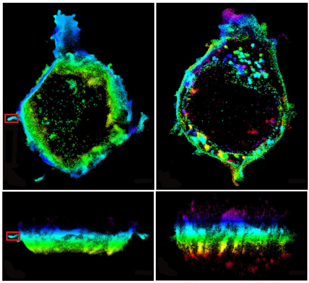
October image of the month is from @Ruby_Peters_ of the
@PaluchLab and shows HeLa cells, labelled with Tubulin-GFP. Taken using our Elyra 7 Lattice SIM and colour coded for depth
1
12
22 Oct 2020
Beautiful!
21 Oct 2020

Excited to process and analyse my first nlsABACUS #lightsheet images, even if it is just the negative control😅! Martin Lenz at @CamMicroscopy and @slcuplants has built a brilliant microscope and is an imaging ninja.
10
Cambridge Advanced Imaging Centre retweeted

8 Oct 2020
how to cover yourself when you are a plant? Just weave your own fabric! Check our collaborative work between @slcuplants @CUBotanicGarden @ChemCambridge @CamMicroscopy on how Dionysia tapetodes extrudes flavones threads from its glandular trichomes. biorxiv.org/content/10.1101/…
6
19
30
Cambridge Advanced Imaging Centre retweeted

2 Oct 2020
Via #OSA_Optica: Single molecule light field microscopy ow.ly/Urg550BEkQ4 #FlatOptics #SpectralFIlters @koholleran @Cambridge_Uni @UniversityLeeds
1
4
13
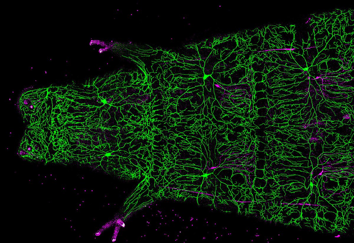
August image of the month: Drosophila larvae expressing green and magenta fluorescent proteins in nociceptive (noxious touch), and proprioceptive (body movement) neurons. Image: @Paul_brooks who researches neuron degeneration, captured by confocal microscope in @CamZoology
1
10
36
24 Jul 2020
We're hiring! Research associate position in advanced photo-manipulation microscopy. Come join us in developing technology critical for cutting edge optical trapping, optogenetic and photoablation experiments. jobs.cam.ac.uk/job/26323/
1
23
15
22 Aug 2020
Deadline is fast approaching, get your applications in by 26th August!
22 Aug 2020
Congratulations to @r_r_sims @sohaib_ar and the whole team for their new 3D super-resolution method, now published in Optica.
1
3
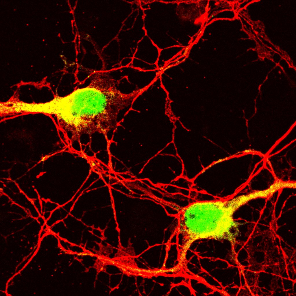
July image of the month. Puromycin assay on cultured hippocampal neurons showing new proteins that are produced within 15 minutes (yellow/green) and microtuble-associated protein 2 staining (red).
Captured on our laser scanning confocal microscope by Dr Tanja Fuchsberger
1
4
17
The EPSRC Research Software Engineer Fellowship call is now live:
epsrc.ukri.org/funding/calls…
1
The @SarrisLab made incredible use of our two-photon microscope in their recently published research on neutrophil migration to sites of tissue damage cell.com/current-biology/ful…
4 Jun 2020

In this video, we capture neutrophil swarming at a site of acute wounding, as the locus is invaded by opportunistic bacteria @CamMicroscopy
1
2
25 May 2020
Hot off the press: our research lab's preprint of their new super-resolution microscopy technology.
4
8
CAIC is starting a monthly series of informal clinics for super-resolution fluorescence microscopy. Come along for advice on getting the most out of our SIM, STED and SMLM systems. First clinic is Friday 21st February - follow link for details caic.bio.cam.ac.uk/news/supe…
1
9
32
Cambridge Advanced Imaging Centre retweeted

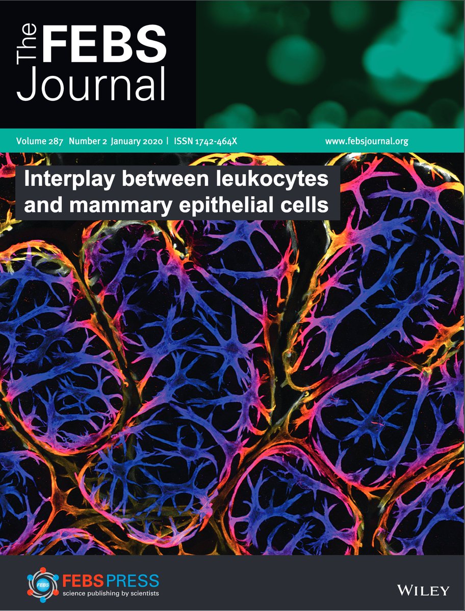
22 Jan 2020
We got the cover! The wonderful @JessieJRH's new paper is out now in @FEBSJournal, with an excellent commentary from @WendyIngman. If you like immune cells and boobs, or nice 3D imaging, this is worth a read. febs.onlinelibrary.wiley.com… with @katevetpath & @cjwhelix
4
7
23
11 Dec 2019
Fantastic opportunity to join the @EmilianiLab and get involved in advanced optogenetics technologies. You'll also get to work alongside the talented @r_r_sims who developed light sheet and light field microscopes in CAIC.
9 Nov 2019

PhD Position in the #interdisciplinary PhD program #zenith_etn. @EmilianiLab is looking for candidates interested in non-linear optics,wavefront shaping and optogenetics( zenith-etn.com/projects/all-… ).Send your application soon,deadline is January 5th 2020! #ITN @MSCActions #zenith

2
Great work by Martin Lenz showcased in his talk about the multi-view light sheet microscope for gentle fast imaging of plants he has developed in the @slcuplants imaging facility.

2
6
25
Flaminia Kaluthantrige Don, a @BBSRC CASE student hosted by CAIC and Meri Huch's lab, used our two-photon microscope to great effect. She optimised imaging conditions in order to observe single cells proliferating into liver organoids over three days.
18
41
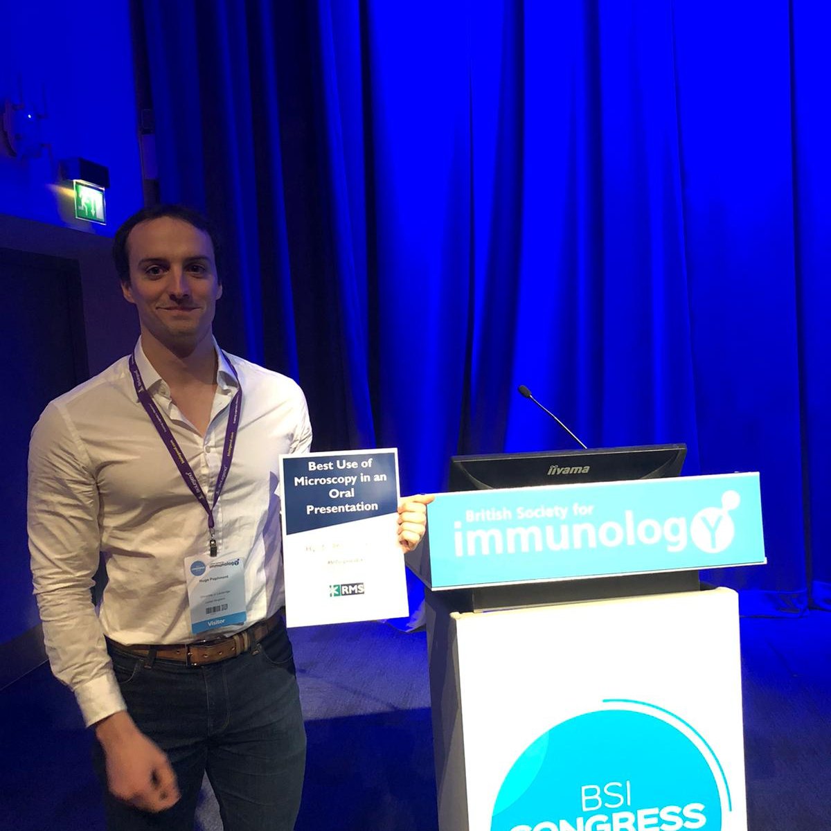
Congratulations to Hugo Poplimont who won a @RoyalMicroSoc prize for best use of microscopy in a presentation at #BSI2019. You can read about his work on neutrophil swarming in damaged tissue using our two-photon microscope in their recent @biorxivpreprint biorxiv.org/content/10.1101/…
1
6