
Assistant Prof. of EM @PittTweet. ER doc @upmcem MD, FAWM. Focus on trauma, TBI, wilderness medicine, prehospital EMS. Lover of the outdoors & hops. Hoya Saxa.
Joined June 2009
- Tweets 3,513
- Following 1,252
- Followers 728
- Likes 5,488
214 Photos and videos


Now accepting Neuro-EM K12 Scholar Applications and Pipeline Program Nominations!
#AWAEM #SAEM #WomeninEM #WomeninMedicine

2
1
205
David Barton retweeted

16 Jul 2025
We’re thrilled to announce #PittMed’s #12 spot in CEOWORLD Magazine’s global rankings for 2025.
This recognition reflects the unwavering commitment of our talented faculty and staff who advance our mission every day.
Thank you for all you do.
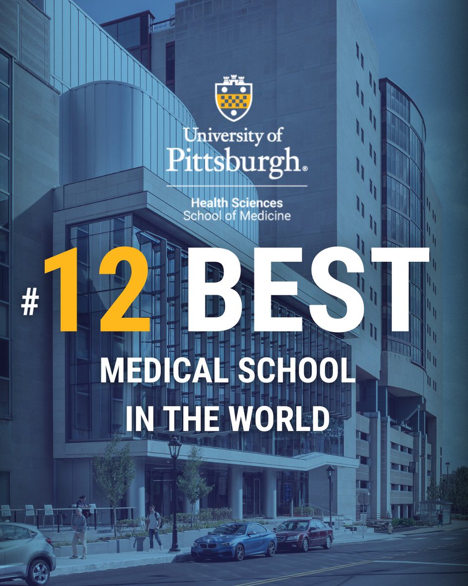
ALT Promotional image featuring the University of Pittsburgh Health Sciences School of Medicine building with a banner stating '#12 Best Medical School in the World.'
14
25
1,817

A new policy from the US National Institutes of Health (NIH) will end billions of dollars of funding to laboratories and hospitals outside the United States, imperiling thousands of projects on topics such as emerging infectious diseases and cancer.
go.nature.com/3Yu1Ddw
21
62
136
21,746
David Barton retweeted

5 Feb 2025
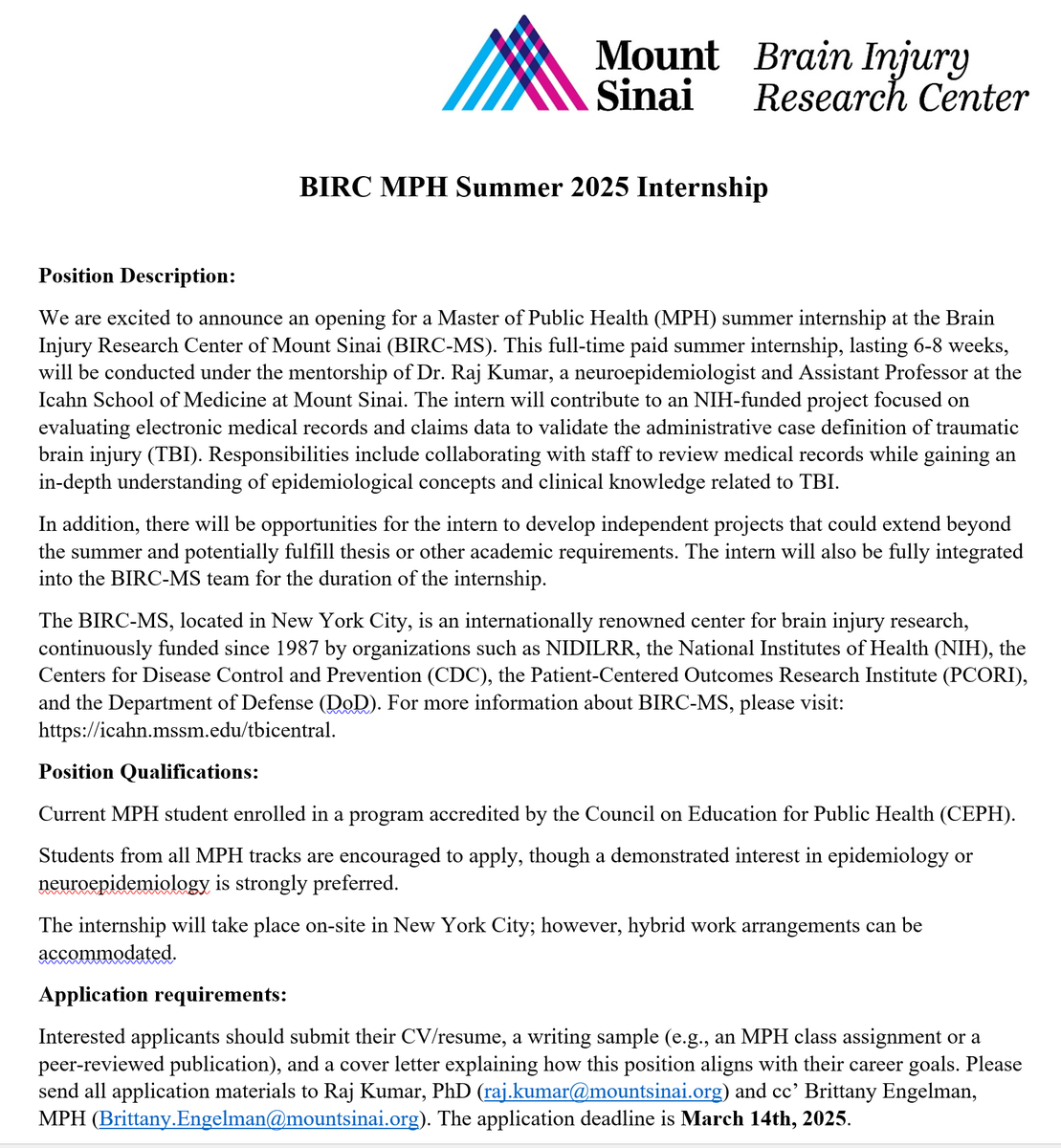
We are excited to announce an opening for a full-time paid MPH summer internship at @SinaiTBI! More information on the position is included below. Please share with your network!
5
15
1,178
David Barton retweeted

27 Jan 2025
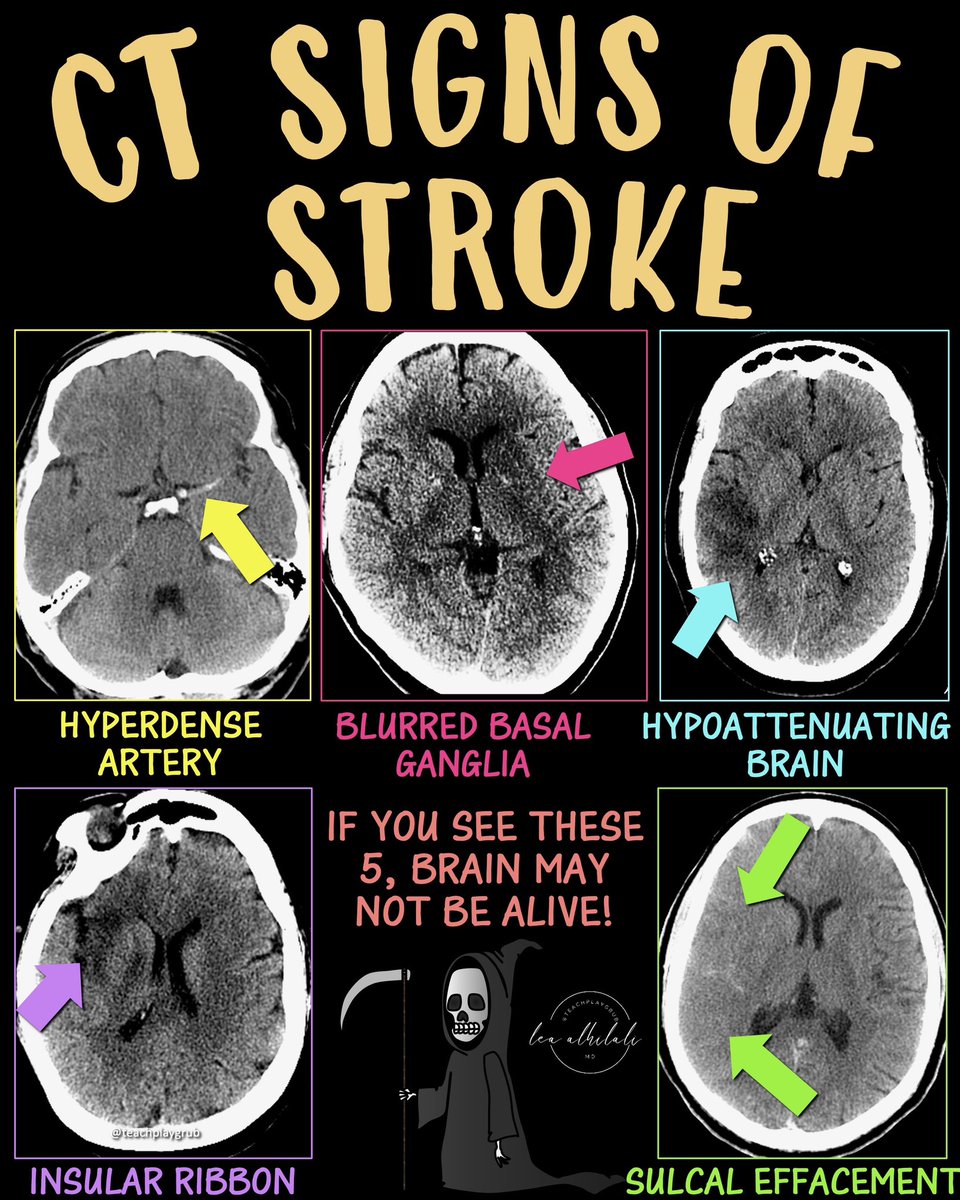
Time is brain!
So you don’t have time to struggle w/that stroke alert head CT.
Here’s are the FIVE main the CT findings in acute stroke.
--Hyperdense artery sign
Occurs when you see the thrombus in the artery.
Thrombus appears hyperdense bc clot is denser than normal flowing blood—& CT is just a measure of density.
--Blurred basal ganglia/lentiform nucleus.
Usually this region is a triangle of low density white matter surrounding the high density lentiform nucleus
In an acute infarct, this triangle becomes blurred, bc the lentiform nucleus becomes edematous & similar in density to white matter.
--Hypodense regions of brain
When O2 & ATP run out, Na/K pump stops working
Osmotic gradient causes Na & H20 rush into the cell.
More water in the cell = lower density. For every 1% increase in H20 there is a 2.5 HU decrease in density
Means damage is irreversible
--Insular ribbon
Insula is an internal MCA watershed between the lenticulostriates & M2 sylvian branches
Infarcts relatively early with low blood supply & becomes a low density ribbon
--Sulcal effacement
Normally, brain has sulci that look like ice cracks/crevasses along its surface.
As water accumulates in dead cells, swelling occurs, & the crevasses are effaced by the swollen brain
So now you know the 5 main signs of acute infarct on CT—remember, if you see these five, soon that brain won’t be alive!
8
170
772
57,867

On this day in 1952, Dr. Paul Zoll reported in NEJM the first description of two patients whose hearts were resuscitated using electric charges. Today, automatic external defibrillators (AEDs) are used everywhere to save lives. Learn more: nej.md/GUw39W
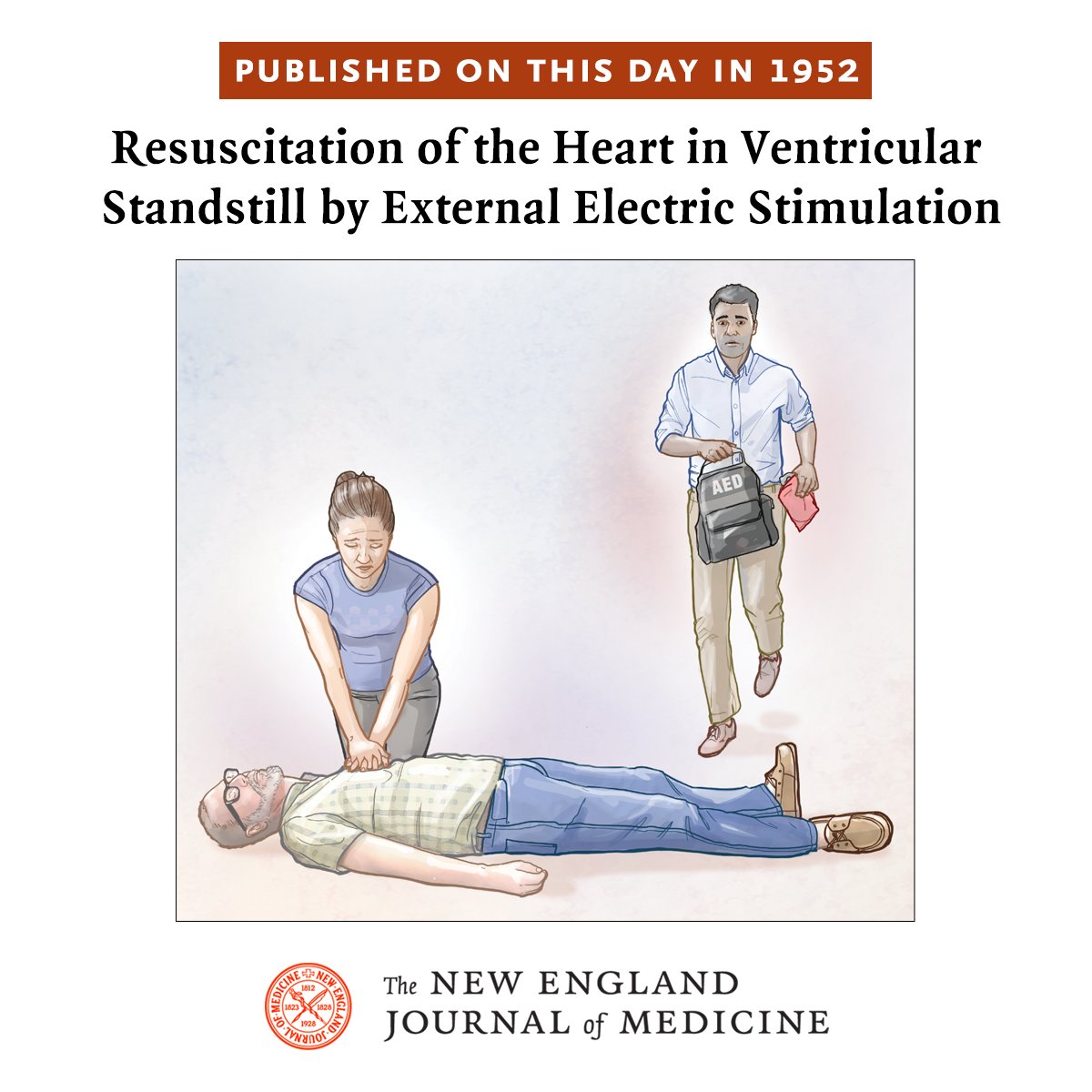
ALT PUBLISHED ON THIS DAY IN 1952 Resuscitation of the Heart in Ventricular Standstill by External Electric Stimulation A patient having an out-of-hospital cardiac arrest is attended to by a lay rescuer performing chest compressions while another lay rescuer is returning to the patient with an automated external defibrillator (AED).
7
174
406
51,636
David Barton retweeted

18 May 2024
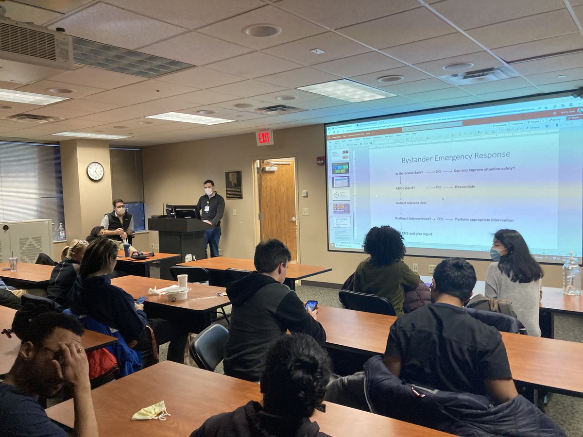
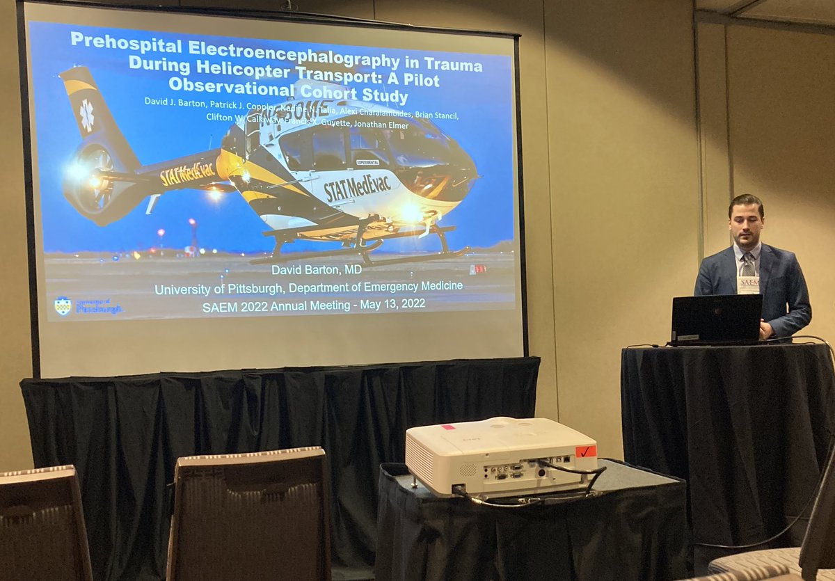
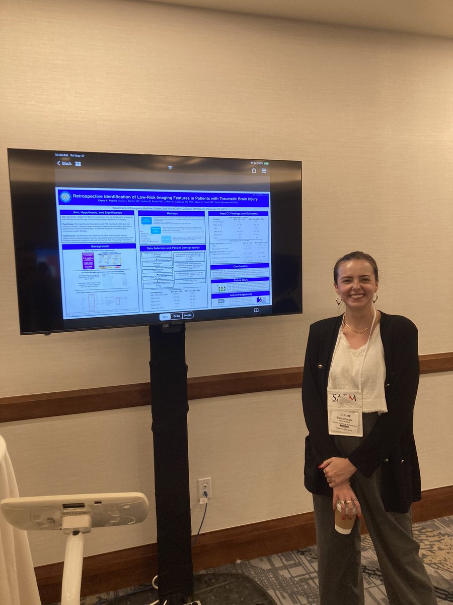
We had a great time and strong representation at #SAEM24! Our upcoming chief residents attended to train in leadership, faculty attended to lecture, and our med stud. presented a research poster leaving us all energized for the upcoming year! #SAEM @SAEMonline

1
7
701
David Barton retweeted

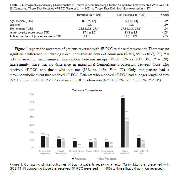
19 Apr 2024
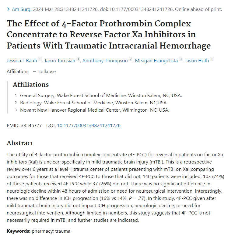
New study looking at mild tICHs (GCS 14-15) on Xa's and reversing (w/ 4PCC) vs not. No difference in hemorrhage progression or neuro outcomes btwn groups (some baseline diff in ISS)
I'll be discussing prior lit @EMPRx24 on this controversial topic
bit.ly/3U3BH5n
4
34
4,209

15 Mar 2024
Happy Match Day!
15 Mar 2024

We are incredibly excited to welcome our new Pitt EM class of 2027! Congratulations! #MATCHDAY

3
261
David Barton retweeted
15 Mar 2024
We are incredibly excited to welcome our new Pitt EM class of 2027! Congratulations! #MATCHDAY
7
36
7,876
David Barton retweeted

4 Feb 2024
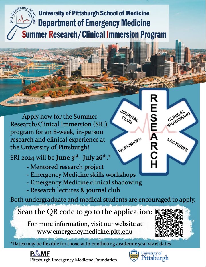
🙌🏽Exciting Opportunity @upmcem!
Summer Research/Clinical Immersion Program
6/3/24-7/26/24
Engage in hands-on research, EM skills workshops, and clinical shadowing. Open to undergrads & med students, including UiM folks
Apply by April 1
🔗 emergencymedicine.pitt.edu/s… @raindancers12
6
5
4,140
David Barton retweeted

24 Jan 2024
That's a wrap! The DEEP (Dsuvia Early Evaluation of Pain) study completed enrollment on 01/21/24. Thanks to everyone who facilitated this work, especially @Jjasonsperrymd, @Guyettef, @David_Barton, @macky_neal, @joshua_b_brown, and @PittCCM. 150 patients in less than 18 months!
3
9
877
David Barton retweeted

15 Jan 2024
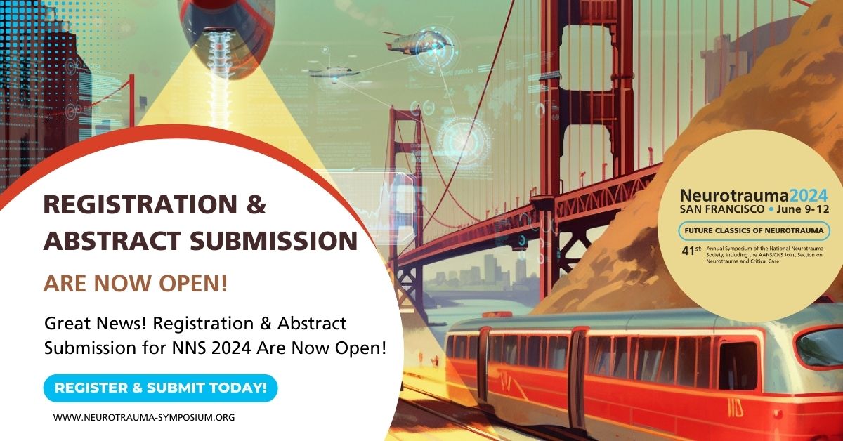
Exciting News! Registration and Abstract Submission for #NNS2024 are Now Open!
Secure your spot at the forefront of neurotrauma research and innovation. Mark your calendars and act fast!
👉 Register at neurotrauma-symposium.org/k6…
👉 Submit your abstract at neurotrauma-symposium.org/ik…
Don't miss the chance to connect with experts, explore groundbreaking research, and be part of the conversation shaping the future of neurotrauma.
#NeurotraumaSymposium
4
6
2,728
David Barton retweeted

2 Nov 2023
Nice article in Neurology Today about the history of concussion management.
journals.lww.com/neurotodayo…
4
8
686
David Barton retweeted

2 Nov 2023
Head injury in older adults presenting to the ambulance service: who do we convey to the emergency department, and what clinical variables are associated with an intracranial bleed? A retrospective case–control study sjtrem.biomedcentral.com/art…
17
56
6,210
David Barton retweeted
31 Oct 2023
Our awesome PGY-3 resident Dr. Annabelle Croskey recently published this great case report!
Ocular injury from saltwater coral palytoxin: A case report. American Journal of Emergency Medicine sciencedirect.com/science/ar… @David_Barton @WillTrautman @joshua_shulman @KatieRatay
2
5
675
David Barton retweeted

14 Oct 2023
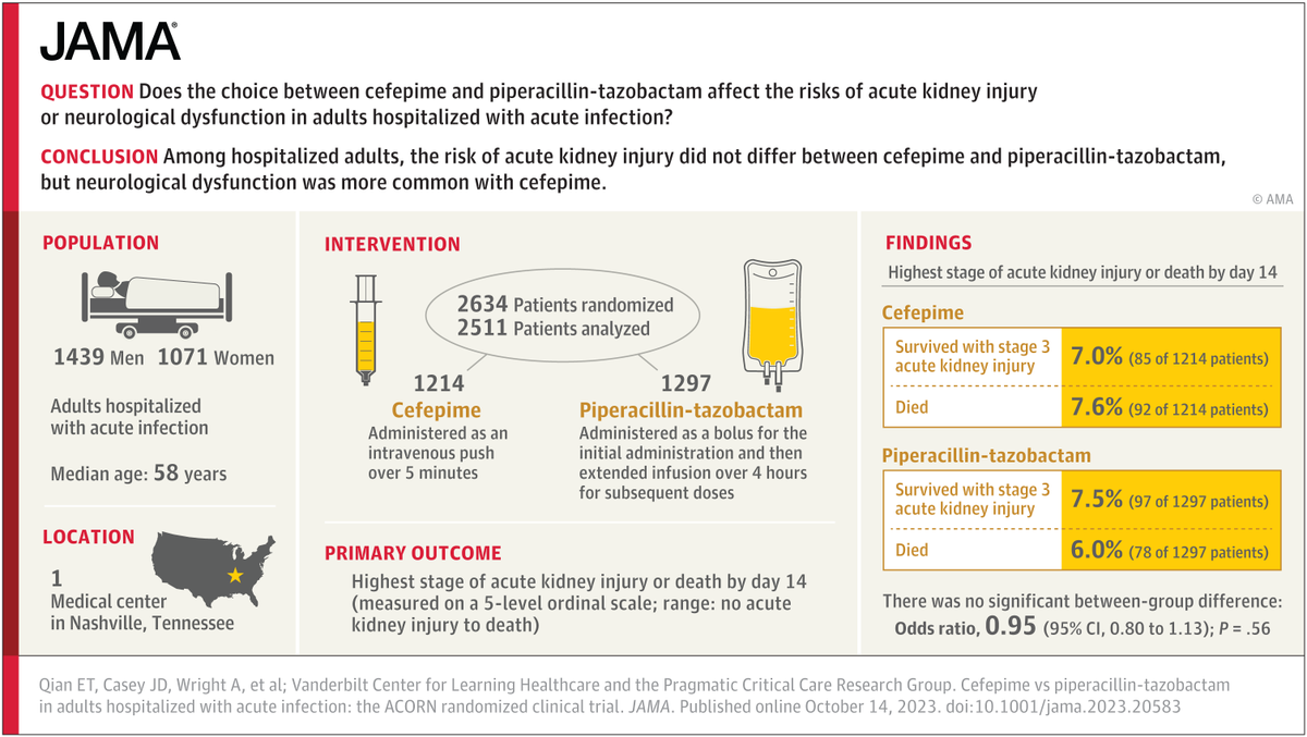
PIPERACILLIN-TAZOBACTAM IS NOT NEPHROTOXIC: exploring the arc of a myth over time
this was clearly a myth in 2016:
emcrit.org/pulmcrit/piperaci…
blog from 2022 exploring the evolution of this myth: emcrit.org/pulmcrit/myth-pip…
fresh RCT to finally settle this:
jamanetwork.com/journals/jam…

18
329
1,059
242,575
David Barton retweeted

3 Oct 2023
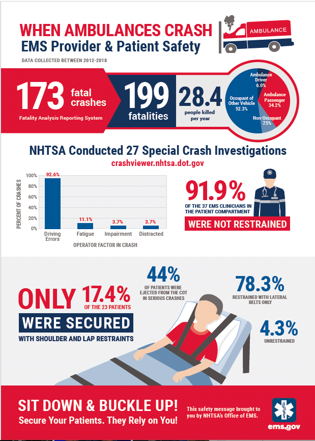
Having spent my career researching and teaching about this, these rates of failire to use seatbelts by EMS clinicians in thr patient compartment and failure to properly restrain patients with shoulder straps are sad. Be safe out there! @NAEMSP @NAEMT_ @NASEMSO @amerambassoc
3 Oct 2023

2
6
28
4,022
David Barton retweeted

3 Oct 2023
Hear @McDonaldStu_ @MonashTrauma talk with Patricia Karvelas @RadioNational re a new biomarkers study for diagnosis in #concussion in acute stages in the ED @Monash_FMNHS @AlfredHealth @Biswadev_M
#emergencymedicine #emergencytest #biomarker #braininjury abc.net.au/listen/programs/r…
1
5
18
2,723
14 Sep 2023
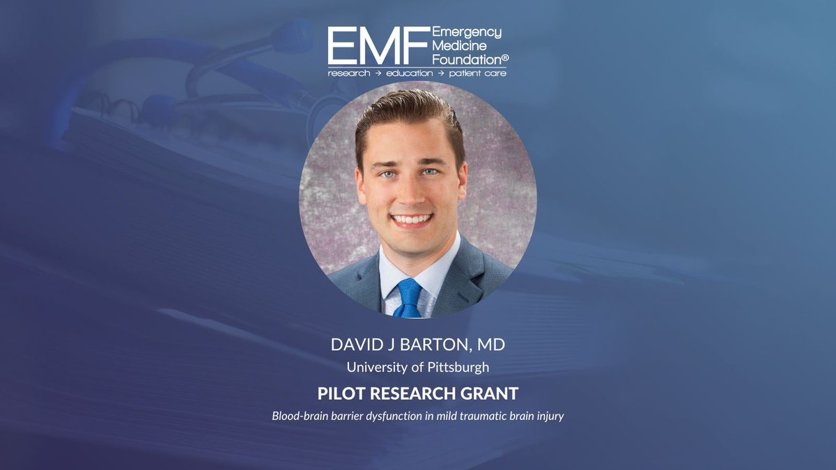
Many thanks to @TheEMFoundation for supporting our work and mission to improve care of patients with traumatic brain injury @upmcem @callaway3 @Rehabilomics4U @AshokPanigrahy6 @PittTweet
13 Sep 2023

EMF is proud to award Dr. David Barton with the Pilot Research Grant! Congratulations!
3
3
20
2,440