
Senior Lecturer in Bioinformatics @cardiffuni I am fascinated about how genomic variation leads to disease & how we can use this knowledge to improve treatment
Joined September 2011
- Tweets 4,248
- Following 420
- Followers 739
- Likes 5,080
364 Photos and videos


Geneticist J. Craig Venter, best known for his role in sequencing the human genome, has died aged 79.
He spoke to Nature in 2023 about AI, sequencing the ocean – and why he had no plans to stop working.
go.nature.com/4tHEf9M
17
251
669
80,920
Hywel Williams retweeted

Mar 13
👋We are hiring a computational PhD student, interested in functional genomics and the non-coding genome. To continue developing our BRAIN-MAGNET algorithm recently published in Cell (cell.com/cell/fulltext/S0092…). See vacancy for details!
werkenbijerasmusmc.nl/en/vac…
1
3
1
296
Hywel Williams retweeted

Jan 10
Another modern miracle in our lifetime

7
101
551
27,889

2 Dec 2025
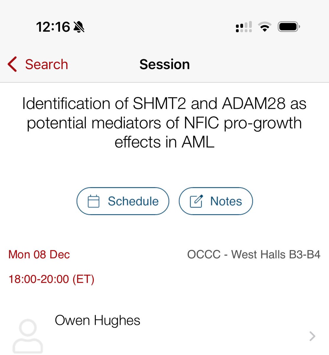
Good luck @alex_tonks and @OwenHughes42 at the #ASH2025 conference. Safe journey & make sure you take your suncream (I’m not jealous at all) 😎
2 Dec 2025

#ASH2025 preparations underway! Excited to share that @OwenHughes42 will be presenting his work from our group! Well done 👏! We are grateful to @ResearchWales for funding the study and American Society of Hematology for selecting his work. Orlando here we come @haematology_CU
118
24 Nov 2025
Amazing story about the power of a new gene-therapy treatment to help a little boy with the rare disease Hunter syndrome.
Still early days in this field but it feels like this will become common practise in the not too distant future?
bbc.co.uk/news/articles/c5y0…
3
4
228
10 Nov 2025
Really looking forward to today’s @GenomicsEngland conference on long-read sequencing & how it’s driving research through to clinical impact.
A fantastic line up of speakers, including the mighty @generoom
Great to see representation from @cardiffuni too.
1
48
Hywel Williams retweeted

1 Nov 2025
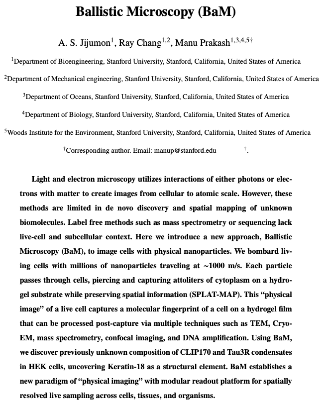
Finally got around to reading the "Ballistic Microscopy" paper, and it is really incredible.
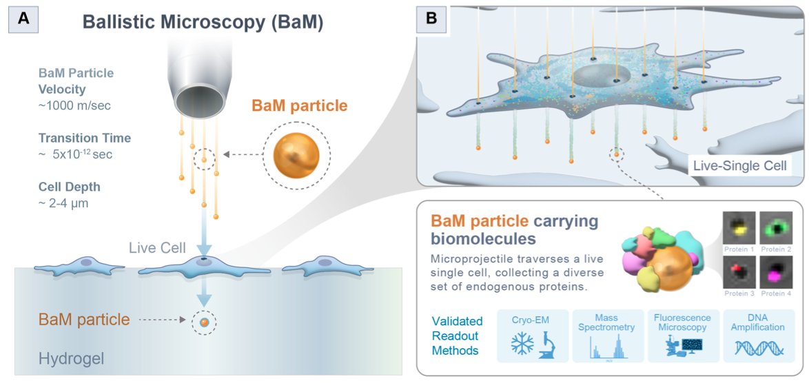
The paper opens with a compelling idea; one I hadn’t explicitly thought about before: “Light and electron microscopy utilizes interactions of either photons or electrons with matter to create images…” In other words, we see small objects by literally hurling things at them. Particles bounce off the object and reflect back into a lens, or scatter into a detector, which we then use to "see."
The question asked in this paper, then, is thus: Can we hurl even larger things at cells to image them? The answer is yes.
The gist of ballistic microscopy is that you first "bombard living cells with millions of nanoparticles traveling at ~1000 m/s." Each particle rips through the cell, picks up a tiny amount of cytoplasm, and comes out the other side.
If you place a hydrogel film underneath the sample, the nanoparticles will crash into it and get stuck there; just like shooting a bullet into a ballistics dummy. Finally, you take out these nanoparticles and study the molecules they carry, like by using mass spectrometry or really anything else.
This method preserves spatial information. The "nano-bullets" rip through the cell in a straight line, meaning that the pattern in the hydrogel corresponds with the nano-bullet's path through the cell. Nano-bullets embedded in the left side of the hydrogel will be carrying proteins, metabolites, and other "pieces" from the same side of the cell. So TL;DR, you're getting SPATIAL and MOLECULAR information, without having to label cells with anything.
"This is akin to a 'physical image' being captures on a hydrogel 'film'," the authors write, "with physical material captured on these nano-bullets."
Each bullet is between 50 to 1,000 nanometers in diameter. This is small but not exceptionally small. A typical E. coli bacterium measures about 2 micrometers long and 1 micrometer wide. Human cells are quite a bit larger.
The next step will be to increase the resolution of this method, perhaps by using smaller nanoparticles. But then there is a tradeoff; if the nanoparticles are TOO small, they need to be accelerated at much higher speeds or they won't penetrate cleanly, or their path of travel will get deflected and mess up the spatial information.
This first paper is just a proof-of-concept, of course. It reminds me a bit of Expansion Microscopy, at least in the narrow sense that it's a super creative, original solution to solving a problem.
In expansion microscopy, you use a swellable polymer gel to physically ENLARGE a biospecimen, rather than try to make a microscope that can see smaller objects. It's an inverse solution to the microscopy resolution problem. In the original expansion microscopy paper (from 2015), samples were only expanded ~4.5x in each dimension. More recent papers have upped this to ~20x in each dimension; a huge improvement.
I expect similar improvements for ballistic microscopy.
17
54
452
34,221
3 Oct 2025
If you're doing any form of research related to rare diseases in Wales this is a must-join network.
We need to work together to make us more than the sum of our parts, that way we can do real life-changing research and lead the way.
3 Oct 2025

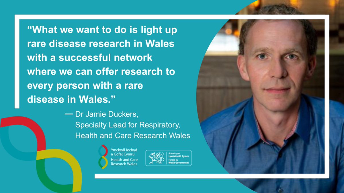
The Wales Rare Diseases Research Network connects patients, researchers, clinicians, policy makers and industry to transform rare disease research in Wales.
@DuckersJamie
Find out more the network and its future plans.
healthandcareresearchwales.o…
3
115
3 Oct 2025
This is a fascinating read about what's lurking in our genome.
A message from the dark genome @TheCrick crick.ac.uk/news/2025-10-02_…
1
2
222
23 Sep 2025
This is a must go conference for anyone working in the #Raredisease field in Wales 🏴
Come & see the breadth of research being done here & find like-minded people to network with.
23 Sep 2025
The Wales Rare Diseases Research Network is holding its first in-person meeting.
When: 26 September 2025
Where: Swansea University Bay Campus
Hear from @JamieDuckers, @SophiePearce and Alan Thomas (@AtaxiaAndMe).
Register now.
healthandcareresearchwales.o…
4
151
Hywel Williams retweeted

26 Jun 2025
25 years ago today, the first draft of the human genome was announced, a breakthrough that changed science forever 🧬
Read more about this achievement and the future of genomics over the next 25 years, here ⤵️
sanger.ac.uk/news_item/25-ye…
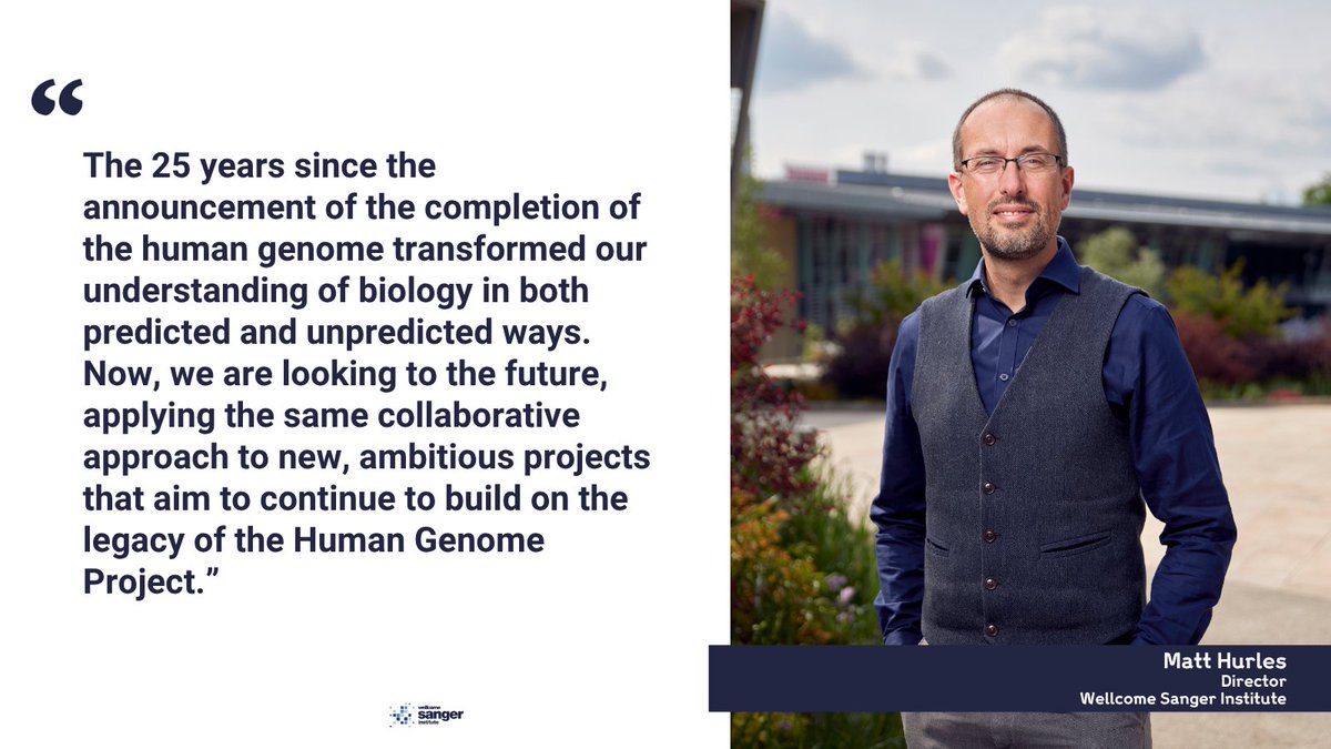
ALT Image of Matt Hurles, Director of the Sanger Institute on the right-hand side. Quote reads: ““The 25 years since the announcement of the completion of the human genome transformed our understanding of biology in both predicted and unpredicted ways. Now, we are looking to the future, applying the same collaborative approach to new, ambitious projects that aim to continue to build on the legacy of the Human Genome Project.”
1
10
38
2,440
Hywel Williams retweeted

18 Jun 2025
TOP STORY🗞️
A clinical-stage spinout company is launching with a $140 million (£107 million) investment – the most significant commercial investment into Welsh research to date.
@cardiffuni spinout Draig Therapeutics will launch with the investment from leading international venture investors to advance the development of novel therapies for major neuropsychiatric disorders such as major depressive disorder.
buff.ly/ZjXcFn5
1
2
2
2,865
Hywel Williams retweeted

16 Jun 2025
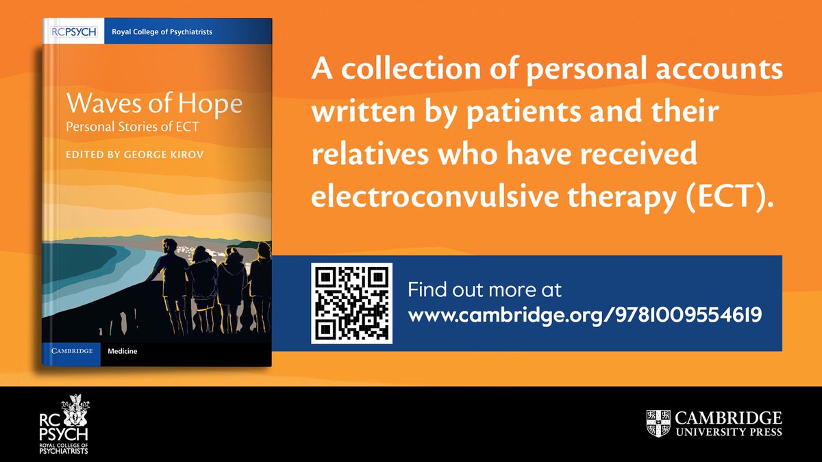
Our book on ECT is out. People who had ECT or their relatives came together to share their journeys through severe depression and ECT. I was amazed by their stories.
@DrAnnieHickox @ProfRobHoward @JunHongLi56447 @Joshua_RSmith @jonathanstea @JacquesSloman @DrT_Gergel
24
26
69
17,947
27 May 2025
Really excited, exhausted and relieved to finally be able to share the results of my analysis of the @GenomicsEngland 100,000 Genomes Project dataset.
Here's a link to free access to the manuscript:
authors.elsevier.com/a/1l8iU…
see below 🧵for main outcomes
1/4
1
4
11
758
27 May 2025
It's important because pre-filtering genomic data to identify these variants is a rapid way to diagnose patients, leaving more time to analyse additional patients.
If you don't have trio data don't worry, we show that 81% of variants in high CCRs are also pathogenic.
3/4
1
3
81
27 May 2025
Finally, we show that there is a significant enrichment (9.5x10⁻⁹) of these pathogenic variants in patients with neurodevelopmental disorders.
The reasons for this are unclear but suggests for such patients this would be a good first approach.
Pls read and RT
4/4
2
73
A baby boy with a devastating genetic disease is thriving after becoming the first known person to receive a bespoke, CRISPR therapy-for-one, designed to correct his specific disease-causing mutation
go.nature.com/4dkFKTV
7
170
563
37,558
Hywel Williams retweeted

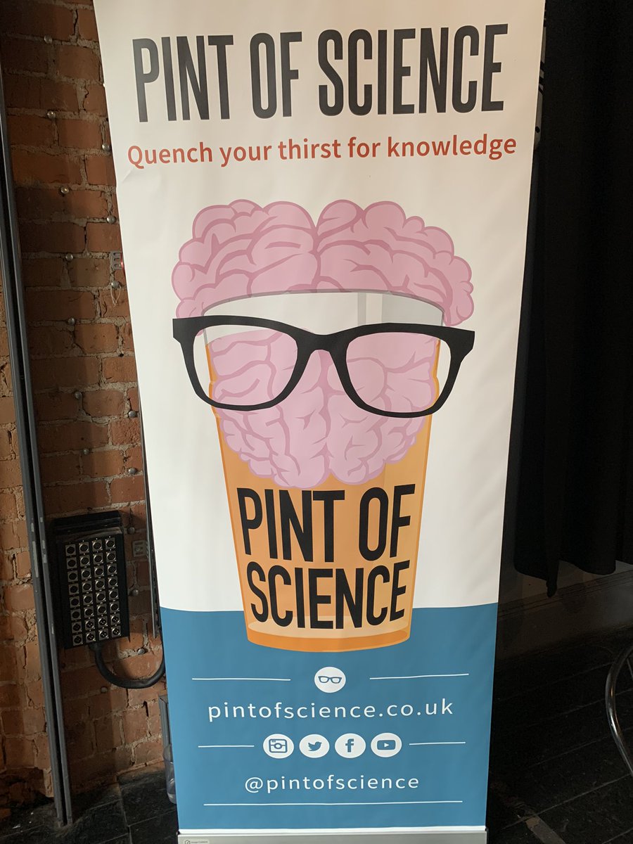
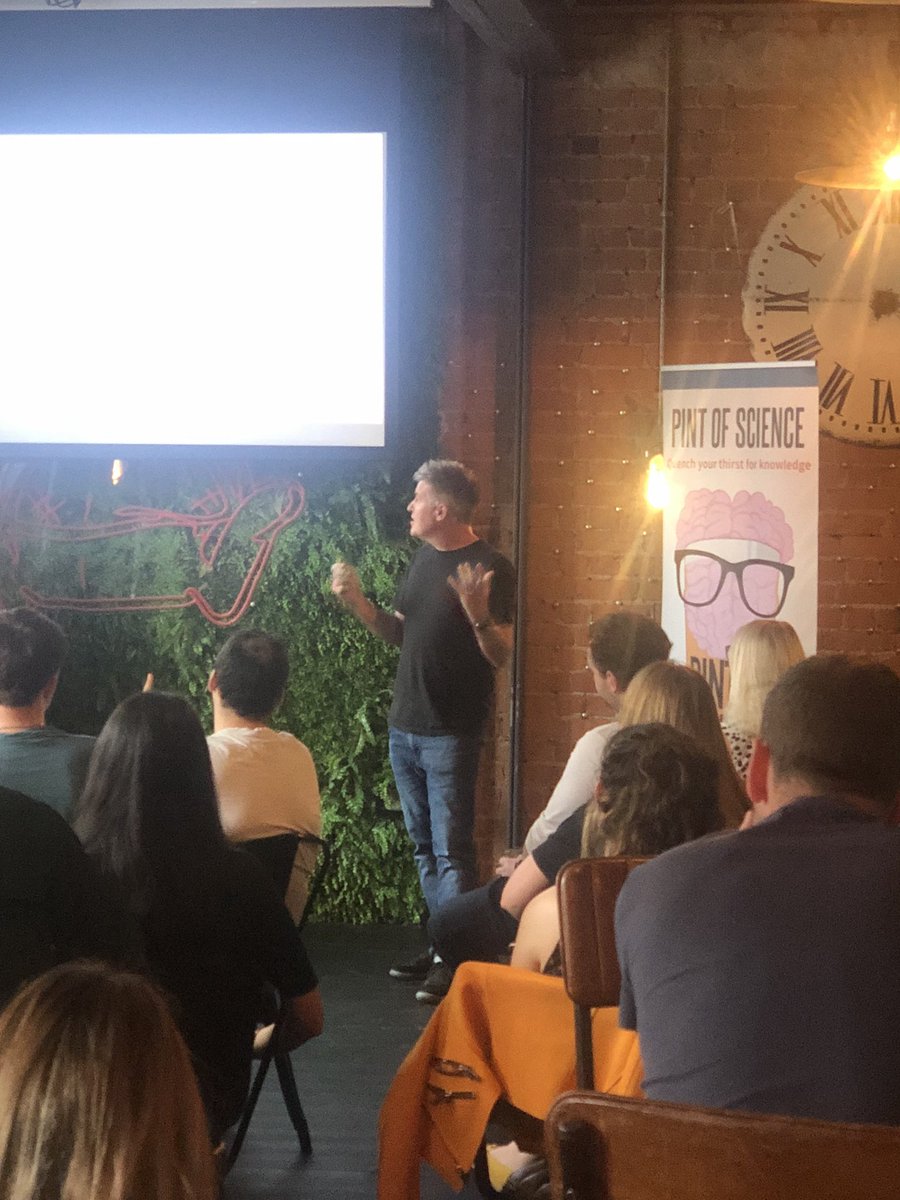
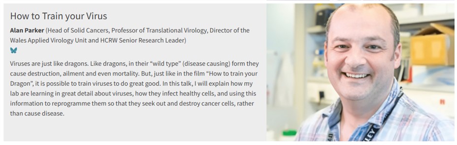
5 May 2025
Looking forward to my favourite #PublicEngagement event @pintofscience! I'll talk "how to train your virus" at @TinyRebelCdff on 20/05, discussing how we turned a disease causing "nasty" into a clinical stage anticancer medicine. Grab a pint, pop along! pintofscience.co.uk/event/fr…
8
11
685
Hywel Williams retweeted

20 Mar 2025
Postdoc position #1 is live. Please apply / spread the word!
lms.mrc.ac.uk/work/vacancies…
1
11
12
2,832