
We are a synthetic biology research group in the Bioengineering Department at Rice University.
Joined June 2018
- Tweets 162
- Following 367
- Followers 2,014
- Likes 788
14 Photos and videos


We are so excited that Jeff was named a 2023 #DoD Vannevar Bush Faculty Fellow, and look forward to participating in this unique and rewarding program!
news.rice.edu/news/2023/rice…
4
1
26
1,314
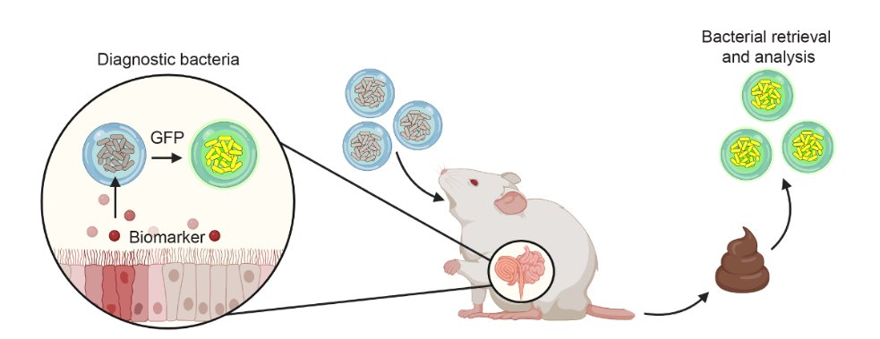
In this new paper, we collaborated with the wonderful @veiseho group to encapsulate engineered bacteria in hydrogel spheres to enhance their survival through the GI tract and ability to diagnose intestinal inflammation. Congrats to the team!
doi.org/10.1016/j.biomateria…
19
83
10,218
Tabor Lab retweeted

17 Jul 2023
Day 6 of great synthetic biology papers.
"A Synthetic Genetic Edge Detection Program," (2009).
Multiple genetic circuits were added to E. coli cells, endowing them with the ability to sense light, communicate with each other, and identify the light-dark edges.
A classic.
***
Yesterday, I told you about the @iGEM competition, and how students from UCSF and UT-Austin helped engineer E. coli into a sort of photographic film.
The truth is more complex, though: That iGEM team was actually working on this paper, the Edge Detector, but they were unable to get it to work.
Jeff Tabor, a member of the team (now a professor at Rice University), worked on the edge detector idea while in Andrew Ellington's lab at UT Austin, but later joined Chris Voigt's lab (then at UCSF) as a postdoctoral fellow to finish up the project. This paper was finally published in Cell in 2009.
The Edge Detector was, at the time, an incredibly complex genetic circuit. It was made by stitching together several smaller circuits. Getting this to work in living cells required the addition of many genes, and I'll do my best to break it down and explain the basic principles.
***
In computer science, edge detection algorithms are used to pick out the boundaries of objects within an image. They work by "scanning for a white pixel" in a digital image, "and then comparing the intensity of that pixel to its eight neighboring pixels," the study authors wrote.
"If any of the neighbors is black, the algorithm classifies those pixels as being part of an edge. The serial nature of this search process results in a computation time that increases linearly with the number of pixels in the image."
The question is, could such an algorithm be implemented in billions of cells at the same time? Could individual bacteria detect light and then communicate with neighbors to "find" an edge?
Yes. And here's how the biological edge detector works:
First, you grow bacteria on an agar plate. The cells divide, grow, and form a lawn.
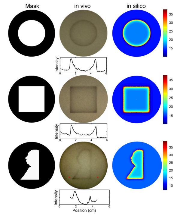
In the next step, a pattern of light is projected onto the cells. This pattern could be a star, or a circle, or an outline of George Washington. Anything with edges, really (see Figure 1 below).
Now, each bacterium carries genetic circuits, which are stitched together, such that a cell IN THE DARK is programmed to produce a diffusible signal.
This signal, a small molecule, exits out from the cells and seeps into the lawn of bacteria. When it encounters a cell IN THE LIGHT, that cell produces a BLACK PIGMENT. The edge appears.
The genetic pseudocode works out to something like:
> IF NOT light, produce signal.
> IF signal AND light, produce pigment.
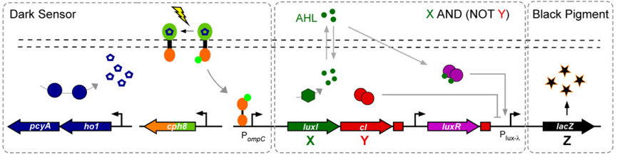
And that's it. This edge detection system was made by piecing together three different "genetic modules" — a dark sensor (which I described how to make yesterday), a form of cell-cell communication (via the diffusible signals), and X AND (NOT Y) genetic logic, to switch on the black pigment.
The full genetic circuit is presented below, as Figure 2. If this is your first time looking at a genetic circuit diagram, then let me explain what all the different components mean! Click on Figure 2 to enlarge it, and follow along...
A big arrow denotes a coding sequence; basically, a sequence of DNA that encodes a protein. The name of the gene is usually written on the arrow. In this case, luxI is a gene that encodes an enzyme that synthesizes acyl-homoserine-lactone, the small diffusible molecule.
Little circles or pentagons denote different types of molecules.
The small arrow next to each coding sequence denotes a promoter, which is a DNA sequence that proteins bind to in order to control gene expression.
Any arrows drawn between two objects denote movement or some kind of positive interaction, such as turning a promoter ON. If you see an arrow with a flat end, that means it is REPRESSING or turning a gene OFF.
The dotted lines near the top of the image represent the cell membrane. The AHL molecules move in and out of cells, through the membrane.
Until next time.
Paper: cell.com/cell/fulltext/S0092…
4
36
195
41,307
Tabor Lab retweeted
17 Jul 2023
Day 5 of great synthetic biology papers.
"Engineering Escherichia coli to see light" (2005).
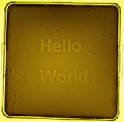
A 1-page paper that reports the "world's first camera that uses bacteria to take photographs."
I love this study because it is simple.
*****
The International Genetically Engineered Machine competition, or @iGEM, began at MIT in 2003. It was originally a one-month course that taught students how to make cells blink (using synthetic gene circuits from Day 3 of this series), but it has since grown into a global competition with more than 350 teams (old.igem.org/Previous_Compet…).
For a now-legendary iGEM project from the 2004 and 2005 seasons, a team from UCSF and the University of Texas - Austin engineered E. coli to detect light. The work was led by a graduate student, Anselm Levskaya.
Here's how the light-detecting bacteria were made:
First, they identified a photoreceptor protein in cyanobacteria, called phytochrome. Microbes use these proteins to control photosynthesis and to regulate the biosynthesis of protective pigments. E. coli don't naturally have them.
A phytochrome protein has two parts: A SENSOR that sits on the cell membrane and detects incoming light and a RESPONSE-REGULATOR, inside the cell, that acts as a sort of signal to tell the cell, "Hey, there's light!"
These response-regulators don't normally bind to DNA; they signal in other ways. But the students really wanted it to bind DNA so that they could use light to directly control genes.
So the team did something really clever. They fused the response-regulator bit of the phytochrome to a protein, called OmpR, that does bind to DNA and does control gene expression.
And that's when they ran into a problem!
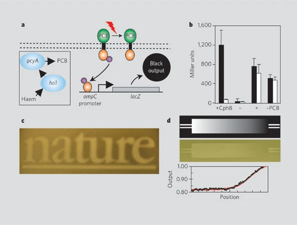
You see, the phytochrome proteins in cyanobacteria are only able to sense light because they have a molecule inside, called phycocyanobilin, that changes its shape when struck by photons. (This is similar to retinal, a form of vitamin A that is used in human opsin proteins to sense light.)
To make phycocyanobilin in the E. coli cells, the students added two additional genes: ho1 and pcyA. These genes, plucked from a freshwater cyanobacterium, encode enzymes that turn heme (yes, the stuff in blood) into phycocyanobilin.
Et voila! Their designs worked. As a final step, all they had to do was add a gene encoding LacZ, an enzyme that turns a molecule, called S-Gal, into a black color.
So now what happens is, if you grow bacteria on a plate of agar, they will spread and divide and slowly form an entire lawn that coats the surface. And if you add some S-gal, the plate will turn black.
But — and here's the fun bit — if you shine light on the cells, the phytochrome protein switches on (ping!), the response-regulator gets activated, and the OmpR protein shuts down the lacZ gene.
This means that cells exposed to light appear lighter. Unexposed cells appear darker.
And that's the whole paper. It's a clever bit of engineering!
If you'd like to try this out for yourself, all the genes and components are entirely open source (parts.igem.org/Featured_Part…). There is even a guide to make your own "Light Cannon" that can project custom images onto lawns of engineered bacteria (openwetware.org/wiki/LightCa…).
Paper: nature.com/articles/nature04…
ALT Image from http://parts.igem.org/File:HelloWorldColiroid.jpg
7
105
569
86,408
Tabor Lab retweeted

20 Jun 2023
Prof Christopher A. Voigt will be the next head of the MIT Department of Biological Engineering. Learn more about Prof Voigt at ow.ly/ZkVT50OSVBp @Geneticdesigner

1
27
252
86,685
Tabor Lab retweeted

28 Jun 2023
We worked with @synlogic to make therapeutic bacteria easier to program. The era of living medicines is coming fast.
13
61
8,326
Tabor Lab retweeted
16 Dec 2022
A marriage between Synthetic Biology and Synthetic Chemistry! Computationally design pathways to molecules by combining enzymatic and chemistry steps.
15 Dec 2022

I'm happy to share our work on computer-aided synthesis planning of hybrid enzymatic/synthetic reaction pathways with @Geneticdesigner and @cwcoley!
nature.com/articles/s41467-0…
4
56
6,658
Congrats Kathryn!
8 Dec 2022

Interested in bacterial peptide sensing? Check out my latest paper! Thanks to all of my amazing co-authors in
@LabTabor lab beyond: @synthetic_queer Andrew Mu @KGroszman @KyVanHoang @kevin_lorch @bhpogostin @DrGunn43 @jeffrey_j_tabor nature.com/articles/s41589-0…
11
Check out our new paper! We use E. coli display to characterize interactions between antimicrobial peptides and bacterial two-component systems. We discover numerous new human AMP activators of PhoPQ! Congrats @kathryn_brink and team. nature.com/articles/s41589-0…
1
9
49
Wonderful review for anyone interested in optogenetics beyond neuroscience and cell biology
14 Oct 2022

Here's our new article -- potentially, everything you ever wanted to know about light-regulated gene expression in bacteria (& much more).
frontiersin.org/articles/10.…
Welcome opportunity to team up once again with @RobertOhlendorf (this guy knows stuff!). Enjoy
7
Tabor Lab retweeted

20 Sep 2022
Please RT: Duke BME is recruiting a tenure track faculty member in #syntheticbiology , delivery of #complexbiologics, or #mechanobiology, each being broadly defined. academicjobsonline.org/ajo/j…
7
12
Tabor Lab retweeted

17 Sep 2022
Still time to apply y'all! In addition to exciting science, Rice and Houston are culturally rich and welcoming to all. We encourage applicants from diverse backgrounds to apply. We also have the best food in the world! #neurotwitter #AcademicTwitter @SfNtweets @FuturePI_Slack
19 Aug 2022

The Department of BioSciences @RiceUniversity is hiring an Assistant Professor in Neuroscience! Beyond the department, there are unique opportunities to engage with Neuroengineers @RiceNeuro and faculty at world class medical institutions @TXMedCenter. apply.interfolio.com/110165 PRT

6
6
Exciting!
8 Sep 2022

1/n PAPER ALERT: In collaboration with @davidrliu, we combined eVOLVER PACE (= ePACE) to facilitate the evolution of compact Nme2Cas9 variants that can target single-nucleotide (previously-inaccessible) PAM sequences for precision genome editing.
go.nature.com/3d1A9Y3
3
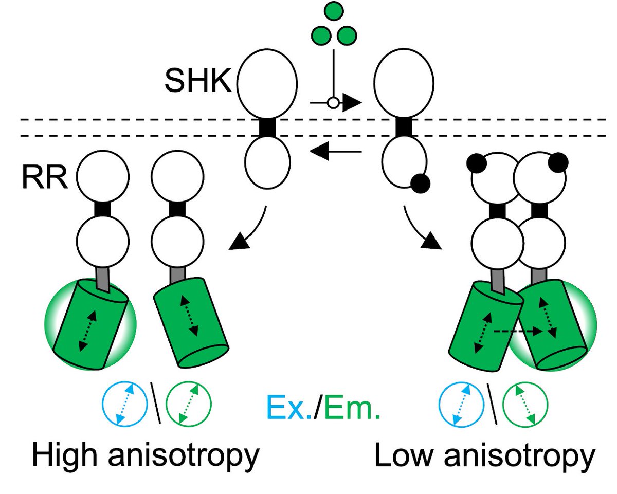
Bacteria phosphorylate response regulator proteins to survive stresses, colonize hosts, and resist antibiotics among other processes. Here, we report an optical method to monitor this process in live cells. Congrats @Rice_BIOE Ph.D. Ryan Butcher.
doi.org/10.1073/pnas.2201204…
4
18
81
Tabor Lab retweeted

29 Jun 2022
🚨Our lab has MULTIPLE funded postdoctoral positions open for different microbiome projects ranging from Cancer, Rare Earth Elements, and Anti-mold applications!! We build & use cool tech for microbiomes.🛠️🧬🦠 DM or email me at hw2429@columbia.edu to apply. Pls RT
2
167
231
Welcome, Reggie! The future is bright. We are so excited.
1 Jul 2022

Today marks my first day as president of Rice University. I started it out with a run around the beautiful Rice campus with my wife, Paula, and daughter, Shelby. I am grateful for the opportunity to serve such a distinguished institution. bit.ly/3OUndRc

12
We are looking for summer undergraduate researchers to work on a new NSF-funded project to develop a synthetic biology/optogenetics teaching module for high school classrooms, starting ASAP. Please reach out to Jeff Tabor if you are interested!
taborlab.rice.edu
6
79
210
Tabor Lab retweeted

9 May 2022
Deadline in 9 days!
22 Apr 2022

#RiceBIOE is looking for postdoctoral fellows and senior PhD students to become future leaders in bioengineering! bit.ly/31Pjth2
🗓️ The deadline to apply is MAY 18! 🗓️
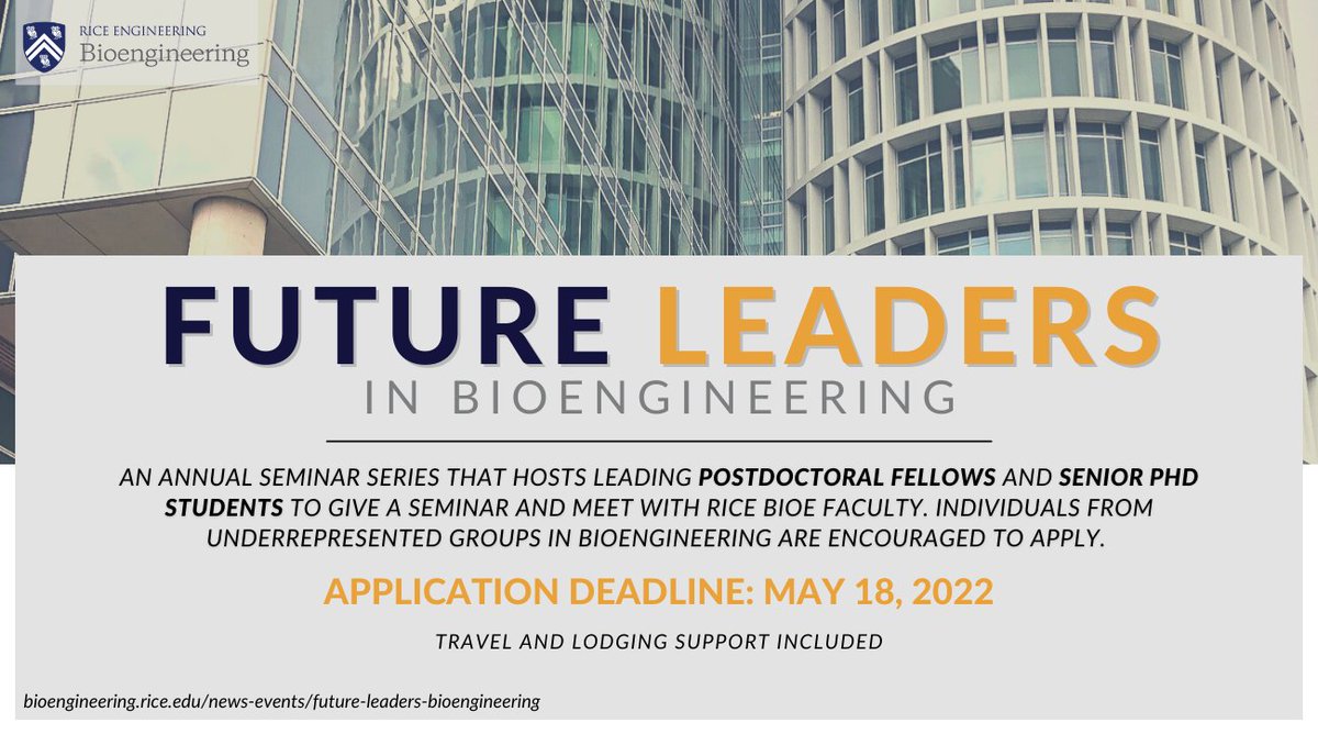
4
8
Tabor Lab retweeted

16 Feb 2022
Great chief of staff opportunity at a microbiome x food startup co-founded by the brilliant @raviusheth! A rare chance to create consumer products through cutting edge biotech. DM me if you're interested.
jobs.ashbyhq.com/kingdomsupe…
4
7