
Immune system formation in development 🧬 LDN-JA @ACRC_UK 🇯🇲 Views mine ✨ she/her
- Tweets 3,589
- Following 1,057
- Followers 3,090
- Likes 8,734


ALT Group of 10 people smiling at the camera, in front of a university building and a lot of greenery.


ALT Middle grade and picture book mentees: Bayley Mae is mentoring Elise Blaauw and Samira Lari. Taylor Pitts is mentoring Megan Donnelly. Rosario Martinez is mentoring Shanelle Webb and Tiffanie Leung Abbott.


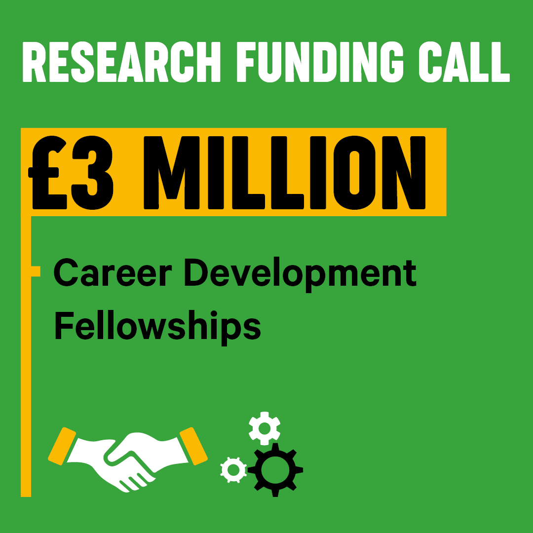
ALT Graphic promoting a research funding call with a budget of £3 million for Career Development Fellowships. The image features icons of a handshake and gears, set against a green background.


ALT Researcher Simone Webb sits with a cheerful expression sitting at a desk in a laboratory. She is wearing a green lanyard and is in front of a laptop and lab equipment.

